
Abstract
Background:
Long noncoding RNA (lncRNA) papillary thyroid carcinoma susceptibility candidate 3 (PTCSC3) inhibits several types of cancer, whereas its role in gastric cancer is unknown.
Materials and Methods:
From May 2010 to May 2015, this study included 68 (36 males and 32 females, 35–69 years, 48.3 ± 7.1 years) gastric cancer patients and 60 healthy volunteers (32 males and 28 females, 34–67 years, 48.8 ± 6.5 years). Transient transfections, QPCR, CCK-8, transwell cell migration, and invasion assay were used for carrying out the research.
Results:
The authors found that plasma PTCSC3 was downregulated in gastric cancer patients. Downregulation of PTCSC3 distinguished early stage gastric cancer patients from healthy controls. LncRNA PTCSC3 expression levels were increased on the day of discharge in gastric cancer patients compared with pretreatment levels. During follow-up, patients with low plasma levels of PTCSC3 showed a significantly lower overall survival rate. PTCSC3 negatively regulated proliferation, invasion, and migration of gastric cancer cells.
Conclusions:
Therefore, lncRNA PTCSC3 may serve as a biomarker for the treatment and prognosis of gastric cancer.
Introduction
Incidence of gastric cancer ranks 4th among all types of malignancies worldwide. It is also the second major cause of cancer mortalities due to its aggressive nature. 1 Developments of cancer therapies, such as endoscopic submucosal dissection (ESD), has significantly improved the treatment outcomes in some cases, however, only a small portion of gastric cancer patients can benefit from the technique due to the existence of metastatic tumor by initial diagnosis. 2,3 Therefore, early diagnosis is still important. 4,5 Accurate prognosis is also critical.
Despite the lack of protein-coding capacity, long (>200 nt) noncoding RNAs (lncRNAs) participate in both physiological processes and disease progression by regulating the expression of genes. 6,7 Altered expression of lncRNAs may lead to expression of downstream oncogenes or tumor suppressors in cancer cells. 8,9 LncRNA expression regulation is a promising therapeutic target for cancer. 10 Therefore, identification of the functions of lncRNAs in cancer is always needed. The tumor suppression role of lncRNA papillary thyroid carcinoma susceptibility candidate 3 (PTCSC3) has been mainly investigated in thyroid cancer and glioma, 11 –13 whereas its role in other types of cancer is unknown. This study was therefore performed to study the function of PTCSC3 in gastric cancer.
Materials and Methods
Research subjects
From May 2010 to May 2015, this study included 68 (36 males and 32 females, 35–69 years, 48.3 ± 7.1 years) gastric cancer patients and 60 healthy volunteers (32 males and 28 females, 34–67 years, 48.8 ± 6.5 years). This study passed the Review Board of Daqing Oilfield General Hospital (no. DOGH20103543). No significant differences in age, gender, and other basic clinical data were found between two groups of participants. Gastric cancer patients were diagnosed by X-rays combined with histopathological examinations.
Inclusion criteria of patients: (1) first time diagnosis; (2) no previous history of malignancy; and (3) no therapy performed before admission; (4) early stages of AJCC stage IA (n = 38), IB (n = 20), and IIA (n = 10). Exclusion criteria: (1) combined with other clinical disorders besides gastric cancer; and (2) who were transferred from other hospitals.
According to Dukes-MAC-like staging, there were 38 cases at A1, 17 cases at A2, and 13 cases at B1. The 68 patents included 28 cases of tubular cancer, 20 cases of papillary cancer, and 20 cases of mucinous cancer. The 60 healthy volunteers were selected to match the age and gender distributions of gastric cancer patients. All healthy controls received routine physiological exams and they showed normal physiological indexes. All healthy volunteers showed normal values of physiological parameters. All patients and healthy volunteers signed informed consent.
Plasma preparation, treatment, and follow-up
All patients were subjected to ESD combined with individualized chemotherapy (5-fluorouracil, platinum, and mitomycin C) and radiation therapy (γ-ray) and fasting blood (5 mL) was extracted from both patients (before treatment and on the day of discharge) and healthy controls. Blood was centrifuged in ethylenediaminetetraacetic acid (EDTA) tubes at 1200g for 12 min to prepare plasma. Patients were followed up for 5 years to record the survival of patients. Follow-up was performed every 2 months through phone call and outpatient visit.
Cells and transfection
SNU-1 and AGS human gastric cancer cell lines (AJCC) were used in this study to perform in vitro cell experiments. PTCSC3-expression pcDNA3.1 vectors were constructed by Sangon (Shanghai, China). Negative control (NC) miRNA and PTCSC3 siRNA were also from Sangon. Lipofectamine 2000 reagent (Invitrogen) was used to perform transient transfection with 10 nM vectors (empty vector as NC group) or 50 nM siRNA (NC siRNA as NC group) into 106 cells. Control (C) cells were untransfected. Lipofectamine 2000 was first mixed with vectors or siRNAs to form transfection mixtures, followed by the incubation with cells for 6 h at 37°C. After that, cells were washed with fresh medium to terminate transfections. Cells were harvested at 24 h after transfection to perform subsequent experiments.
Total RNA extraction and real-time quantitative polymerase chain reaction
Expression of PTCSC3 was detected through following steps: 1) total RNA extraction using TRIzol reagent (Invitrogen); 2) High-Capacity cDNA Reverse Transcription Kit (Bio-Rad) was used to perform reverse transcriptions to prepare cDNA using RNA samples with a ratio of 260/280 between 1.8 and 2.0 and a ratio of 260/230 between 2.0 and 2.2; 3) Applied Biosystems™ Power SYBR™ Green PCR Master Mix (Life Sciences) was used to prepare polymerase chain reaction (PCR) systems with cDNA as template and 18S rRNA as endogenous control; and 4) 2-ΔΔCT (14) method was used to perform data normalizations. Primer sequences were: 5’-TCAAACTCCAGGGCTTGAAC-3’ and 5’-ATTACGGCTGGGTCTAC-3’ for PTCSC3; 5’-GTAACCCGTTGAACCCCAT-3’ and 5’-CCATCCAATCGGTAGTAGC-3’ for 18S rRNA.
CCK-8 assay
Cells of both SNU-1 and AGS cell lines were collected at 24 h after transfection to prepare single cell suspensions (5 × 10⁴ cells/mL), followed by cell culture in a 96-well plate 1 (100 μL cell suspension per well) in an incubator (37°C, 5% CO2). CCK-8 solution (10 μL; Sigma-Aldrich) was added at 4 h before the end of cell culture. Finally, optical density (OD) values at 450 nm were measured.
Transwell assays
Cells of both SNU-1 and AGS cell lines were collected at 24 h after transfection and serum-free single-cell suspensions (5 × 10⁴ cells/mL) were prepared. Before invasion assay, Transwell upper chamber was coated with Matrigel (356234; Millipore). Single-cell suspensions (100 μL) were transferred to the upper chamber, and the lower chamber was filled with 100 μL mixture of 20% fetal bovine serum and 80% RPMI-1640 medium. Cells were cultivated for 3 h, followed by upper chamber membrane staining for 20 min with 0.5% Crystal Violet (Sigma-Aldrich) at room temperature. Finally, invading and migrating cells were observed under optical microscope.
Statistical analysis
Three biological replicates were included in each experiment. Data distribution is normal. Comparisons between two groups were performed by unpaired t-test. Comparisons between two time points were performed by paired t-test. One-way analysis of variance (ANOVA) and Tukey's test were performed for multigroup comparisons. Receiver operator characteristic (ROC) curve analysis was carried out to analyze the potentials of PTCSC3 in the diagnosis of early stage gastric cancer. Based on Youden's index and pretreatment PTCSC3 level, patients were divided into high (n = 33) and low (n = 35) PTCSC3 level groups. Survival curves were plotted and compared by K–M method and log rank t-test, respectively. Correlation between PTCSC3 expression and patients' clinical data was analyzed by chi-squared test. Differences with p-value <0.05 were statistically significant.
Results
Downregulation of plasma PTCSC3 in early gastric cancer patients had diagnostic potentials
Plasma levels of circulating PTCSC3 in both gastric cancer patients (n = 68) and healthy controls (n = 60) were measured by real-time quantitative PCR. The results were compared by unpaired t-test.
It was observed that plasma PTCSC3 was significantly downregulated in gastric cancer patients compared with the control group (Fig. 1A, p < 0.05). ROC curve analysis was carried out to analyze the potentials of PTCSC3 in the diagnosis of early stage gastric cancer. In ROC curve, patients were true positive cases and healthy controls were true negative cases. As shown in Figure 1B, area under the curve was 0.92 (standard error: 0.023; 95% confidence interval: 0.88–0.97). Chi-squared test showed that PTCSC3 was not significantly associated with patients' age, gender, and histological subtypes (Table 1), but was significantly correlated with AJCC stage (p = 0.03) and Dukes-MAC-like stage (p = 0.03).
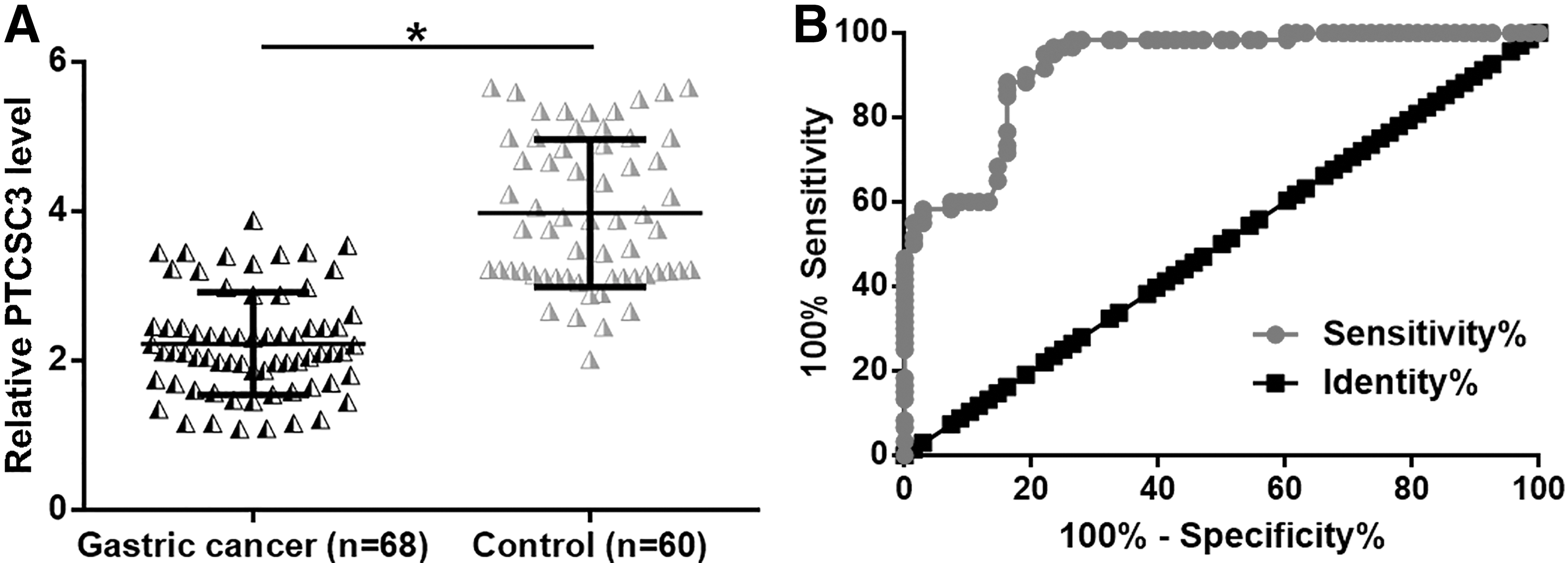
Downregulation of plasma PTCSC3 in early gastric cancer patients had diagnostic potentials. ROC curve analysis showed that PTCSC3 was downregulated in plasma of gastric cancer patients than in healthy controls
Correlation Between PTCSC3 Expression and Patients' Clinical Data
PTCSC3, papillary thyroid carcinoma susceptibility candidate 3.
Plasma levels of PTCSC3 were increased on the day of discharge
Plasma levels of circulating PTCSC3 in gastric cancer patients (n = 68) were also measured on the day of discharge. Pre and posttreatment levels of PTCSC3 were compared by paired t-test. Comparing pretreatment levels, lncRNA PTCSC3 expression levels were increased on the day of discharge (Fig. 2, p < 0.05).
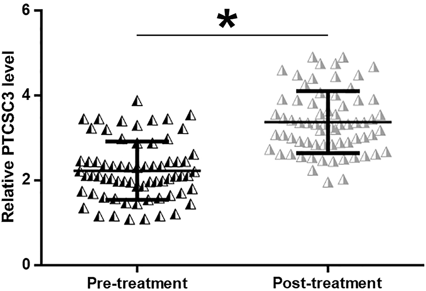
Plasma levels of PTCSC3 were increased on the day of discharge. RT-qPCR results showed that long noncoding RNA PTCSC3 expression levels were increased on the day of discharge in gastric cancer patients compared with pretreatment levels (*p < 0.05). RT-qPCR, real-time quantitative polymerase chain reaction.
Low plasma levels of PTCSC3 predicted poor survival
Based on Youden's index and pretreatment PTCSC3 level, patients were divided into high (n = 33) and low (n = 35) PTCSC3-level groups. No significant differences in clinical stages and therapies were found between two groups. The authors found that patients with low plasma levels of PTCSC3 showed a significantly lower overall survival rate compared with patients in the high plasma PTCSC3-level group (Fig. 3, p < 0.05).
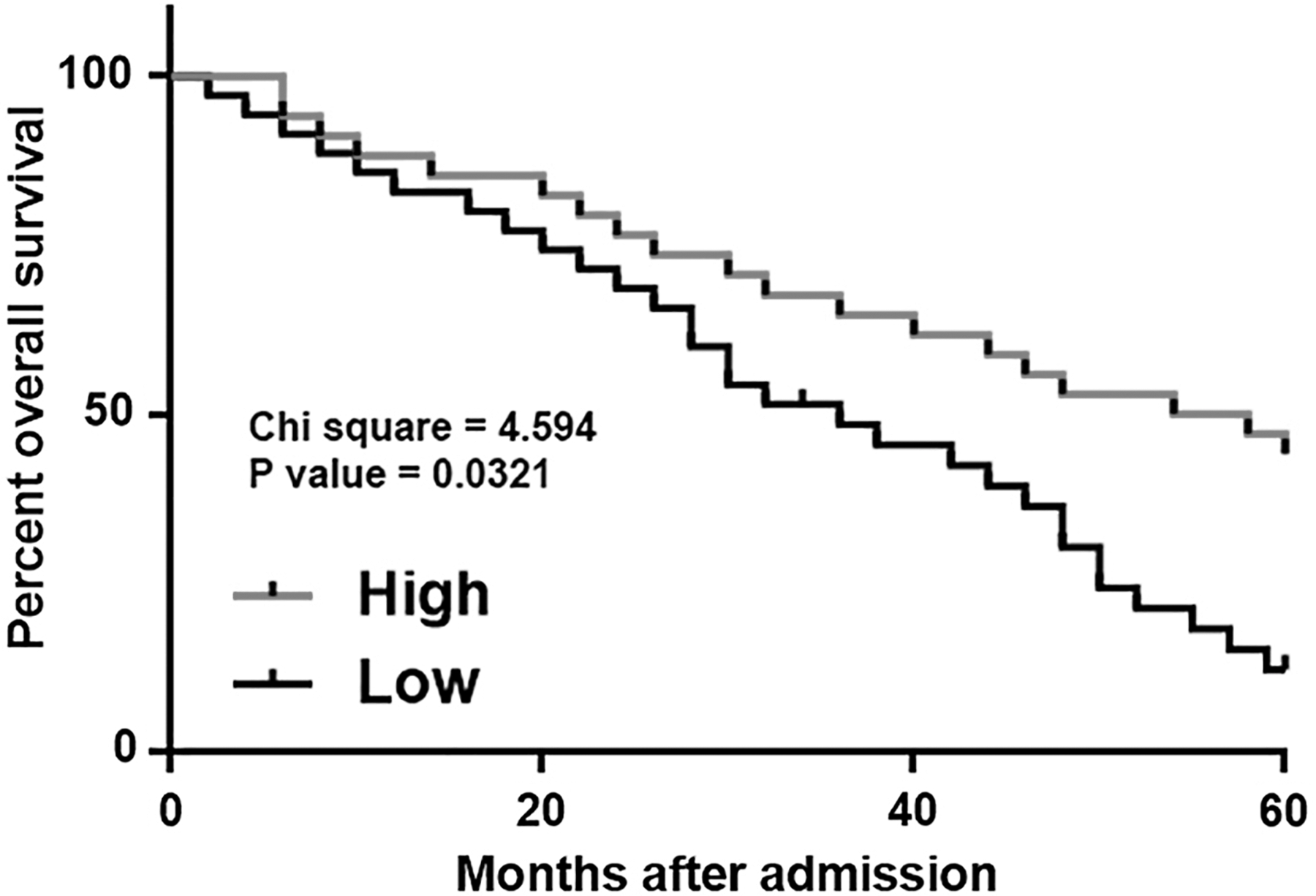
Low plasma levels of PTCSC3 predicted poor survival. Survival curve analysis showed that patients with low plasma levels of PTCSC3 had a significantly lower overall survival rate compared with patients with high plasma levels of PTCSC3.
PTCSC3 negatively regulated the behaviors of gastric cancer cells
To investigate the role of PTCSC3 in gastric cancer, CCK-8 assay was performed to study cell proliferation and Transwell migration, and invasion assays were performed to study cell migration and invasion after PTCSC3 overexpression and siRNA silencing in cells of both SNU-1 and AGS cell lines. Compared with two control groups (control, C; negative control, NC), PTCSC3 overexpression led to inhibited, whereas PTCSC3 siRNA silencing led to enhanced proliferation (Fig. 4), migration (Fig. 5A), and invasion (Fig. 5B) of gastric cancer cells (p < 0.05).
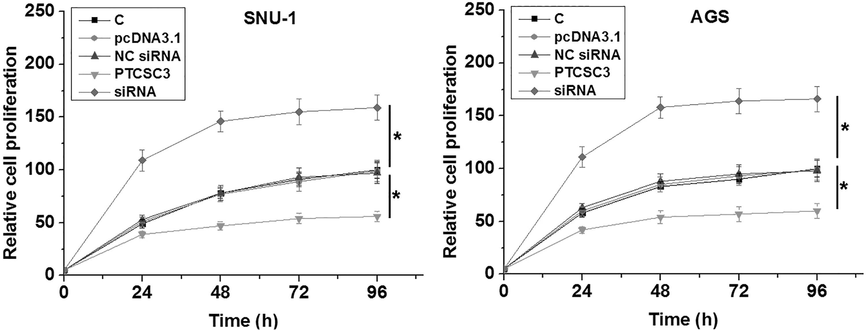
PTCSC3 overexpression caused the inhibited proliferation of gastric cancer cells. Compared with two control groups (control, C; negative control, NC), PTCSC3 overexpression led to inhibited, whereas PTCSC3 siRNA silencing led to enhanced proliferation of gastric cancer cells (*p < 0.05).

PTCSC3 overexpression caused the inhibited migration and invasion of gastric cancer cells. Compared with two control groups C, control; NC, negative control. PTCSC3 overexpression led to inhibited, whereas PTCSC3 siRNA silencing led to enhanced migration
Discussion
Previous studies have shown that PTCSC3 is downregulated in thyroid cancer and glioma and overexpression of PTCSC3 inhibits the cancer growth and metastasis, 11 –13 suggesting its role as a tumor suppressor. The authors' study first reported the involvement of PTCSC3 in gastric cancer and revealed its diagnostic and prognostic potentials.
Due to the lack of radical treatment approaches for metastatic cancer, early diagnosis is always critical for the treatment of different types of cancer, including gastric cancer. 14 At present, a considerable number of biomarkers have been developed to improve the early diagnosis of gastric cancer, such as CA 19-9, CEA, and imaging features. 4 However, application of these biomarkers is usually challengeable, owing to the insufficient sensitivity and/or specificity. With the advantage of noninvasive nature, plasma circulating lncRNAs are emerging novel biomarkers for cancer diagnosis. 15 This study showed that downregulated plasma levels of PTCSC3 in early stages gastric cancer patients distinguished them from healthy controls, indicating the clinical potentials of PTCSC3 in early diagnosis of gastric cancer.
Besides that, follow-up study also revealed that low PTCSC3 level is closely correlated with the high mortality rate after treatment. It is suggested that detection of the pretreatment levels of PTCSC3 may also has prognostic values for gastric cancer patients. The authors observed that PTCSC3 expression was not associated with patients' age, gender, and histological subtypes, but was significantly associated with patients' cancer stages. Therefore, PTCSC3 is likely downregulated with increase in clinical stages and the measurement of PTCSC3 expression may assist the prognosis of gastric cancer by clinical stages.
After treatment, plasma levels of PTCSC3 were reduced in gastric cancer patients. Although lncRNAs are usually expressed in certain types of cells, 16 they can also be released into the circulating system to participate in systemic gene expression regulation. 15 The downregulated expression of PTCSC3 cannot be explained by the resection of tumors because PTCSC3 may also be downregulated in tumor tissues. Therefore, the reduced plasma levels of PTCSC3 may be caused by the downregulated PTCSC3 in tissues after therapies.
Their in vitro cell experiments revealed the inhibitory effects of PTCSC3 overexpression on cancer cell proliferation, migration, and invasion, whereas the molecular mechanism is unclear. It has been reported that PTCSC3 can interact with multiple downstream oncogenic or tumor suppression pathways, such as STAT3/INO80 pathway and Wnt/β-catenin signaling pathway, to regulate cancer cell behaviors, and similar interactions may exist in gastric cancer. It is known that Wnt/β-catenin signaling is a critical player in the endothelial/mesenchymal transition (EMT) in solid tumors. 17 Therefore, PTCSC3 may also participate in the EMT of gastric cancer.
The authors' study is limited by the small number of patients included. In addition, no animal model experiments were performed. Their future study will try to establish animal models to further verify their conclusions.
Conclusions
In conclusions, PTCSC3 is downregulated in gastric cancer and has diagnostic and prognostic values. PTCSC3 overexpression can inhibit gastric cancer cell proliferation, migration, and invasion.
Ethics Approval and Consent
Ethics approval was obtained from the Ethics Committee of Daqing Oilfield General Hospital. The study followed the tenets of the Declaration of Helsinki, and informed written consent was obtained from all patients of the study.
Author Contributions
G.Z.: experiments work, data analysis, and writing the article. N.C., Q.L., and D.Z.: part of the laboratory work, clinical research, and article preparation. All authors reviewed and approved the final article.
Footnotes
Disclosure Statement
No competing financial interests exist.
Funding Information
No funding was received for this article.