
Abstract
Background:
MACC1-AS1 is an oncogenic lncRNA in gastric cancer, which interacts with AMP-activated protein kinase (AMPK) to promote cancer development. AMPK is known to interact with phosphatase and tensin homolog (PTEN). Therefore, MACC1-AS1 may also have associations with PTEN. This study aimed to investigate the interactions between MACC1-AS1 and PTEN in lung adenocarcinoma (LUAD).
Materials and Methods:
This study recruited 64 LUAD patients admitted to The First People's Hospital of Wenling City. Gene and protein expression levels were determined by qPCR and western blot, respectively. Cell transfections were performed to assess gene interactions. Cell proliferation was evaluated by CCK-8 assay.
Results:
MACC1-AS1 was upregulated in LUAD and inversely correlated with the expression of PTEN. High expression levels of MACC1-AS1 in LUAD tissues were closely correlated with poor survival rate of LUAD patients. In LUAD cells, overexpression of MACC1-AS1 led to decreased expression of PTEN and increased proliferation rate of LUAD cells, while MACC1-AS1 silencing led to increased expression of PTEN and decreased proliferation rate of LUAD cells. Furthermore, overexpression of PTEN attenuated the effects of overexpressing MACC1-AS1.
Conclusions:
The authors' results demonstrated that MACC1-AS1 promoted cell proliferation by downregulating PTEN in LUAD cells.
Introduction
The mortality rate of lung cancer is one of the highest among all types of malignancies. 1 Nonsmall cell lung cancer (NSCLC) is one of the major subtypes of lung cancer. With the application of targeted therapies and immunotherapies, the survival rate of NSCLC patients has been improved significantly during the past several decades. However, only less than 1/3 of NSCLC patients with metastatic cancer can live longer than 5 years after the initial diagnosis. 2 Most patients are diagnosed at advanced stages due to less obvious clinical symptoms during the early stage of NSCLC. 3,4 In addition, the pathogenesis of NSCLC is still unclear, 5 which limits the development of novel diagnostic and therapeutic approaches.
Phosphatase and tensin homolog (PTEN) is a critical regulator of cell cycle progression. 6 The main role of PTEN is to prevent cells from dividing and growing too rapidly. 7 In effect, mutations of PTEN are closely correlated with the occurrence and development of various types of cancer. 8 Therefore, activation of PTEN is a promising approach to suppress cancer by inhibiting cancer cell proliferation. 9
It has been reported that PTEN in cancer biology can be regulated by long (>200 nt) noncoding RNAs (lncRNAs), 10,11 which do not encode proteins but regulate gene expression. 12 lncRNAs are spatially and temporarily expressed to regulate the expression of tumor suppressors and oncogenes at multiple levels, thereby can either promote or inhibit cancer development. 12 MACC1 antisense RNA 1 (MACC1-AS1) is an oncogenic lncRNA involved in gastric cancer, 13 in which MACC1-AS1 interacts with AMP-activated protein kinase (AMPK) to promote cancer development, 13 Accumulative evidence suggested that AMPK can interact with PTEN. 14 Therefore, the authors hypothesized that MACC1-AS1 may also have crosstalk with PTEN. This study aimed to investigate the interaction between MACC1-AS1 and PTEN in lung adenocarcinoma (LUAD), a major subtype of NSCLC.
Materials and Methods
LUAD patients and follow-up
Research subjects of the present study include 64 LUAD patients (all were adenocarcinoma; 40 males and 24 females, with the age range of 22 to 68 years old and the mean age of 44.1 ± 12.0 years old). These patients were selected from the 217 LUAD patients admitted to the First People's Hospital of Wenling City between January 2011 and January 2014. These patients were diagnosed by histopathological biopsy. Inclusion criteria: (1) new LUAD cases; and (2) patients who were willing to participate in a 5-year follow-up. Exclusion criteria: (1) other severe clinical disorders were observed; (2) recurrent cases; (3) with unclear medical record; and (4) therapies were initiated before this study. The 64 patients included 18, 23, and 23 cases at clinical stage II, III, and IV, respectively. All the 64 patients were followed up for 5 years after admission. Follow-up was performed in a monthly manner through telephone and/or outpatient visits. Patients who were lost during follow-up period were excluded. Patients died of causes other than LUAD were also excluded. Before the recruitment of patients, the study was approved by the Ethics Committee of the First People's Hospital of Wenling City. All patients were educated with the details of experimental design, and the informed consent was obtained.
LUAD cells and tissues
Human LUAD cell lines H23, A549, and H1299 (adenocarcinoma; ATCC) were used in this study. RPMI-1640 medium (containing 10% fetal bovine serum [FBS]) was used for cell culture with the culture conditions of 37°C and 5% CO2. Biopsy was performed on all patients to collect noncancer (lung tissue within 2 cm around tumors) and LUAD tissues (0.012 to 0.016 g) before therapies. All tissues were tested by histopathological examination and all tissue specimens were correct.
Transient cell transfections
MACC1-AS1 (NCBI Accession: NR_046756.1) and PTEN (NCBI Accession: U93051.1) expression vectors were constructed using pcDNA3.1 vector (RIBOBIO, Guangzhou, China). Negative control (NC) siRNA (5′-UAAGGCUAGGAAGAGAUAC-3′) and MACC1-AS1 siRNA (5′-AUUGUAAAUGGUGUACUGAGG-3′) were also obtained from RIBOBIO. Cells were harvested and counted, followed by transfection of 10 nM vector (empty vector as a NC) or 35 nM siRNA (NC siRNA as NC) into 106 cells using Lipofectamine 2000 reagent (Sangon, Shanghai, China). The interval between the following experiments and transfections was 24 h.
RNA extractions and qPCR
Total RNAs in 106 H23 cells and 0.01 g tissue were extracted using TRIzol reagent (Invitrogen). All extracted RNA samples were digested with DNase I for 80 min at 37°C to remove genomic DNA. The Tetro Reverse Transcriptase (Bioline) was used for reverse transcriptions. The qPCR mixtures were prepared using QuantiTect SYBR Green PCR Kit (Qiagen, Shanghai, China). GAPDH was used as an endogenous control. Primer sequences were: 5′-CAGTCAGAAAATGAGGAACA-3′ (forward) and 5′-TTCTTGCTTCGATGCAACCA-3′ (reverse) for MACC1-AS1; 5′-CAGTCAGAAAATGAGGAACA-3′ (forward) and 5′-TTTACAGCCCCGATTGG-3′ (reverse) for PTEN; and 5′-GTCTCCTCTGACTTCAACAGC-3′ (forward) and 5′-CCACCCTGTTGCTGTAGCCAA-3′ (reverse) for GAPDH. It was noted that multiple endogenous controls, such as β-actin were used and similar results were obtained. All reactions were repeated three times and Ct values were calculated using the 2−ΔΔCT method.
Western blot
Total proteins in 106 H23 cells were extracted using RIPA solution (Sangon). All protein samples were first denatured in boiled water for 10 min, followed by electrophoresis using 10% SDS-PAGE gel (30 μg protein per well). Following gel transfer (to polyvinylidene difluoride membranes) and blocking (1 h in phosphate-buffered saline containing 5% nonfat milk at room temperature), first blotting was performed using rabbit primary antibodies of PTEN (ab31392, 1:900; Abcam) and GAPDH (ab9485, 1:900; Abcam), and the incubation was performed at 4°C overnight. Secondary blotting was performed using IgG-HRP secondary antibody (MBS435036, 1:900; MyBioSource), and the incubation was performed at room temperature for 2 h. CL (Sigma-Aldrich) was used for signal production, and gray values were normalized using the ImageJ v1.46 software.
Cell proliferation analysis
H23, A549, and H1299 cells were harvested and counted. A total of 3 × 10⁴ cell were mixed with 1 mL RPMI-1640 medium (containing 10% FBS) to prepare single-cell suspensions. In a 96-well plate, H23 cells were cultivated at 37°C with 0.1 mL cell suspension per well (three replicate wells were set for each experiment), followed by the addition of 10 μL CCK-8 solution at 4 h before the end of cell culture. After the termination of cell culture, 10 μL DMSO was added into each well, and optical density (OD) values were measured at 450 nm wavelength. OD values of control (C) group at 96 h were set to 100%, and all other groups were normalized to C groups.
Statistical analysis
All experiments, including qPCR, western blot, and cell proliferation assay, were performed in at least three biological replicates. Analysis of variance (one-way) combined with Tukey's test and paired t-test were used to explore differences among multiple cell groups or between two different types of tissues (noncancer and LUAD), respectively. All correlation analyses were performed using linear regression. With the median expression levels of MACC1-AS1 and PTEN as cutoff values, the 64 patients were divided into high and low groups (n = 32). K-M plotter was used to draw survival curves, which were compared by the log-rank test. p < 0.05 was statistically significant.
Results
Inverse correlation between the expression of MACC1-AS1 and PTEN in LUAD
The expression levels of MACC1-AS1 and PTEN in noncancer and LUAD tissues were evaluated by qPCR. The expression levels of MACC1-AS1 were significantly higher (Fig. 1A), while the expression levels of PTEN were significantly lower (Fig. 1B) in LUAD tissues compared to that in noncancer tissues (p < 0.05). Linear regression showed inverse correlation between the expression of MACC1-AS1 and PTEN in LUAD tissues (Fig. 1C), while the correlation between them was not significant in noncancer tissues (Fig. 1D).

Inverse correlation between the expression of MACC1-AS1 and PTEN in LUAD. Expression levels of MACC1-AS1
MACC1-AS1 and PTEN mRNA expression predicted poor survival
With the median expression levels of MACC1-AS1 and PTEN as cutoff values, the 64 patients were divided into high and low groups (n = 32). K-M plotter was used to draw survival curves. Compared to patients in the low MACC1-AS1 level group, patients in high MACC1-AS1 level group showed a significantly lower overall survival rate (Fig. 2A). In contrast, the overall survival rate of patients in the high PTEN group was significantly higher than patients in the low PTEN group (Fig. 2B).

MACC1-AS1 and PTEN mRNA expression predicted poor survival. With the median expression levels of MACC1-AS1
MACC1-AS1 negatively regulated PTEN in H23 and A549 cells
MACC1-AS1 expression vector and siRNA were transfected into H23 cells. Overexpression of MACC1-AS1 was confirmed by qPCR. As shown in Figure 3A, compared to the NC and C group, expression level of MACC1-AS1 was significantly increased after transfection with MACC1-AS1 expression vector, and it was significantly decreased after siRNA transfection (p < 0.05). Moreover, compared to C and NC groups, expression level of PTEN mRNA and protein were significantly decreased after transfection with MACC1-AS1 expression vector, and these were significantly increased after siRNA transfection. Furthermore, A549 cells were also transfected with MACC1-AS1 expression vector and siRNA. Transfections were confirmed by qPCR (Supplementary Fig. S1A). Similarly, MACC1-AS1 negatively regulated expression levels of PTEN mRNA and protein in A549 cells (Supplementary Fig. S1B).
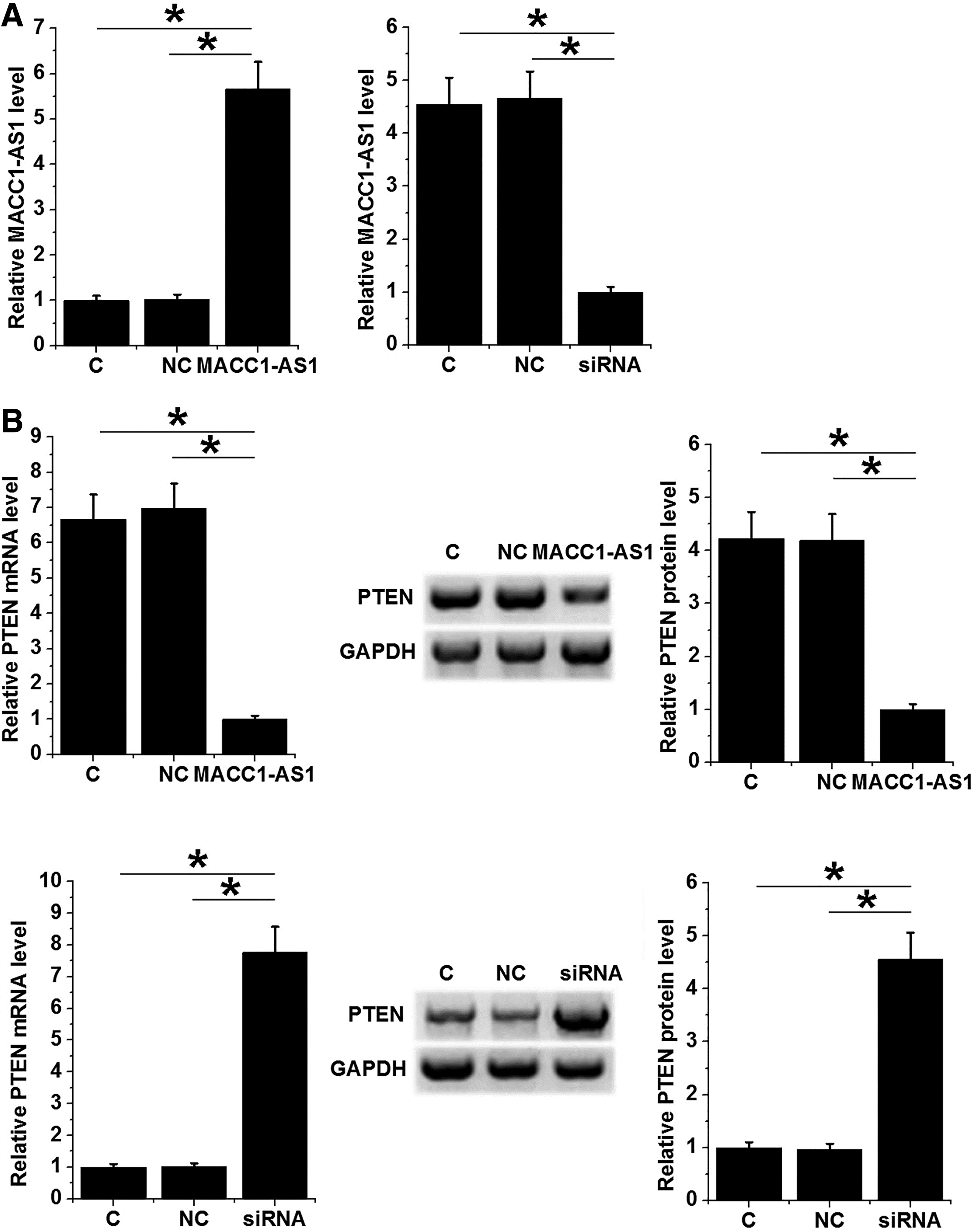
MACC1-AS1 negatively regulated PTEN in H23 cells. MACC1-AS1 expression vector and siRNA were transfected into H23 cells. Overexpression of MACC1-AS1 was confirmed by qPCR
MACC1-AS1 promoted proliferation of H23, A549, and H1299 cells through PTEN
Cell proliferation was evaluated after transfections. Compared to C and NC group, overexpression of MACC1-AS1 led to increased, while silencing of MACC1-AS1 led to decreased proliferation rate of H23 (Fig. 4A, p < 0.05), A549 (Fig. 4B, p < 0.05) and H1299 (Fig. 4C) cells (p < 0.05). In addition, overexpression of PTEN played the opposite role and attenuated the effects of overexpressing MACC1-AS1 (p < 0.05).
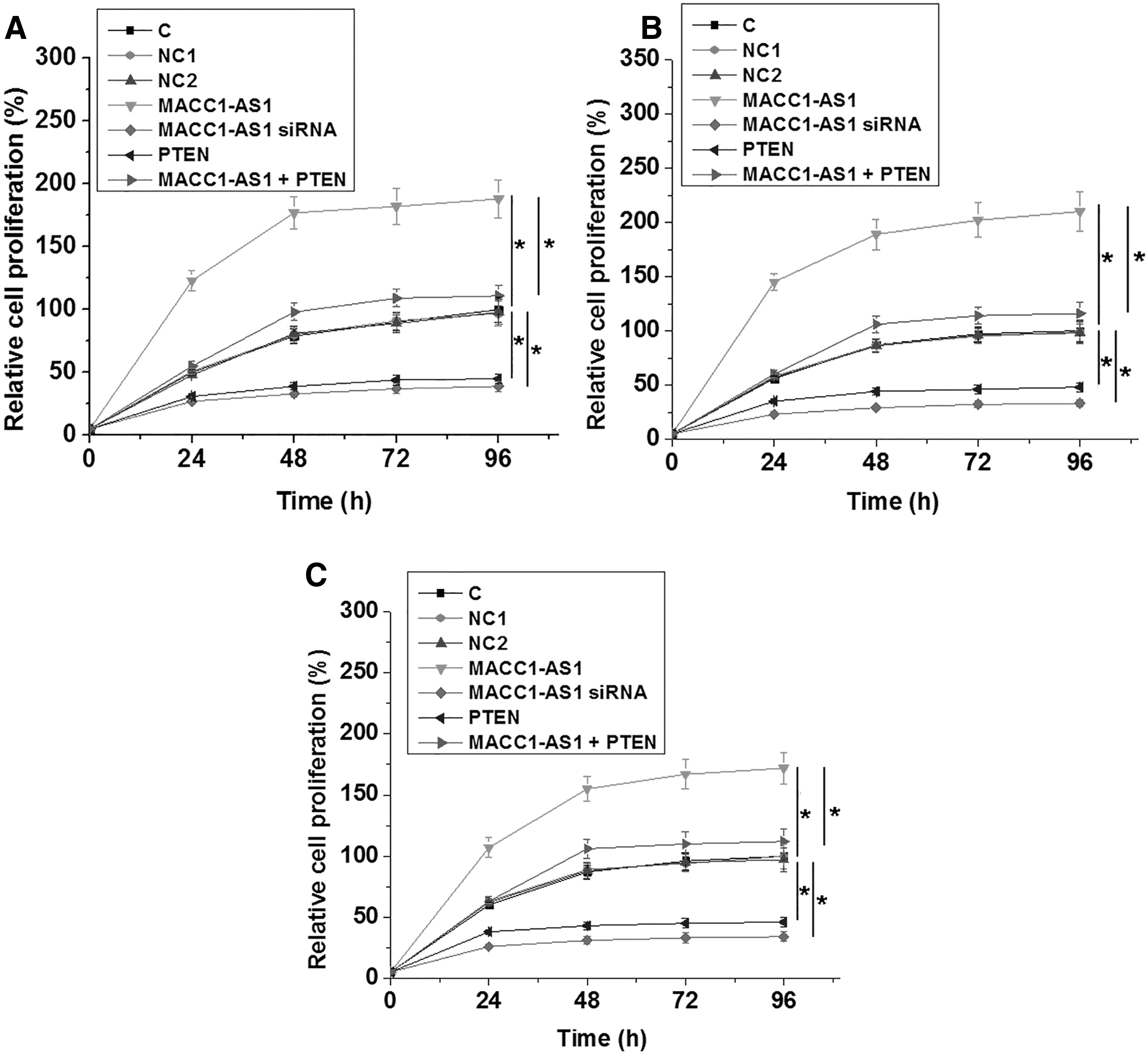
MACC1-AS1 promoted the proliferation of LUAD through PTEN. Cell proliferation was analyzed after transfections by performing cell proliferation assay. The effects of MACC1-AS1 and PTEN overexpression as well as MACC1-AS1 siRNA on H23
Discussion
The function of MACC1-AS1 in LUAD has been investigated in this study. The authors' results showed that MACC1-AS1 was upregulated in LUAD and promoted the proliferation of LUAD cells by downregulating PTEN, which is a critical player in cell cycle regulation.
The functions of MACC1-AS1 have only been investigated in gastric cancer, 13,15 in which MACC1-AS1 interacts with AMPK, Lin28, and MACC1 to promote cell metabolic plasticity. 13 MACC1-AS1 also regulates fatty acid oxidation in cancer cells to promote cancer cell stemness and chemoresistance. 15 The development of LUAD also requires the involvement of multiple lncRNAs. For instance, AFAP1-AS1 is upregulated in LUAD and promotes the invasion and migration of cancer cells by upregulating AFAP1-AS1. 16 To the best of the author's knowledge, this study is the first report to upregulated expression pattern of MACC1-AS1. The authors also showed the positive regulation of LUAD cell proliferation by MACC1-AS1. Therefore, MACC1-AS1 is an oncogenic lncRNA in LUAD. It is worth noting that their preliminary data showed that MACC1-AS1 had no significant effects on LUAD cell invasion, migration, and apoptosis (data not shown). Therefore, MACC1-AS1 may have different functions in different types of cancer cells.
In this study, the authors proved that MACC1-AS1 can negatively regulate PTEN in LUAD cells. Through bioinformatics analysis, they observed no promising binding site of MACC1-AS1 on the promoter region of PTEN (data not shown). In addition, they found that expression levels of MACC1-AS1 and PTEN were only significantly and inversely correlated in LUAD tissues but not in noncancer tissues. Therefore, the interaction between MACC1-AS1 and PTEN is likely to be indirect. It is known that both MACC1-AS1 and PTEN have interactions with AMPK. 13,14 Therefore, AMPK may mediate the interaction between MACC1-AS1 and PTEN in lung cancer. The authors' future studies will test this possibility and explore other possible mechanisms.
LUAD patients are suffering for the low survival rate. 17 Due to the low specificity and sensitivity of early diagnostic markers, early diagnostic rate of LUAD is low and this situation may not be changed within short-term. 18 Therefore, an accurate prognostic assignment may be an alternative approach to improve patients' survival. In this study, they showed that high levels of MACC1-AS1 and low levels of PTEN in LUAD were closely associated with the poor survival of LUAD patients. Their future studies will further analyze their prognostic values for LUAD.
Conclusion
In conclusion, MACC1-AS1 was upregulated in LUAD and may promote LUAD cell proliferation by downregulating PTEN.
Ethical Approval and Informed Consent
Ethical approval was obtained from the Ethics Committee of The First People's Hospital of Wenling City and it conforms to the provisions of in accordance with the Declaration of Helsinki as revised in 2013. Written informed consent was obtained from all individual participants included in the study.
Footnotes
Authors' Contributions
W.W.M.: Conception and design, Collection and assembly of data, Data analysis and interpretation, Article writing and final approval of the article; T.J.L.: Administrative support, Conception and design, Research design, Article writing and final approval of the article. All authors read and approved the final article.
Disclosure Statement
No competing financial interests exist.
Funding Information
No funding was received for this article.
Supplementary Material
Supplementary Figure S1
References
Supplementary Material
Please find the following supplemental material available below.
For Open Access articles published under a Creative Commons License, all supplemental material carries the same license as the article it is associated with.
For non-Open Access articles published, all supplemental material carries a non-exclusive license, and permission requests for re-use of supplemental material or any part of supplemental material shall be sent directly to the copyright owner as specified in the copyright notice associated with the article.