
Abstract
Cancer Biotherapy and Radiopharmaceuticals
officially retracts the paper entitled, “Long Noncoding RNA HOTAIR Contributes to Progression in Hepatocellular Carcinoma by Sponging miR-217-5p,” by Ximing Gong and Zhenya Zhu (Cancer Biother Radiopharm 2020;35(5):387–396; doi: 10.1089/cbr.2019.3070) due to the discovery that the paper was submitted from a paper mill, which is a violation of the journal's standard protocols.
The Editor and Publisher of Cancer Biotherapy and Radiopharmaceuticals are committed to preserving the scientific literature and the community it serves and does not tolerate any violations of scientific misconduct.
Introduction
Hepatocellular carcinoma (HCC) is an aggressive primary hepatic cancer with high malignancy and poor prognosis. 1 Hepatocarcinogenesis is associated with multiple etiological factors, such as alcohol abuse, epigenetic alterations, hepatitis cirrhosis, and virus infection. 2 –4 Till now, surgical resection and liver transplantation are still the preferred and effective therapies for early stage of HCC patients, but the recurrence rate is >65%. 5,6 In addition, most patients are diagnosed at advanced stage that vitiates the therapeutic outcomes. 7 Thus, explication of the underlying molecular mechanism and development of novel diagnostic and therapeutic strategies against HCC is of urgent need.
Long noncoding RNAs (lncRNAs) are transcripts with >200 nucleotides and involved in tumorigenesis, epithelial-mesenchymal transition (EMT), and post-transcriptional processing. 8 –10 HOX transcript antisense intergenic RNA (HOTAIR), derived from locus intergenic region, was reported to regulate epigenetic programming and facilitate tumorigenesis and metastasis by interacting with the histone demethylation enzyme lysine-specific demethylase 1. 11,12 HOTAIR, a novel 158-bp lncRNA resided in chromosome 12q13.13 region, is involved in the progression of many diseases as essential regulators, such as diabetes and cardiovascular diseases. 13,14 HOTAIR is frequently observed in different cancer types and closely related to increased malignancy and poor prognosis due to the acceleration of tumor metastasis. 15 For example, HOTAIR elevated ZEB1 protein expression by sponging miR-217 to promote cell development in osteosarcoma. 16 What is more, HOTAIR knockdown led to reduction of cell viability and improvement of apoptosis by upregulating miR-148b in human lymphoma. 17 Typically, lncRNA acts as competing endogenous (ceRNA) to regulate cell behavior by sponging the target microRNAs (miRNAs).
miRNAs refer to highly conserved small RNAs comprising 17–25 endogenous nucleotides in length. 18 Despite with limited protein-coding potential, they can induce gene regulation, mRNA degradation, and protein expression suppression by base-pairing with 3′UTR of mRNAs. 19 Therefore, as oncogene or suppressor, miRNAs play pivotal roles in diverse biological and pathological processes such as cell cycle, differentiation, inflammation, proliferation, and invasion. 20 –22 Typically, miR-217-5p, a member of miR-217, is involved in tumorigenesis of many cancer types. For example, miR-217-5p strongly induced apoptosis in colorectal cancer cells by regulating its targets PRKCI, BAG3, ITGAV, and MAPK1. 23 Similarly, miR-217 suppressed cell progression by regulating JAK3/STAT3 signaling pathway and inhibiting tumor-induced M2 macrophage polarization in ovarian cancer. 24 Thus, the role of miR-217-5p in HCC tumorigenesis requires further exploration.
In this study, HOTAIR exerted its oncogenic function by targeting miR-217-5p to contribute to cell progression in HCC. HOTAIR promoted cell development, whereas HOTAIR knockdown suppressed cell growth in vitro and in vivo. Moreover, rescue experiments revealed that miR-217-5p inhibitor attenuated HOTAIR silencing induced inhibition on cell proliferation, migration, invasion, and EMT. Our results represent HOTAIR can be a promising biomarker for targeted therapy.
Materials and Methods
Patient tissues
A total of 35 HCC patients were recruited from New Area People's Hospital of Pudong and all the patients were informed about the research. Fresh HCC tumor tissues and the corresponding adjacent tissues were collected by surgery from the HCC patients. All the patients have not received preoperative treatment and claimed no family history. The volunteers have signed informed consents and the experiments have been approved by the Ethics Committee of New Area People's Hospital of Pudong.
Quantitative real-time polymerase chain reaction
Total RNA was extracted from HCC tissues and cells using of TRIzol reagent (Invitrogen). Quantitative real-time polymerase chain reaction (qRT-PCR) was performed by SYBR green (Applied Biosystems, Foster City, CA) under the standard procedure. The primers for HOTAIR, miR-217-5p, and GAPDH were listed as follows: HOTAIR, (forward, 5′-GGTAGAAAAAGCAACCACGAAGC-3′; reverse, 5′-ACATAAACCTCTGTCTGTGAGTGCC-3′); miR-217-5p, (forward, 5′-TACTGCATCAGGAACTGATTGGA-3′; reverse, 5′-CATCAGTTCCTAATGCATTGCCT-3′); GAPDH, (forward, 5′-CCCACTCCTCCACCTTTGAC-3′; reverse, 5′-GGATCTCGCTCCTGGAAGATG-3′).
Cell transfection
HCC cell lines Hep3B and Huh-7 and human normal liver cell line MIHA were incubated in DMEM medium (Gibco, Grand Island, NY) containing 10% FBS and 0.05% penicillin/streptomycin (Invitrogen) at 37°C in 5% CO2 incubator. Small interfering RNA (siRNA) targeting HOTAIR (si-HOTAIR), siRNA negative control (si-NC), pcDNA-HOTAIR, and pcDNA negative control (pcDNA-NC) were synthesized by Genepharma (Shanghai, China). The miR-217-5p mimics, miR-195-5p inhibitor (AntagomiR-195-5p), and miRNA negative control (miR-NC) were purchased from RIBOBIO (Guangzhou, China). The relative plasmids were transfected in Huh-7 and Hep3B cells using Lipofectamine 2000 (Invitrogen).
Detection of cell proliferation, apoptosis, migration, and invasion
Cell proliferation, apoptosis, migration, and invasion ability were detected using CCK8, flow cytometry, and transwell migration and invasion assays, respectively. For CCK8 assay, transfected Huh-7 and Hep3B cells were resuspended, seeded in 96-well plate, and incubated for another 24, 48, and 72 h. After reacting with 10 μL CCK8 reagent (Beyotime, Shanghai, China) for 2 h, the optical density (OD) value at 450 nm was measured using microplate reader (Bio-Rad, Hercules, CA). For flow cytometry, cells were harvested, resuspended, and stained with Annexin V-FITC/PI Apoptosis Detection Kit I (Ruibo, Guangzhou, China). The apoptosis rate was analyzed by a flow cytometer (BD Biosciences, San Jose, CA). For transwell assay, cells were seeded in the upper chamber pretreated with Matrigel (Becton Dickinson, Franklin Lakes, NJ) and continuously incubated for 48 h. The cells at the lower chamber were stained with 0.1% crystal violet (Sigma, St. Louis, MO) for 10 min and counted using a microscope.
Murine xenograft assay
Male Balb/c nude mice of 5 weeks old were purchased from Vital River Laboratory Animal Technology (Beijing, China). All the animal experiments were approved by the Animal Care Committee of New Area People's Hospital of Pudong. Mice were divided into three groups (n = 6), including lv-sh-HOTAIR, lv-sh-NC, and control group. In brief, the mice were subcutaneously injected with Huh-7 cells (3 × 10⁶ cells, 100 μL) stably transfected with lv-sh-HOTAIR or lv-sh-NC. Tumor volumes were measured every week until 4 weeks. Then, tumor tissues were harvested after 4 weeks' observation for the detection of HOTAIR expression.
Luciferase reporter assay
Wild-type and mutant-type luciferase vectors (wt-HOTAIR and mut-HOTAIR) were constructed. Then the vectors were cotransfected with miR-217-5p mimics or miR-NC in Huh-7 and Hep3B cells using Lipofectamine 2000 transfection reagent. Subsequently, luciferase activities were determined by dual-luciferase assay kit (Promega).
RNA pull-down assay
Biotinylated miR-217-5p (miR-217-5p wt), input and control group were transfected into Huh-7 and Hep3B cells, followed by incubation with Dynabeads M-280 Streptavidin (Invitrogen) for 10 min. The expression of HOTAIR was measured by qRT-PCR.
Western blot
Total protein was obtained by lysing transfected cells with cell lysis buffer (Beyotime, Shanghai, China) containing protease inhibitors and quantified by BCA protein assay kit (Pierce, Rockford, IL). Then, the proteins were separated by SDS-PAGE gels and transferred on polyvinylidene fluoride membranes (PVDF; Millipore, Billerica, MA). After blocking with 5% nonfat milk for 1 h, the membranes were incubated with primary antibodies against Vimentin, N-cadherin, E-cadherin, P13K, p-P13K, AKT, p-AKT, MMP-2, and MMP-9 (Abcam, Cambridge, MA) overnight at 4°C, followed by reacting with horseradish peroxidase-conjugated secondary antibody (Santa Cruz Biotechnology, Santa Cruz, CA) for 2 h. Then, protein level was analyzed by Image Lab software (Bio-Rad).
Statistical analysis
All data were presented as means ± standard deviation (SD) and all experiments were performed at least in triplicate. One-way analysis of variance was used to compare between two or more groups. All statistical analyses were performed using SPSS 13.0 software (Chicago, IL) and GraphPad Prism 7 (GraphPad, Inc., San Diego, CA). The correlation between HOTAIR and miR-217-5p was analyzed by Pearson's correlation coefficient analysis. p-Value <0.05 was considered statistically significant.
Results
Upregulation of HOTAIR whereas downregulation of miR-217-5p in HCC
Increasing studies have identified that HOTAIR is closely implicated with various cancer progression; thus, we assumed HOTAIR participates in the tumorigenesis of HCC. To better understand the role of HOTAIR in HCC, we tested the expression of HOTAIR and miR-217-5p in HCC tumor tissues and cell lines (Hep3B and Huh-7) using qRT-PCR. As illustrated in Figure 1A and B, HOTAIR expression level was upregulated distinctly in HCC tumor tissues and cell lines in comparison with the corresponding normal tissue and liver cell line MIHA. By contrast, miR-217-5p was downregulated significantly in tumor tissues and cells, implicating HOTAIR may be associated with miR-217-5p inversely (Fig. 1C, D). In addition, HOTAIR was negatively correlated with miR-217-5p calculated using Pearson's correlation coefficient analysis (Fig. 1E). Collectively, HOTAIR might function as oncogenic role by modulating miR-217-5p in HCC.
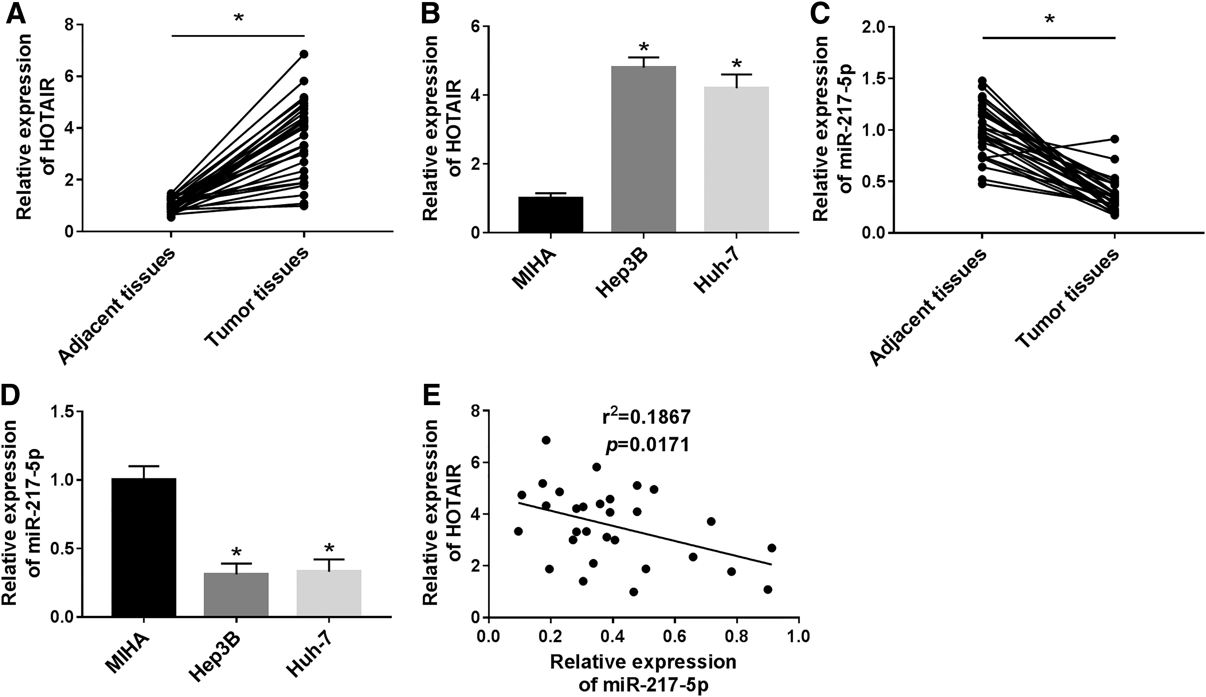
HOTAIR was upregulated and miR-217-5p was downregulated in HCC tumor tissues and cell lines.
Inhibition of HOTAIR attenuates cell progression in HCC
Given the aforementioned results, we hypothesized that HOTAIR exhibited promotive effects on HCC cell growth. Therefore, we transfected si-HOTAIR and si-NC plasmids in Huh-7 and Hep3B cells to evaluate the regulatory effects of HOTAIR on HCC cell growth. The expression of HOTAIR was decreased dramatically after si-HOTAIR transfection, whereas unchanged in control group, representing great transfection efficiency (Fig. 2A). Afterward, cell proliferation, migration, invasion, and apoptosis ability were assessed by CCK8, flow cytometry, and transwell assay. As illustrated in Figure 2B and C, cell viability was reduced markedly, whereas apoptotic rate was enhanced after HOTAIR knockdown compared with the control group. Consistently, migration and invasion cell number was decreased in Huh-7 and Hep3B cells transfected with si-HOTAIR compared with si-NC (Fig. 2D, E). In addition, HOTAIR silencing blocked protein expression of Vimentin, N-cadherin, and boosted protein expression of E-cadherin (Fig. 2F). Those data clarified that HOTAIR silencing reduced cell development and enhanced cell apoptosis in HCC.
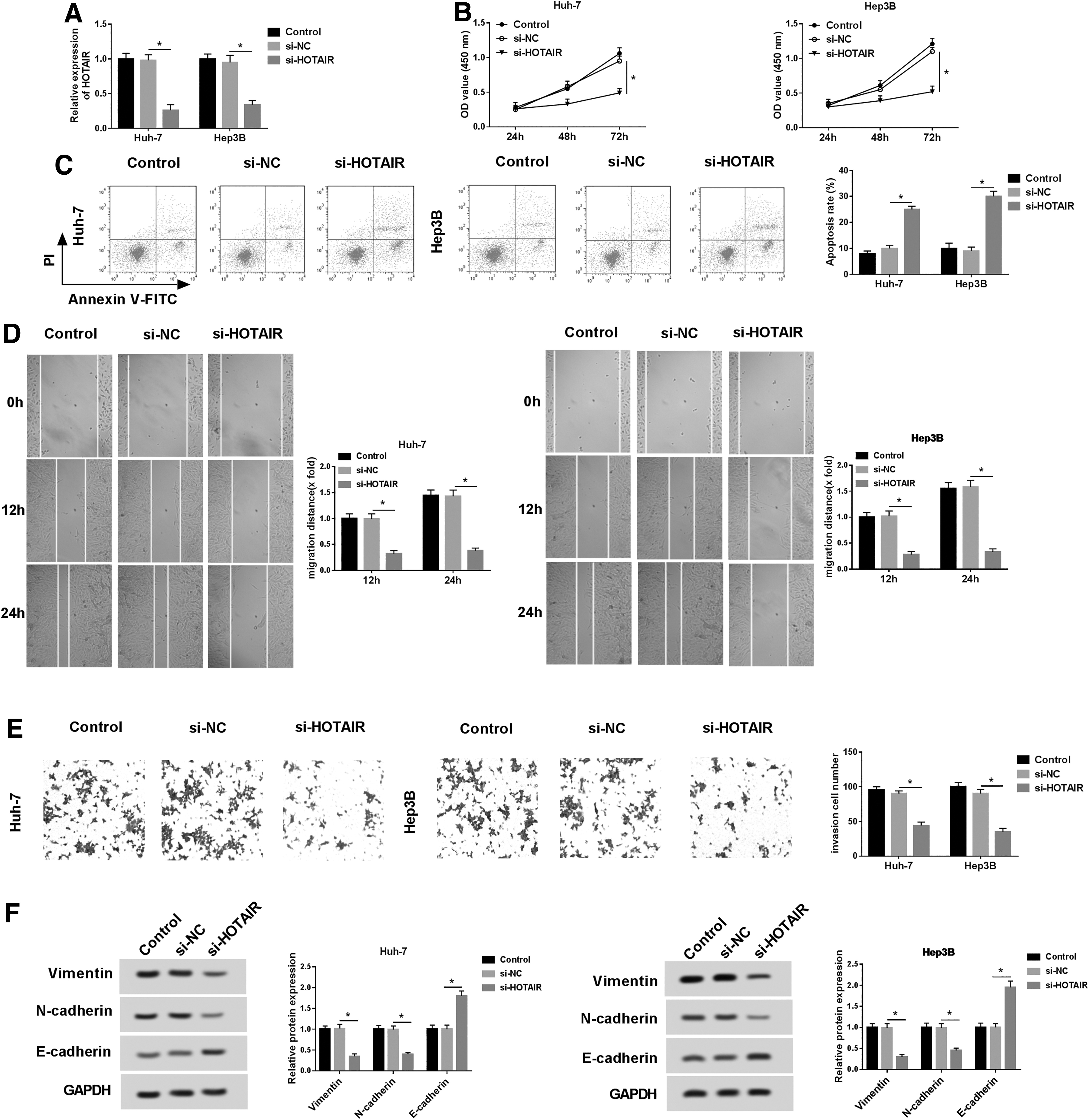
HOTAIR knockdown repressed proliferation, migration, invasion, and promoted apoptosis in HCC.
Interference of HOTAIR inhibits HCC xenograft tumor growth
Subsequently, we constructed xenograft mice by subcutaneous injection of Huh-7 cells stably transfected with lv-sh-HOTAIR and lv-sh-NC to elucidate the effects of HOTAIR on cell growth in vivo. As displayed in Figure 3A, tumor growth was suppressed remarkably in lv-sh-HOTAIR xenograft mice compared with lv-sh-NC group in 4 weeks. After 4 weeks, xenograft mice were dissected and tumor tissues were collected to detect the relative biological characteristics. We observed an obvious reduction of tumor weight in lv-sh-HOTAIR xenograft mice (Fig. 3B). In addition, HOTAIR expression was decreased significantly in lv-sh-HOTAIR group in comparison with lv-sh-NC group (Fig. 3C). Hence, the interference of HOTAIR significantly suppressed tumor growth in vivo.
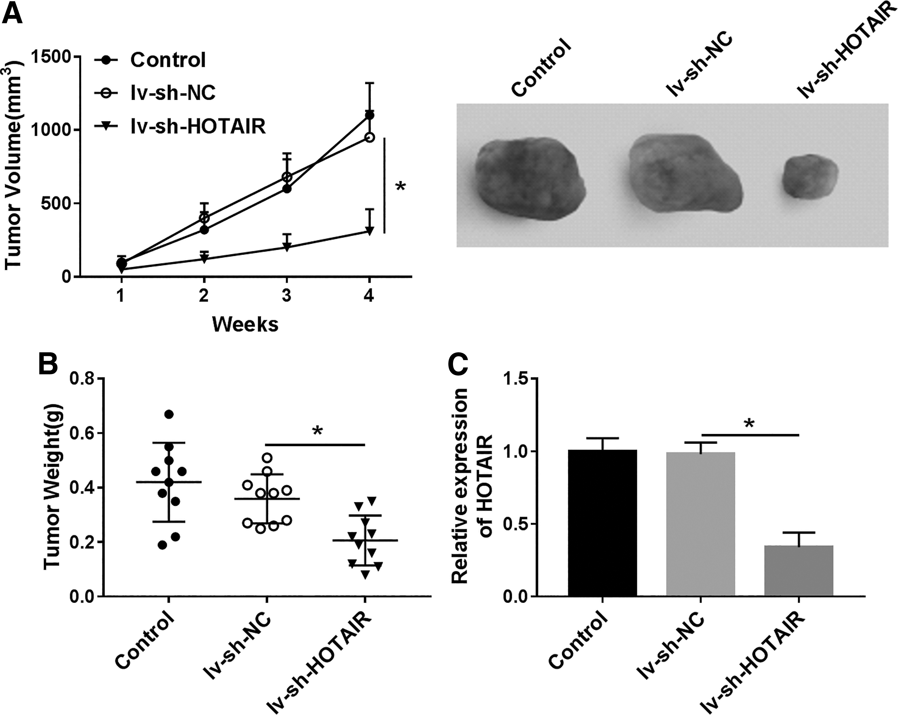
HOTAIR knockdown suppressed tumor growth in vivo. Xenograft mice were constructed by subcutaneous injection of Huh-7 cells stably transfected with lv-sh-HOTAIR and lv-sh-NC.
HOTAIR is a sponge of miR-217-5p
Based on bioinformatics prediction by Starbase v2.0, miR-217-5p comprised the potential binding sites of HOTAIR (Fig. 4A). To validate the interaction between HOTAIR and miR-217-5p, wild-type HOTAIR (wt-HOTAIR) or mutant-type HOTAIR (mut-HOTAIR) vectors were constructed and cotransfected with miR-217-5p mimics or miR-NC in Huh-7 and Hep3B cells. Conspicuous reduction of luciferase activity was observed in wt-HOTAIR and miR-217-5p cotransfection cells compared with mut-HOTAIR group, indicating HOTAIR directly interacted with miR-217-5p (Fig. 4B). Furthermore, RNA pull-down results exhibited that HOTAIR expression was reduced by miR-217-5p wt compared with input group (Fig. 4C). To investigate whether miR-217-5p is modulated by HOTAIR, pcDNA-HOTAIR, pcDNA-NC, si-HOTAIR, and si-NC were transfected in Huh-7 and Hep3B cells. As expected, miR-217-5p expression was decreased in cells transfected with pcDNA-HOTAIR, whereas increased after HOTAIR knockdown (Fig. 4D, E). Taken together, HOTAIR acted as a sponge of miR-217-5p in HCC.

HOTAIR directly targeted miR-217-5p.
HOTAIR modulates cell progression by sponging miR-217-5p in HCC
Next, to clarify the biological function of HOTAIR in HCC cell development, Huh-7 and Hep3B cells were transfected with si-HOTAIR+AntagomiR-217-5p and si-HOTAIR+AntagomiR-NC. We noticed the expression of miR-217-5p was suppressed largely in si-HOTAIR+AntagomiR-217-5p transfection cells compared with si-HOTAIR+AntagomiR-NC group (Fig. 5A). Furthermore, miR-217-5p inhibitor reversed HOTAIR silencing induced suppressive effect on cell proliferation (Fig. 5B). In addition, improvement of apoptosis in si-HOTAIR+AntagomiR-NC group and reduction of apoptosis in si-HOTAIR+AntagomiR-217-5p group was displayed in Figure 5C. Similarly, transwell assay results validated that miR-217-5p inhibitor abrogated the inhibition of HOTAIR silencing on cell migration and invasion (Fig. 5D, E). As expected, miR-217-5p inhibitor counteracted HOTAIR silencing mediated inhibition of EMT (Fig. 5F). All the findings represented that HOTAIR modulated cell progression by sponging miR-217-5p in HCC.
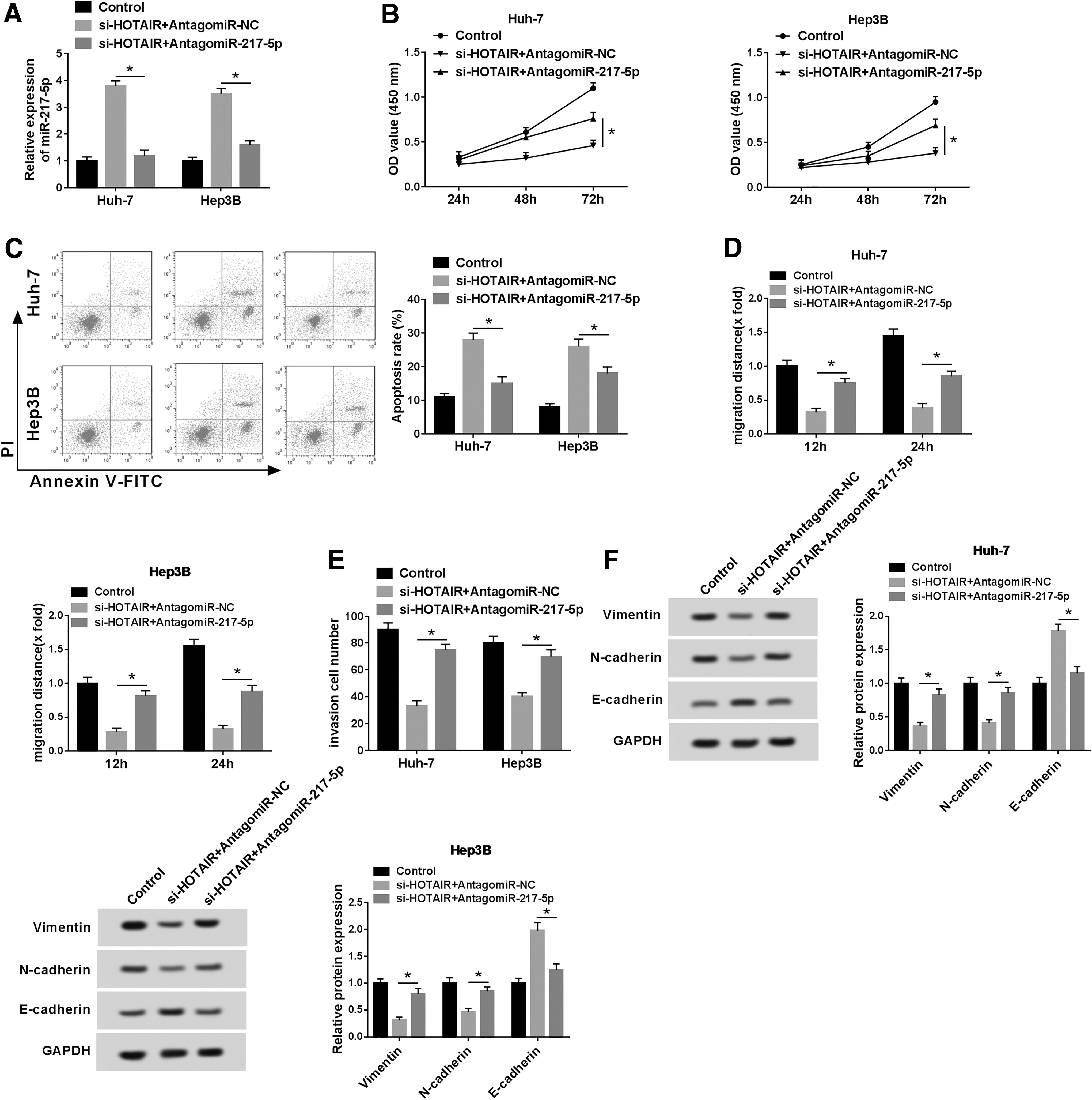
miR-217-5p inhibitor attenuated the suppression of HOTAIR on cell proliferation, migration, and invasion. Huh-7 and Hep3B cells were transfected with si-HOTAIR+AntagomiR-217-5p and si-HOTAIR+AntagomiR-NC.
HOTAIR regulates p-PI3K/p-AKT/MMP-2/9 signaling pathway by sponging miR-217-5p
Furthermore, we detected the expression of the relative protein P13K, p-P13K, AKT, p-AKT, MMP-2, and MMP-9 by western blot assay to evaluate the influence of HOTAIR/miR-217-5p axis on cell development-related protein level. As illustrated in Figure 6A and B, the expression of protein p-P13K, p-AKT, MMP-2, and MMP-9 was distinctly decreased in cells transfected with si-HOTAIR and increased by miR-217-5p inhibitor. Activation of p-PI3K/p-AKT/MMP-2/9 signaling could promote cancer cell migration, invasion, and metastasis. Thus, our results represented that miR-217-5p inhibitor could restore the inhibition of HOTAIR silencing on HCC progression by activating p-PI3K/p-AKT/MMP-2/9 signaling pathway.
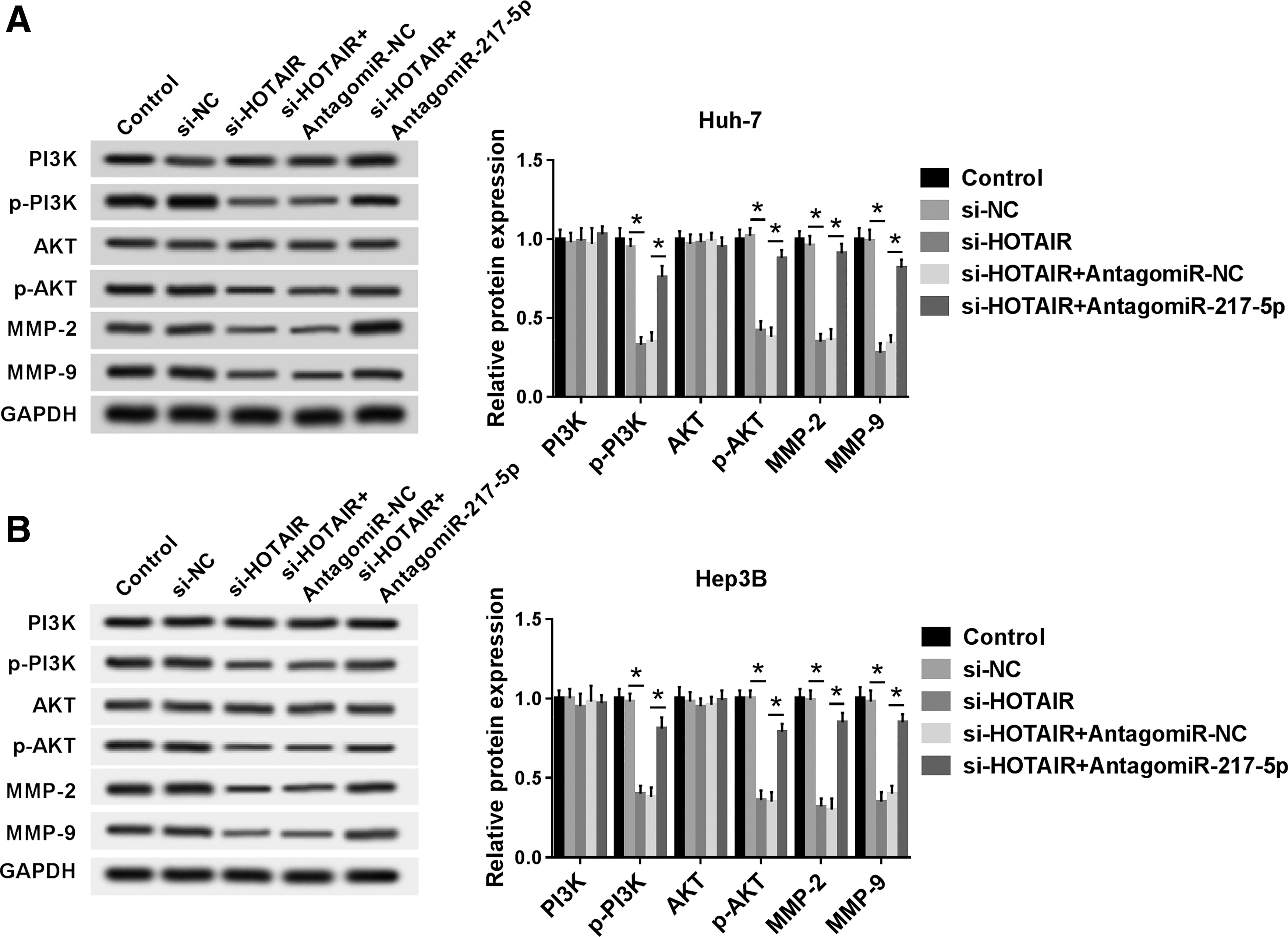
miR-217-5p inhibitor restored the inhibition of HOTAIR silencing mediated p-PI3K/p-AKT/MMP-2/9 protein expression.
Discussion
In our study, we demonstrated that HOTAIR serves as an oncogene to contribute to HCC proliferation, migration, invasion, and EMT by sponging miR-217-5p. First, we observed HOTAIR expression was upregulated, whereas miR-217-5p expression was downregulated in tumor tissues and cells compared with normal tissues and cells. Particularly, HOTAIR knockdown significantly suppressed cell proliferation, migration, invasion, EMT, as well as enhanced apoptosis in HCC. The further in vivo experiments indicated interference of HOTAIR could inhibit HCC xenograft tumor growth.
It was reported that HOTAIR could interact with PRC2 and expedite cancer cell metastasis by reprogramming chromatin state. 25 In addition, HOTAIR promoted renal interstitial fibrosis; however, silencing of HOTAIR repressed EMT process and blocked renal interstitial fibrosis development by upregulating miR-124 to inactive the Notch1 pathway. 26 In recent years, accumulating evidences have clarified that differentially expression of HOTAIR is validated in various carcinomas and correlated with lower survival probability of cancer patients. 27 –29 For instance, HOTAIR expression was boosted during bufalin treatment and abundance of HOTAIR obviously inhibited cell migration and invasion by sponging miR-520b in prostate cancer. 30 In addition, HOTAIR was enhanced after ionizing irradiation and its knockdown suppressed cell survival and facilitated cell apoptosis by improving the radiosensitivity of breast cancer through activating miR-218. 31 Besides, HOTAIR was reported to regulate cell growth and EMT in esophageal cancer or head and neck squamous cell carcinoma by acting as a miRNA sponge. 32,33 Therefore, we suggested HOTAIR functions as sponger of specific miRNA in HCC.
Previous studies have demonstrated that miRNAs are fundamental regulators in human genome and aberrantly expression of miRNAs is the leading cause of tumorigenesis and metastasis. 34 Recently, miR-217 has been identified as biomarker for diagnosis as well as predictor of recurrence and progression in a variety of diseases. 35 –37 For example, overexpression of miR-217 improved sensitivity of chronic myelogenous leukemia cells to dasatinib by targeting pro-oncogenic anterior gradient 2. 38 Xiang Nie et al. elucidated that miR-217 contributed to cardiac hypertrophy and fibrosis processes by targeting PTEN. 39 Currently, by Pearson's correlation coefficient analysis, HOTAIR was inversely correlated with miR-217-5p, suggesting HOTAIR might modulate miR-217-5p expression negatively. Luciferase reporter assay confirmed the interaction between HOTAIR and miR-217-5p. Moreover, rescue experiments showed miR-217-5p inhibitor abrogated the inhibition of HOTAIR on cell proliferation, migration, invasion, and EMT. Based on previous study, p-PI3K/p-AKT could induce MMP-2/9 expression and p-PI3K/p-AKT/MMP-2/9 signaling pathway is closely associated with cancer cell migration, invasion, and metastasis. We observed that miR-217-5p inhibitor restored the suppressive effect of HOTAIR silencing on HCC cell progression by activation of p-PI3K/p-AKT/MMP-2/9 signaling pathway.
In conclusion, our study revealed the regulatory mechanism of HOTAIR/miR-217-5p axis in cell progression and apoptosis in HCC. The results obviously manifested HOTAIR functioned as an oncogene to accelerate cell proliferation, migration, invasion, EMT, and inhibit apoptosis in HCC by regulating miR-217-5p to activate p-PI3K/p-AKT/MMP-2/9 signaling pathway, indicating HOTAIR may be a potential biomarker for HCC.
Limitations
Our study revealed that HOTAIR promoted HCC survival, migration, invasion, and EMT by interacting with miR-217-5p through activation of p-PI3K/p-AKT/MMP-2/9 pathway. However, the current data only deepened our understanding of HOTAIR in HCC, and HOTAIR/miR-217-5p axis may be a hopeful biomarker for HCC. The main results were well organized and fully illustrated the role of HOTAIR in HCC, but comprehensive population experiments were needed in next research. As the sample size of this study was small, the results of this study was not enough to extend to the general population.
Footnotes
Disclosure Statement
Authors declare that they have no conflict of interest.
Funding Information
No funding was received for this article.