
Abstract
The Editor-in-Chief of Cancer Biotherapy and Radiopharmaceuticals officially retracts the article entitled, “Mortal Obligate RNA Transcript Inhibits Cancer Cell Invasion and Migration in Lung Adenocarcinoma by Downregulating miRNA-223,” by Yunjian Huang, Qingyang Zhuang, and Wu Zhuang. (Cancer Biother Radiopharm 2020;35(5):345–350; doi: 10.1089/cbr.2019.3244).
The corresponding author, Wu Zhuang, contacted the Editor-in-Chief to request a correction to Figures 5A and 5B and stated:
“Recently, during a self-examination of the paper, I discovered errors in Figures 5A and 5B. Further investigation revealed that the H522 cells presented in Figure 5A were mistakenly used to represent the H23 cells in Figure 5B. This mistake occurred as a result of confusion among team members during the classification and selection of cell images, given the involvement of multiple individuals and the presence of various scattered images in the study. Consequently, the H522 image was erroneously duplicated in Figure 5A.”
Based on the authors' mishandling of image preparation, classifications, and presentation, the editorial leadership of the journal no longer has confidence in the validity of the study and denied the request to publish an erratum and determined an editorial retraction is necessary.
After being notified via email of the determination to retract the article, Dr. Wu Zhuang appealed the decision but was denied based on lack of certainty in the work.
Introduction
Lung cancer has been a common human malignancy and a major cause of deaths among cancer patients worldwide. 1 Lung cancer affects about 2 million new cases and causes about 1.5 million deaths every year. 2 Treatment and prevention of lung cancer has become a heavy burden on public health. Lung adenocarcinoma (LUAD) is one of the major subtypes of lung cancer named non-small cell lung cancer. 3 Surgical resection is the only radical option for LUAD. However, the application of surgical resection is challenged by the fact that more than 70% of patients with LUAD are diagnosed with locally advanced or metastatic tumors, 4 which are usually not appropriate for surgical operations.
There are a large set of noncoding RNAs (ncRNAs) that play key roles in human diseases. 5,6 Despite the lack of protein coding ability, ncRNAs participate in biological processes mainly by regulating the expression of downstream genes. 5,6 Literatures have shown that long noncoding RNAs (lncRNAs, longer than 200 nt) are frequently dysregulated during the progression of various human diseases including different types of cancer. 7 And, regulations of certain key lncRNAs have been proven to be promising approaches to treat cancer. 8,9 It has been observed that lncRNAs may interact with microRNAs (miRNAs), 10 which are another subgroup of ncRNAs that play pivotal roles in cancer, to regulate cancer development. 11 Mortal obligate RNA transcript (MORT) has been reported as a tumor suppressor in various types of cancer. 12,13 Results in the present study showed that MORT may inhibit invasion and migration of LUAD cells by downregulating miRNA-223, which is an oncogenic miRNA in lung cancer. 14
Materials and Methods
Human tissues and LUAD cells
Paired normal tissue and tumor tissue samples were obtained from 67 patients with LUAD (adenocarcinoma, 42 males and 25 females, with the age of 29–65 years and 47.1 ± 5.1 years, respectively). These patients were admitted to Fujian Cancer Hospital and Fujian Medical University Cancer Hospital from January 2015 to March 2018. Inclusion criteria were as follows: (1) histopathologically diagnosed cases; (2) newly diagnosed cases; (3) patients signed informed consent. Exclusion criteria were as follows: (1) other diseases were observed; (2) treatments were performed before admission. There were 28 cases with nonmetastasis (NM), 18 cases with lymph node metastasis (LNM), and 20 cases with distant metastasis (DM). This study passed the review of aforementioned hospital ethics committee. H23 and H522 human LUAD (adenocarcinoma) cell lines (ATCC) were used. Cells were cultivated at 37°C with 5% CO2. RPMI-1640 medium (10% fetal bovine serum [FBS]) was used as cell culture medium.
Quantitative reverse transcription–polymerase chain reaction
The PureLink miRNA Isolation Kit (Thermo Fisher Scientific) and Trizol reagent (Thermo Fisher Scientific) were used for the extraction of miRNAs and total RNAs, respectively. The RevertAid RT Reverse Transcription Kit (Thermo Fisher Scientific) and TaqMan MicroRNA Reverse Transcription Kit (Thermo Fisher Scientific) were used for reverse transcription with total RNAs and miRNAs as templates, respectively. Polymerase chain reaction (PCR) systems were prepared to detect the expression of MORT (endogenous control: 18S rRNA) and miRNA-223 (endogenous control: U6) using the Luna® Universal One-Step RT-qPCR Kit (NEB) and miScript SYBR Green PCR Kit (QIAGEN), respectively. Primer sequences were as follows: 5′-ATGTTTTCATTCCTCATCTC-3′ (forward) and 5′-AACTGCACATTTCAACATGG-3′ (reverse) for MORT; 5′-CTACCACATCCAAGGAAGCA-3′ (forward) and 5′-TTTTTCGTCACTACCTCCCCG-3′ (reverse) for 18S rRNA. Forward primer of miRNA-223 was: 5′-CGUGUAUUUGACAAGCUGAG-3′. Reverse primer of miR-223 and U6 primers were included in the kit. The ABI PRISM 7500 qRT-PCR machine (Applied Biosystems, Rockford, IL) was used to carry out all PCRs. Data were calculated using the 2−ΔΔCT method.
Cell transfection
Empty pcDNA3 vector and MORT-expressing pcDNA3 vector were obtained from Sangon (Shanghai, China). Negative control miRNA and miR-223 mimic were purchased from Sigma–Aldrich. H23 and H522 cells were collected at confluence of 70%–80% and were transfected with 10 nM empty pcDNA3 vector (negative control, NC) or 10 nM MORT-expressing pcDNA3 vector, 40 nM negative control miRNA (NC), or 40 nM miR-223 mimic using the Lipofectamine 2000 (Sangon). Cells without transfections were used as control (C). Cells were harvested 24 h after transfections.
Transwell migration and invasion assay
Cells were harvested 24 h after transfections, and 3 × 10⁴ cells were mixed with 1 mL RPMI-1640 medium (no FBS) to prepare single cell suspensions. For upper Transwell chamber, 3 × 10³ cells in 0.1 mL cell suspension were added, and the lower chamber was added with RPMI-1640 medium, followed by the addition of 20% FBS. Matrigel-coated membranes were used for invasion assay. Chambers were kept at 37°C with 5% CO2 for 12 h, followed by staining with 1% crystal violet (Sangon) for 20 min at 25°C. Finally, invading and migrating cells were counted under an optical microscope.
Statistical analysis
All mean values used in comparisons were calculated using data from three biological replicates. The GraphPad Prism 6 software was used for analyses. Correlations were analyzed by linear regression. Paired t-test was used to explore the differences between two types of tissues. Analysis of variance (one-way) and the Tukey test were used to compare differences among multiple groups. Differences with p < 0.05 were statistically significant.
Results
MORT was downregulated in LUAD
The expression levels of MORT were detected by reverse transcription–polymerase chain reaction (RT-qPCR). Compared with healthy tissues, the expression levels of MORT were significantly decreased in tumor tissues of LUAD patients (Fig. 1). These data suggested that downregulation of MORT was likely involved in LUAD.
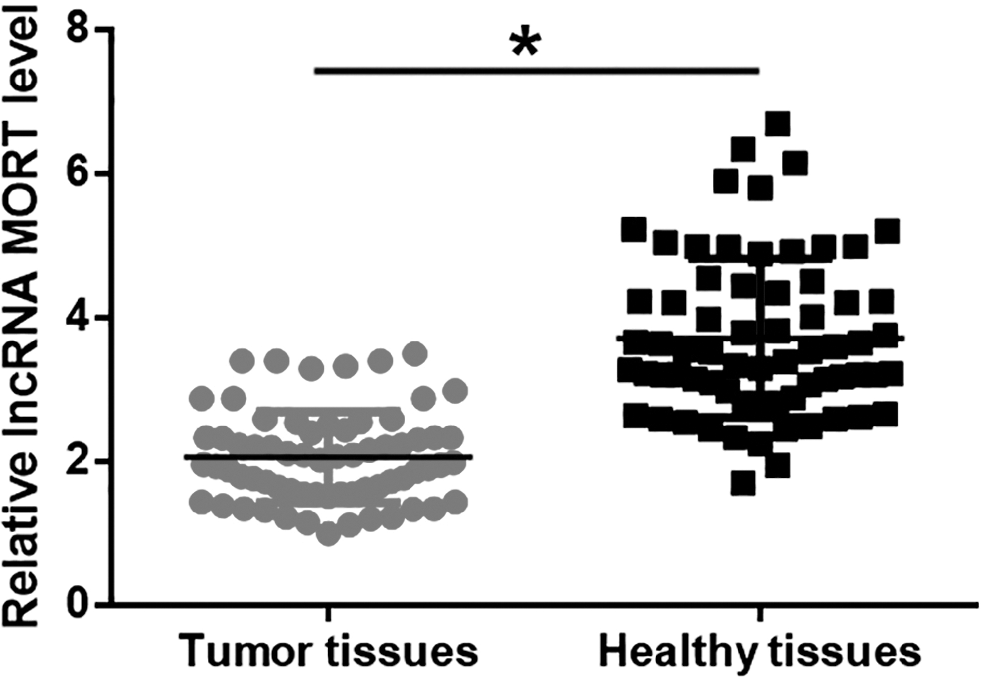
MORT was downregulated in LUAD. Compared with adjacent healthy tissues, the expression levels of MORT were significantly decreased in tumor tissues of LUAD patients (*p < 0.05). LUAD, lung adenocarcinoma; MORT, mortal obligate RNA transcript.
Expression of MORT was affected by tumor metastasis but not by the tumor size
Among the 67 patients with LUAD, there were 20 cases with a diameter of primary tumor < 2 cm, 23 cases between 2 and 4 cm, and 24 cases > 4 cm. No significant differences in expression of MORT were found among patients with different diameters of primary tumors (Fig. 2A). Compared with the NM group, the expression of MORT was significantly downregulated in the LNM and DM groups (p < 0.05, Fig. 2B). Compared with the LNM group, the expression of MORT was also significantly downregulated in the DM group (p < 0.05, Fig. 2B).
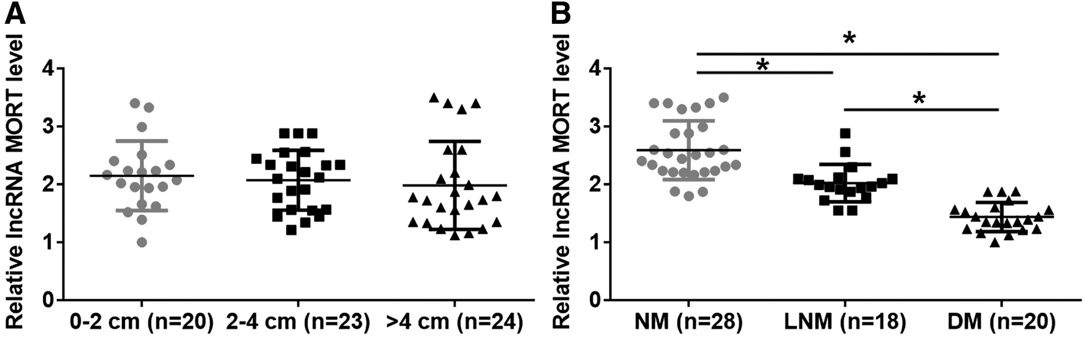
Expression of MORT was affected by tumor metastasis but not by the sizes of tumors. No significant differences in expression of MORT were observed among patients with different diameters of primary tumors
Expression of miRNA-223 was inversely correlated with MORT
Expression of miRNA-223 was detected by RT-qPCR. Compared with healthy tissues, the expression levels of miRNA-223 were significantly increased in tumor tissues of LUAD patients (Fig. 3A). Correlations between the expression of MORT and miRNA-223 were analyzed by linear regression. The expression levels of MORT and miRNA-223 were inversely correlated across tumor tissues (Fig. 3B) but not across adjacent healthy tissues (Fig. 3C).
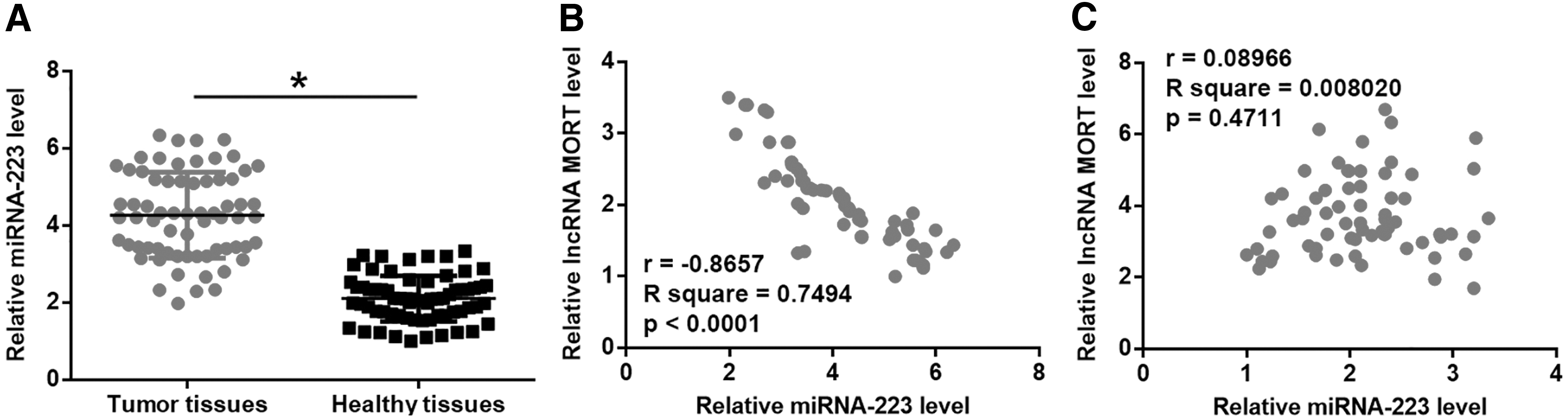
miRNA-223 was upregulated in tumor tissues and was inversely correlated with the expression of MORT. Compared with adjacent healthy tissues, the expression levels of miRNA-223 were significantly increased in LUAD tissues
Overexpression of MORT mediated the downregulation of miRNA-223 in LUAD cells
Overexpression of MORT and miRNA-223 was achieved 24 h after transfections in H23 and H522 cell lines (Fig. 4A, p < 0.05). Compared with the control (C) and negative control (NC) groups, cells with overexpressed MORT showed inhibited expression of miRNA-223 (Fig. 4B, p < 0.05). However, the expression of MORT was not significantly affected by overexpression of miRNA-223 (Fig. 4C, p < 0.05).

Overexpression of MORT mediated the downregulation of miRNA-223 in LUAD cells. Overexpression of MORT and miRNA-223 was achieved at 24 h after transfection in cells of H23 and H522 cell lines
Overexpression of MORT inhibited LUAD cell invasion and migration through miRNA-223
Overexpression of MORT inhibited (Fig. 5A, p < 0.05), whereas overexpression of miRNA-223 promoted the migration and invasion of LUAD cells (Fig. 5B, p < 0.05), respectively. Besides, overexpression of miRNA-223 partially rescued the inhibited cancer cell behaviors after overexpression of MORT.
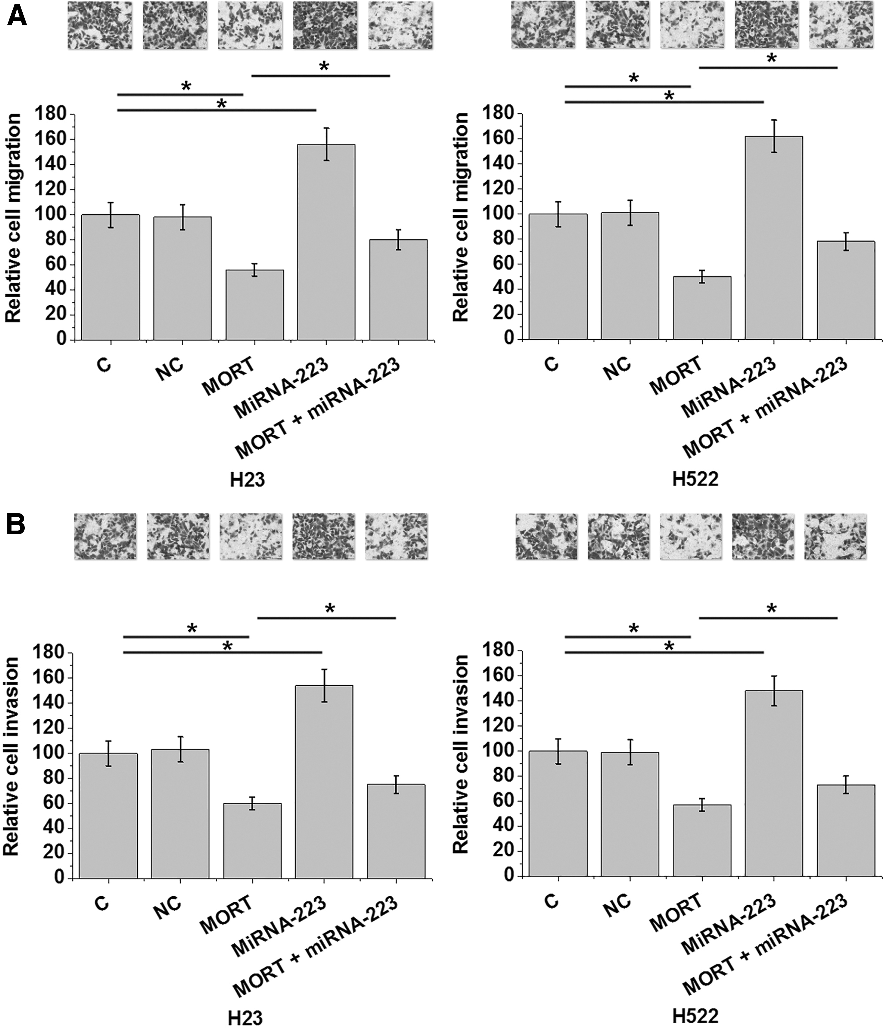
Overexpression of MORT inhibited LUAD cell migration and invasion through miRNA-223. Overexpression of MORT inhibited
Discussion
A recent study reported silencing of MORT in 16 TCGA cancer types, 12 indicating the potential role of MORT as a tumor suppressor in cancer biology. However, the functionality of MORT in cancer is still unknown. We found that MORT was downregulated in LUAD, and overexpression of MORT may inhibit cancer cell migration and invasion through the interaction with miRNA-223, which is an oncogenic miRNA in lung cancer. 14
The progression of LUAD is affected by various internal and external factors including differentially expressed lncRNAs. 15 However, altered expression of those lncRNAs is usually observed in the whole development procedure of LUAD. 16,17 We observed the downregulation of MORT in LUAD. Interestingly, the expression of MORT was not affected by tumor size. In contrast, tumor metastasis significantly affected the expression of MORT. Our in vitro experiments using LUAD cell lines also showed that overexpression of MORT inhibited LUAD cancer cell invasion and migration. Therefore, these data support that downregulation of MORT is specifically involved in the metastasis of LUAD.
miRNA-223 has been reported as an oncogenic miRNA involved in lung cancer. 14 Consistently, we showed that miRNA-223 was upregulated in LUAD. Overexpression of miRNA-223 promotes cancer cell migration and invasion in different types of cancer including lung cancer. 14,18,19 Consistent results were also obtained in the present study. However, the upstream regulator of miRNA-223 in this process is still unknown. Our results demonstrated for the first time that the expression of miRNA-223 could be inhibited by MORT. In addition, our data showed that the regulation of miRNA-223 by MORT can regulate LUAD cell migration and invasion. Furthermore, the MORT/miRNA-223 pathway is likely disease-specific due to the lack of obvious correlation between MORT and miRNA-223 in normal healthy tissues. Our speculation is that certain dysregulated pathogenic factors involved in LUAD might mediate the interactions between MORT and miRNA-223 in LUAD. However, those factors are not dysregulated under normal conditions. Therefore, MORT may not interact with miRNA-223 to participate in physiological processes.
It is worth noting that numerous ncRNAs have been shown with dysregulation in lung cancer and have critical roles in cancer development. However, clinical studies are still needed to explore their roles in the clinical treatment of lung cancer. 20 –22
Conclusions
In conclusion, MORT is downregulated in LUAD. MORT plays a role as tumor suppressor in LUAD by inhibiting cancer cell migration and invasion through the downregulation of miRNA-223.
Ethics Approval and Consent to Participate
Ethical approval was obtained from Fujian Cancer Hospital and Fujian Medical University Cancer Hospital Research Ethics Committee, and it conforms to the provisions of in accordance with the Declaration of Helsinki as revised in 2013. All participants signed informed consent.
Footnotes
Authors' Contributions
Y.H.: data analysis, experimental work, article writing, and literature search; Q.Z.: data analysis and statistical analysis; W.Z.: study design, article writing, and literature search. The article was approved by all authors.
Disclosure Statement
There are no existing financial conflicts.
Funding Information
We are thankful for the financial support from Fujian Cancer Hospital and Fujian Medical University Cancer Hospital.