




Abstract
Background:
Oral squamous cell carcinoma (OSCC) is the most common malignant tumor of the oral cavity. It was determined that circular RNAs were related to the development and progression of various cancers, including OSCC. The purpose of our study was to define the role and potential mechanism of hsa_circ_0072387 in OSCC progression.
Materials and Methods:
Thirty-five patients with OSCC were involved in this study. Real-time quantitative polymerase chain reaction was used to evaluate the expression levels of hsa_circ_0072387 and microRNA (miR)-503-5p. Cell proliferation, migration, and invasion abilities were assessed by Cell Counting Kit-8 (CCK-8) and Transwell assays, respectively. The abundance of cell proliferation marker Ki-67, epithelial–mesenchymal transition (EMT) markers E-cadherin, N-cadherin, and vimentin was analyzed by Western blot assay. Glycolysis was evaluated using commercial kits. The interaction between hsa_circ_0072387 and miR-503-5p was confirmed by bioinformatics analysis, RNA immunoprecipitation (RIP) assay, and dual-luciferase reporter assay.
Results:
hsa_circ_0072387 expression was significantly downregulated, and miR-503-5p was upregulated in OSCC cells and tissues. Gain of hsa_circ_0072387 or knockdown of miR-503-5p suppressed the cell proliferation, migration and invasion, EMT, and glycolysis in OSCC SCC-4 and HSC-3 cells. hsa_circ_0072387 targeted miR-503-5p and inversely regulated miR-503-5p expression. Moreover, upregulation of miR-503-5p could partially revert the tumor-suppressive effects of hsa_circ_0072387 on OSCC cells.
Conclusion:
hsa_circ_0072387 inhibited OSCC progression by downregulating miR-503-5p, explicating that hsa_circ_0072387 could function as a novel potential therapeutic target for OSCC.
Highlights:
hsa_circ_0072387 level is decreased, whereas miR-503-5p level is increased in OSCC tissues and cells.
Upregulation of hsa_circ_0072387 or downregulation of miR-503-5p inhibits proliferation, metastasis, and glycolysis of OSCC cells.
hsa_circ_0072387 targets miR-503-5p.
hsa_circ_0072387 inhibits OSCC progression by altering miR-503-5p expression.
Introduction
Oral cancer is a prevalent malignant tumor, with the 11th most frequent malignancy worldwide, showing the highest morbidity in all head and neck cancers. 1,2 Oral squamous cell carcinoma (OSCC) is the major histologic type of the oral cavity, accounting for ∼90% of oral cancer cases. 2 Although great advances have been made in the treatment of OSCC, the therapeutic efficacy and survival rate of OSCC patients remain unsatisfactory.
Epithelial–mesenchymal transition (EMT), conversion of epithelial cells to mesenchymal cells, not only participates in embryo implantation, embryogenesis, and organ development, but also affects the progression, invasion, and metastasis of malignant tumors. 3,4 Glycolysis is defined as the transformation of glucose to lactate along with the generation of adenosine triphosphate under anaerobic condition, 5 a preferable way for cancer cells to obtain energy. 6 Hence, there is a great interest in clarifying the mechanism of OSCC progression for diagnosis or treatment, especially the EMT and glycolysis.
Circular RNAs (circRNAs) are a novel group of noncoding RNAs and have gained significant attention because of their significant functions in diagnosis and progression of human diseases. 7,8 circRNAs are characterized by their closed circular form without a 5′ cap or a 3′poly (A), generating from covalently closed continuous loops. 7,9
Many circRNAs are involved in the occurrence and progression of OSCC. For instance, hsa_Circ_0001874 and hsa_Circ_0001971 were clearly decreased in salivary samples of postoperative patients with OSCC, with potential serving as biomarkers for OSCC diagnosis. 10 Besides, circDOCK1 participated in the apoptotic process of OSCC cells by sponging as a competing endogenous RNA to modulate baculoviral IAP repeat containing 3 (BIRC3). 11 Gao and his partners identified circ-PKD2 as a suppressor gene in OSCC, shown as the inhibition of proliferation, migration, and invasion, and the contribution to apoptosis and cell cycle arrest of OSCC cells. 12 hsa_circ_0072387 was reported to be downregulated in OSCC and could serve as a valuable predictor of OSCC. 13
However, the explicit role of hsa_circ_0072387 in OSCC progression and the potential mechanism remain to be further elucidated, which is the focus of this study.
As is known, circRNAs could alter the development of human cancers by working as sponges of microRNA (miRNA). 14 miRNAs are a large family of short noncoding RNAs, with ∼22 nucleotides in length. 15 miRNAs exert their functions in the regulation of cellular processes of human cancers, including cell growth and proliferation, differentiation and apoptosis by targeting messenger RNAs. 16 –18
Abnormal expression of miRNAs was reported to affect cell proliferation, migration, or invasion abilities in OSCC. For example, miR-211 promoted proliferation, migration, and invasion abilities of OSCC cells by targeting the bridging integrator 1 protein. 19 miR-300 was reported to suppress metastasis of OSCC by inhibiting EMT. 20 It was suggested that miR-194 inhibited cell proliferation of OSCC by suppressing PI3K-Akt-FoxO3a signaling pathway. 21 In addition, ectopic expression of miR-503-5p was observed in several types of tumors, including ovarian cancer, 22 hepatocellular carcinoma (HCC), 23 and OSCC, 24 indicating its crucial role in tumorigenesis.
It is worth mentioning that miR-503-5p was predicted to be a potential target of hsa_circ_0072387 by Starbase. However, the exact role of miR-503-5p in hsa_circ_0072387-mediated OSCC progression needs further investigation.
As indicated previously, this study aimed to explore the expression and biological functions of hsa_circ_0072387 and miR-503-5p in OSCC, and the probable mechanism.
Materials and Methods
Clinical specimens and cell culture
A total of 35 pairs of tumor tissues and normal oral mucosa tissues were obtained postoperatively from The Third Hospital Affiliated to Qiqihar Medical College. The study was permitted by Ethics Committee of The Third Hospital Affiliated to Qiqihar Medical College. None of the patients had received radiotherapy or chemotherapy before operation. All patients signed written informed consent. Specimens were snap-frozen in liquid nitrogen immediately after resection and stored at −80°C.
The immortalized human oral keratinocytes (RT7) and OSCC cell lines (HSC-3 and HSC-4) were purchased from Institute of Biochemistry and Cell Biology of the Chinese Academy of Sciences (Shanghai, China), and OSCC cell lines (SCC-25 and SCC-4) were obtained from American Type Culture Collection (ATCC, Manassas, VA). All cells were maintained in Dulbecco's modified Eagle's medium (DMEM; Thermo Fisher Scientific, Inc., Waltham, MA) supplemented with 10% (v/v) fetal bovine serum (FBS; HyClone, Logan, UT) at 37°C under 5% CO2.
Reagent and cell transfection
The overexpression plasmid of hsa_circ_0072387 (hsa_circ_0072387), small interference RNA targeting hsa_circ_0072387 (si-hsa_circ_007238) and its negative control (si-NC), miR-503-5p mimic (miR-503-5p), miR-503-5p inhibitor (anti-miR-503-5p), and their negative controls (NC and anti-NC) were obtained from (GenePharma Co. Ltd., Shanghai, China). The pcDNA 3.1 plasmid (circ-NC) was purchased from Thermo Fisher Scientific, Inc. The above-mentioned oligonucleotides or plasmids were transfected into SCC-4 and HSC-3 cells using Lipofectamine™ 3000 reagent (Invitrogen, Carlsbad, CA) according to manufacturer's instructions.
Reverse transcription–quantitative polymerase chain reaction assay
Total RNA was extracted from culture cells and clinical tissues using mirVana miRNA Isolation kit (Life Technology, Gaithersburg, MD). Reverse transcription reaction was performed using 500 ng total RNA as template and using a PrimeScript™ RT Reagent Kit (Promega, Madison, WI). Quantitative polymerase chain reaction (qPCR) was performed using an SYBR Green mix (Takara, Tokyo, Japan) on Applied Biosystems 7500 thermocycler (Thermo Fisher Scientific, Inc.). Glyceraldehyde-3-phosphate dehydrogenase (GAPDH) and U6 were used as the internal reference for hsa_circ_0072387 and miR-503-5p, respectively.
The primers for hsa_circ_0072387, miR-503-5p, GAPDH, and U6 were listed as follows: hsa_circ_0072387, 5′-TCTTCAGTATATGGTTCCCTTGCA-3′ (sense) and 5′-AAGCCCCGGTGTAGCATCTT-3′ (antisense); miR-503-5p, 5′-CCTATTTCCCATGATTCCTTCATA-3′ (sense) and 5′-GTAATACGGTTATCCACGCG-3′ (antisense); GAPDH, 5′-AAGCCACCCCACTTCTCTCTAA-3′ (sense) and 5′-AATGCTATCACCTCCCCTGTGT-3′ (antisense); U6, 5′-CTCGCTTCGGCAGCACA-3′ (sense) and 5′-AACGCTTCACGAATTTGCGT-3′ (antisense). The qPCR for each gene was performed in triplicate. The expression levels of hsa_circ_0072387 and miR-503-5p were calculated using the threshold cycle 2−▵▵Ct method.
Cell Counting Kit-8 assay
Cell Counting Kit-8 (CCK-8) assay was carried out to detect cell proliferation. SCC-4 and HSC-3 cells were seeded in 96-well plates with DMEM, achieving a density of 5000 cells per well. After different treatments, CCK-8 solution (Dojindo Molecular Technologies, Kyushu, Japan) was added into each well (10 μL per well) and incubated for 2 h at 37°C. The absorbance of each well was measured using a Microplate Reader (Bio-Rad, Hercules, CA) at 450 nm.
Transwell assay
An invasion assay was performed using Transwell chamber (8 μm pores; BD Biosciences, San Jose, CA) with Matrigel (BD Biosciences). In brief, 5 × 10⁴ SCC-4 and HSC-3 cells were suspended in 300 μL DMEM (serum-free) and plated in the upper chamber. The complete medium (700 μL) supplemented with 10% FBS was added into the lower chamber, incubated for 24 h at 37°C. Then the cells on the upper surface of the membrane were removed with a cotton swab, and the cells on the lower surface were fixed using 4% paraformaldehyde, stained with 0.1% crystal violet at room temperature for 20 min, washed with phosphate-buffered saline (PBS) three times, photographed, and counted under an optical microscope (magnification, × 100; Olympus, Tokyo, Japan) in five fields at random.
Cell migration assay was conducted in similar procedures, without the Matrigel on the membrane.
Western blot
SCC-4 and HSC-3 cells were harvested and then lysed with ice-cold radioimmunoprecipitation assay (RIPA) buffer (Thermo Fisher Scientific, Inc.) supplemented with 1 mmol/L PMSF (Sigma-Aldrich, St. Louis, MO). The protein concentration was quantified using a bicinchoninic acid assay (BCA) protein assay kit (Pierce Biotechnology, Rockford, IL). Then equal amounts of the proteins (20 μg) were separated by 12% sodium dodecyl sulfate–polyacrylamide gels, transferred onto polyvinylidene fluoride membranes (Bio-Rad).
After blockage in 5% nonfat dried milk for 2 h, the membranes were incubated overnight at 4°C with primary antibody against Ki-67 (1:500 dilution), E-cadherin (1:1000 dilution), N-cadherin (1:1000 dilution), vimentin (1:500 dilution), or GAPDH (1:2000 dilution). After washing using PBS, the membranes were incubated with HRP-conjugated secondary antibodies (1:2000 dilution) at room temperature for 2 h. Signals were visualized by enhanced chemiluminescence procedure (Thermo Fisher Scientific, Inc.). All antibodies were purchased from Santa Cruz Biotechnology (Santa Cruz, CA).
Detection of lactate production and glucose level
Glycolysis was assessed by analyzing the glucose consumption and lactate production. After 48 h transfection, SCC-4 and HSC-3 cells (about 1 × 10⁶) in equal medium were collected, and the levels of glucose and lactate in medium were monitored utilizing specific assay kits (KeyGen, Nanjing, China) referring to the protocols supplied by the manufacturer.
RNA immunoprecipitation
RNA immunoprecipitation (RIP) was conducted according to the manufacturer's instructions with EZ-Magna RIP Kit (Millipore, Billerica, MA). SCC-4 and HSC-3 cells were gathered and resuspended in RIP lysis buffer. Next, the supernatant of whole cell lysate was incubated with magnetic beads and with human anti-Argonaute2 (Ago2) antibody (Abcam, Cambridge, MA) and IgG (Cell Signaling Technology, Boston, MA) as a negative control. After incubation for 24 h, magnetic beads were rinsed and digested with proteinase K to remove the protein. After purification, RNAs were examined by real-time quantitative polymerase chain reaction (RT-qPCR) assay.
Dual-luciferase reporter assay
The target miRNAs of hsa_circ_0072387 were predicted using Starbase. miR-503-5p was identified as a potential target of hsa_circ_0072387. To construct the luciferase reporter gene plasmid, the hsa_circ_0072387 sequence containing the putative binding site of miR-503-5p or its mutant were amplified and subcloned into a pGL3 luciferase promoter vector (pGL3-empty; Promega) to generate hsa_circ_0072387-wt or hsa_circ_0072387-mut. SCC-4 and HSC-3 cells were cotransfected with hsa_circ_0072387-wt or hsa_circ_0072387-mut, and miR-503-5p mimic or NC using Lipofectamine 3000 reagent (Invitrogen) for 48 h. Quick Change Site-Directed Mutagenesis kit (Agilent Technologies, Inc., Santa Clara, CA) was exploited for mutating the binding sites to generate the mutant version.
Then luciferase activities were evaluated using a Dual-Luciferase Reporter detection System (Promega) according to the manufacturer's instructions. Each experiment was repeated three times.
Statistical analysis
All statistical analyses were performed using SPSS 22.0 software (SPSS, Chicago, IL). All data were given as mean ± standard error of the mean. Student's t-test was used to analyze difference between data from two groups, and one-way analysis of variance (ANOVA) was applied to process data from three or more groups. OSCC patients were divided into low or high expression groups of hsa_circ_0072387 and miR-503-5p according to the corresponding median value. Kaplan–Meier curves and log-rank test were performed to analyze the survival rate of OSCC patients with different levels of hsa_circ_0072387 or miR-503-5p. Spearman's correlation analysis was implemented to validate the correlation between the levels of hsa_circ_0072387 and miR-503-5p in OSCC tissues. p < 0.05 was considered statistically significant.
Results
hsa_circ_0072387 was underexpressed in OSCC tissues and cell lines
To figure out the role of hsa_circ_0072387 in the progression of OSCC, RT-qPCR assay was first performed to test the hsa_circ_0072387 level in OSCC tissues and cell lines. It was obvious that the expression of hsa_circ_0072387 was downregulated in OSCC tissues with reference to the corresponding normal oral mucosa (Fig. 1A, B). As given in Figure 1C, OSCC patients in High hsa_circ_0072387 level group had higher survival rate than those in Low hsa_circ_0072387 level group. Evident diminishment of hsa_circ_0072387 expression was detected in four OSCC cell lines (SCC-25, HSC-3, HSC-4, and SCC-4) compared with RT7 cells, especially in SCC-4 and HSC-3 cells (Fig. 1D). Hence, SCC-4 and HSC-3 cells with lower expression level of hsa_circ_0072387 were used for the following investigation.
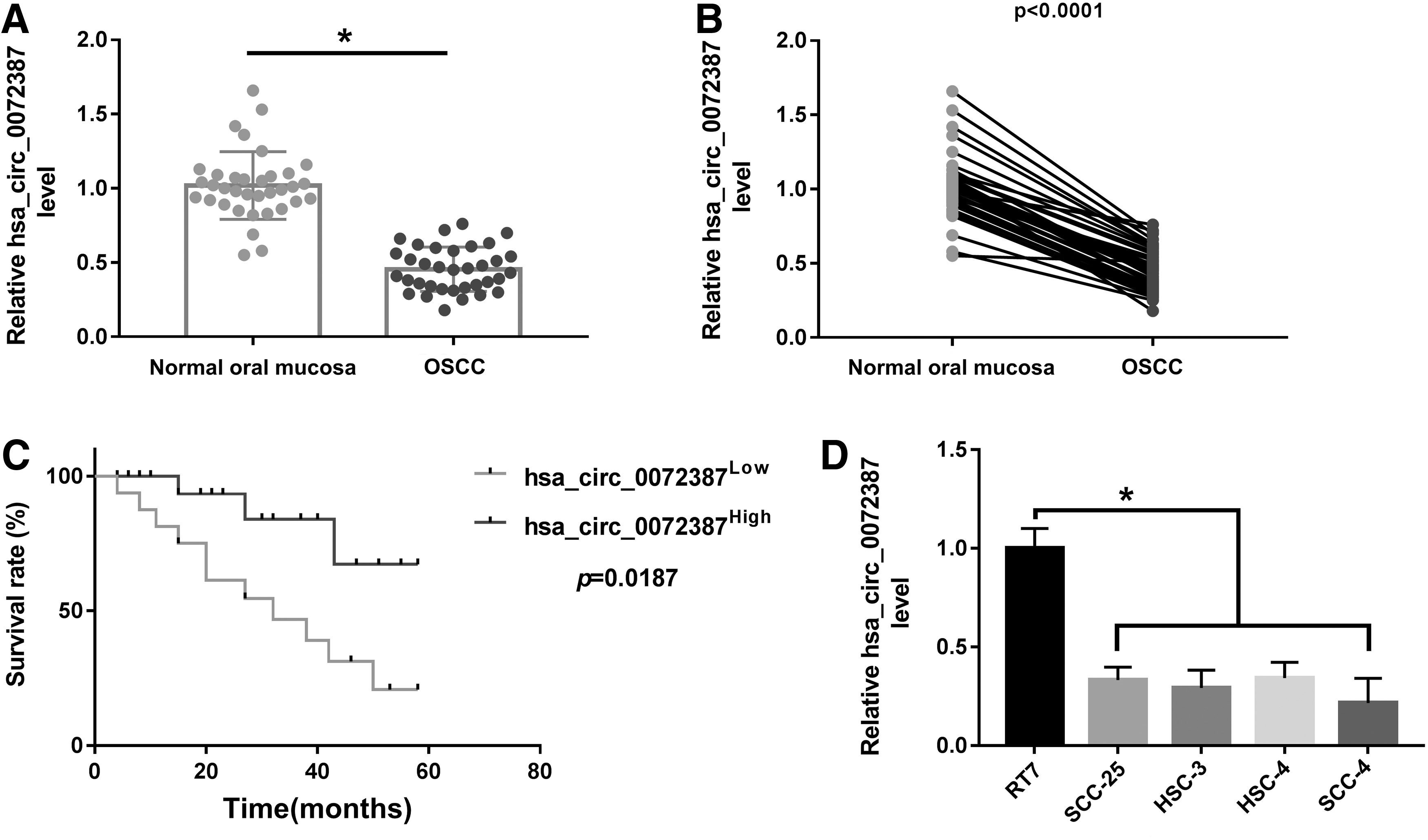
hsa_circ_0072387 was underexpressed in OSCC tissues and cell lines.
Overexpression of hsa_circ_0072387 suppressed cell proliferation, metastasis, and glycolysis in OSCC cells
To clarify the role of hsa_circ_0072387 on proliferation, metastasis, and glycolysis in OSCC cells, hsa_circ_0072387 or circ-NC was transfected into SCC-4 and HSC-3 cells. Next RT-qPCR assay verified that the hsa_circ_0072387 level in SCC-4 and HSC-3 cells was significantly elevated, and was a little higher than that in RT7 cells (Fig. 2A). The cell proliferation was detected through CCK-8 assay, and results indicated that overexpression of hsa_circ_0072387 triggered an obvious repression of cell proliferation in the two cell lines (Fig. 2B).

Overexpression of hsa_circ_0072387 suppressed cell proliferation, metastasis, and glycolysis in OSCC cells in vitro. SCC-4 and HSC-3 cells were transfected with circ-NC or hsa_circ_0072387.
In addition, Transwell chamber assay indicated that the number of both migrated cells and invaded cells was notably decreased in SCC-4 and HSC-3 cells transfected with hsa_circ_0072387 compared with circ-NC-transfected cells (Fig. 2C, D). The levels of Ki-67, N-cadherin and vimentin in SCC-4 and HSC-3 cells transfected with hsa_circ_0072387 were markedly decreased, whereas the level of E-cadherin was elevated (Fig. 2E, F). As given in Figure 2G and H, upregulated hsa_circ_0072387 reduced the lactate production while it facilitated glucose level compared with cells transfected with circ-NC, meaning that glycolysis was repressed.
miR-503-5p was a direct target of hsa_circ_0072387 and was negatively regulated by hsa_circ_0072387
Starbase was applied to conduct bioinformatics analysis, and we found that the hsa_circ_0072387 contained a putative binding region (5′-3′, CCGCUGCUA) for miR-503-5p (5′-3′, GGCGACGAU); hence, it had the condition to absorb miR-503-5p. The predicted binding sites and the mutant are given in Figure 3A. RIP assay demonstrated that the enrichment of hsa_circ_0072387 and miR-503-5p was obviously increased in Anti-Ago2 group of SCC-4 and HSC-3 cells with respect to Anti-IgG group (Fig. 3B). To further confirm the target interaction between hsa_circ_0072387 and miR-503-5p, we performed dual-luciferase activity assay and observed that the luciferase activity of hsa_circ_0072387-wt was decreased owing to miR-503-5p mimic in SCC-4 and HSC-3 cells. However, there was no obvious change in the luciferase activity of hsa_circ_0072387-mut (Fig. 3C).
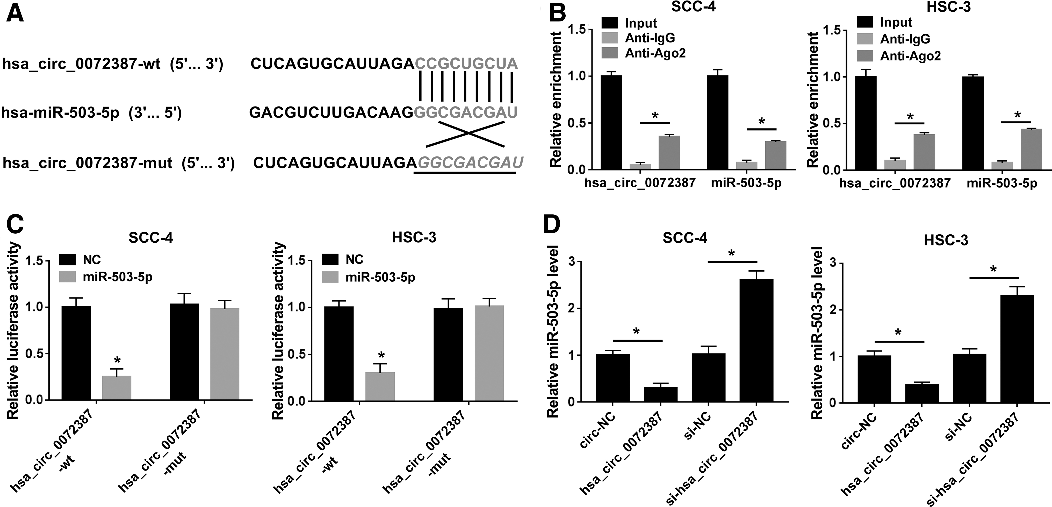
miR-503-5p was a direct target of hsa_circ_0072387 and was negatively regulated by hsa_circ_0072387.
Relative expression of miR-503-5p in transfected SCC-4 and HSC-3 cells was determined by RT-qPCR, and the results showed that miR-503-5p level was downregulated in cells transfected with hsa_circ_0072387 versus cells transfected with circ-NC; In contrast, its level was clearly increased in cells transfected with si-hsa_circ_0072387 (with hsa_circ_0072387 knockdown, Supplementary Figure S1) in comparison with cells transfected with si-NC (Fig. 3D).
miR-503-5p was upregulated in OSCC, miR-503-5p interference restrained proliferation, metastasis, and glycolysis in OSCC cells
To explore the expression level of miR-503-5p in OSCC tissues and cell lines, we conducted RT-qPCR assay. Data suggested that the level of miR-503-5p in OSCC tissues was much higher than that in the corresponding normal oral mucosa (Fig. 4A, B). Following Spearman's analysis indicated an apparent inverse correlation of hsa_circ_0072387 and miR-503-5p expression in OSCC tissues (Fig. 4E). The data from Kaplan–Meier curves suggested that OSCC patients with low miR-503-5p expression harbored higher survival rate in contrast to those with high miR-503-5p expression (Fig. 4C). In addition, the enrichment of miR-503-5p in OSCC SCC-4 and HSC-3 cells was elevated relative to that in RT7 cells (Fig. 4D).
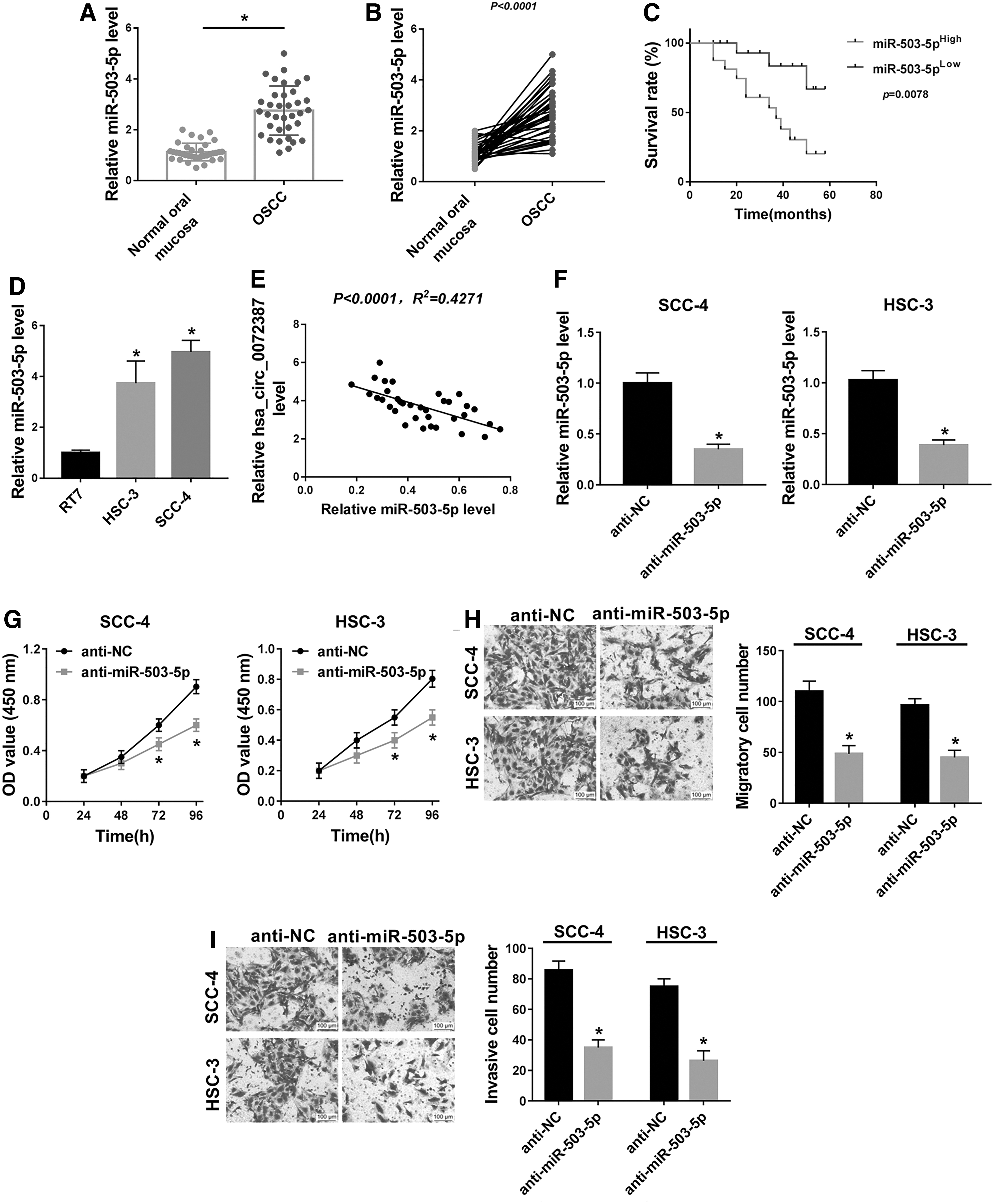
miR-503-5p was upregulated in OSCC, miR-503-5p interference restrained proliferation, metastasis, and glycolysis in OSCC cells.

To investigate the effects of miR-503-5p on proliferation, metastasis, and glycolysis of OSCC cells, SCC-4 and HSC-3 cells with silenced miR-503-5p were constructed. The transfection efficiency was validated by RT-qPCR assay (Fig. 4F). CCK-8 assay revealed that downregulation of miR-503-5p resulted in an obvious decrease of cell proliferation in SCC-4 and HSC-3 cells (Fig. 4G). As given in Figure 4H and I, knockdown of miR-503-5p significantly inhibited the migration and invasion in SCC-4 and HSC-3 cells. The levels of Ki-67, N-cadherin and vimentin were notably reduced, whereas the level of E-cadherin was markedly elevated in miR-503-5p silencing SCC-4 and HSC-3 cells (Fig. 4J, K). In addition, downregulated miR-503-5p efficaciously blocked glycolysis in SCC-4 and HSC-3 cells (Fig. 4L, M) relative to cells transfected with anti-NC.
Overexpression of miR-503-5p partially alleviated hsa_circ_0072387-mediated inhibition on OSCC cell proliferation, metastasis, and glycolysis
To further clarify mechanism of hsa_circ_0072387 in OSCC progression, we co-transfected SCC-4 and HSC-3 cells with circ-NC, hsa_circ_0072387, hsa_circ_0072387+NC, or hsa_circ_0072387+miR-503-5p. Compared with cells cotransfected with hsa_circ_0072387+NC, the miR-503-5p distinctly abolished hsa_circ_0072387-mediated miR-503-5p expression inhibition in cells cotransfected with hsa_circ_0072387+miR-503-5p (Fig. 5A). As given in Figure 5B, hsa_circ_0072387 inhibited the cell proliferation of SCC-4 and HSC-3 cells, whereas reintroduction of miR-503-5p mostly abated this inhibitory impact.
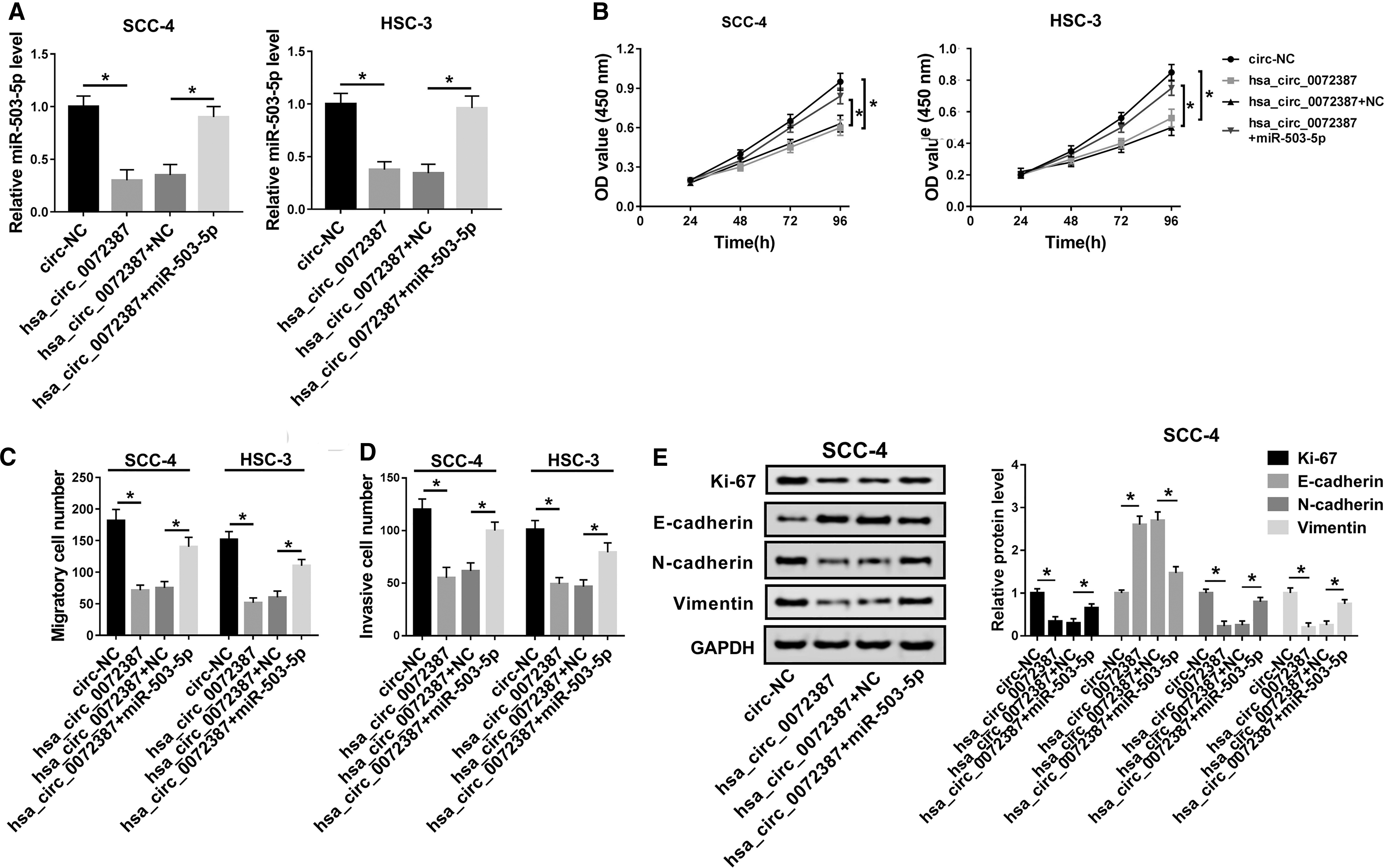
Overexpression of miR-503-5p partially alleviated hsa_circ_0072387-mediated inhibition on OSCC cell proliferation, metastasis, and glycolysis. SCC-4 and HSC-3 cells were cotransfected with circ-NC, hsa_circ_0072387, hsa_circ_0072387+NC, or hsa_circ_0072387+miR-503-5p.

In addition, the Transwell assay indicated that miR-503-5p reversed the suppressed cell migration and invasion in SCC- 4 and HSC-3 cells caused by hsa_circ_0072387 (Fig. 5C, D). Using Western blot analysis, we found that levels of Ki-67, N-cadherin, and vimentin were reduced and that the level of E-cadherin was raised in the hsa_circ_0072387+NC group of SCC-4 and HSC-3 cells, whereas miR-503-5p could partially recover the repression of expression of Ki-67, N-cadherin and vimentin and promotion of E-cadherin expression in the hsa_circ_0072387+miR-503-5p group (Fig. 5E, F). Besides, additional miR-503-5p also reverted hsa_circ_0072387-mediated glycolysis reduction in SCC-4 and HSC-3 cells compared with cells co-transfected with hsa_circ_0072387+NC (Fig. 5G, H).
Discussion
OSCC mainly results from excessive intake of alcohol and tobacco. 25 At present, the treatment of OSCC consists of surgery and/or radiation with chemotherapy. 26 Therefore, a deeper understanding of molecular mechanism of OSCC pathologies provides a new insight to promote effective therapeutic approaches for OSCC.
Previous studies have suggested that circRNAs play dual roles in tumor progression, including OSCC. Zhao et al. reported that circUHRF1 (hsa_circ_0002185) sponged miR-526b-5p to accelerate OSCC tumorigenesis in vitro and in vivo. 27 Circular matrix metalloproteinase 9 (hsa_circ_0001162), a circRNA derived from matrix metalloproteinase 9, was deemed as an effective diagnostic and prognostic biomarker for patients with OSCC, acting as a metastasis-promoting gene. 28
On the contrary, quantities of evidences demonstrated that certain circRNAs also exerted tumor suppressor function in OSCC progression. Circ-PKD2 was distinguished to be a sponge of miR-204-3p to upregulate adenomatous polyposis coli 2 (APC2) level, so as to suppress proliferation, migration, and invasion and trigger apoptosis and cell cycle of OSCC SCC15 cells. 12 Su et al. pointed out that hsa_circ_0005379 impeded migration, invasion, and proliferation of OSCC cells in vitro and hindered OSCC tumor growth in vivo. 29 hsa_circ_0055538 expression was low in OSCC tumor tissues and cell lines, and high expression of hsa_circ_0055538 caused malignant biological behavior of OSCC cells and tumor growth inhibition. 30
As for hsa_circ_0072387, only one related literature was searched, and the report suggested that this circRNA was significantly downregulated in OSCC. 13 Similarly, the present data manifested that expression level of hsa_circ_0072387 was downregulated in OSCC tissues and cell lines. Subsequently, gain-of-function assays were carried out to explore the role of hsa_circ_0072387 in OSCC progression. After CCK-8 assay, Transwell assay, Western blot assay, and commercial kits were used, the results indicated that overexpression of hsa_circ_0072387 suppressed cell proliferation, migration, and invasion abilities, EMT, and glycolysis of OSCC SCC-4 and HSC-3 cells in vitro. Data in this study, taken together with foregoing studies revealed that hsa_circ_0072387 played a tumor-suppressor role in OSCC progression.
As is well known, CircRNAs are well known to be able to act as sponges of miRNAs to regulate miRNA expression and then modulate tumorigenesis. 31,32 Therefore, the authors tried to search the downstream miRNAs of hsa_circ_0072387 to investigate the mechanism by which hsa_circ_0072387 was involved in the OSCC progression. Bioinformatics analysis with Starbase was conducted, and it was observed that miR-503-5p could bind to hsa_circ_0072387. Both RIP assay and dual-luciferase reporter assay confirmed the targeted relationship between hsa_circ_0072387 and miR-503-5p.
To date, the functional role of miR-503-5p in modulating biological processes through regulating target gene expression was reported, acting as an inhibitory miRNA. 33 The study of Su et al. demonstrated that miR-503-5p inhibition could partially reverse silence of metastasis-associated lung adenocarcinoma transcript 1 (MALAT1)-mediated proliferation inhibition and apoptosis in ovarian cancer SKOV3 and OVCAR3 cells serving as tumor suppressor. 22 Overexpression of miR-503-5p suppressed migration, invasion, and EMT of HCC HCCLM3 cells by targeting WEE1, a prognosis predictor of HCC. 23
On the contrary, the oncogenic role of miR-503-5p was also validated. Unbiased miRNA array screening revealed that miR-503-5p was elevated in two oxaliplatin (OXA)-resistant colorectal carcinoma cell lines, and miR-503-5p conferred resistance to OXA-mediated cell apoptosis and tumor growth repression in vitro and in vivo by downregulating PUMA. 34 Besides, in OSCC, using the deep sequencing and bioinformatics analysis, miR-503-5p was observed to be upregulated in OSCC tissues and directly target RORA, indicating its potential in OSCC treatment. 24
In this study, miR-503-5p abundance was increased in OSCC tissues and cell lines, and miR-503-5p level was inversely correlated with hsa_circ_0072387 level in OSCC tissues. In addition, miR-503-5p was negatively regulated by hsa_circ_0072387 in OSCC SCC-4 and HSC-3 cells. The authors further investigated the effect of miR-503-5p on the proliferation, metastasis, and glycolysis in OSCC cells, and found that the knockdown of miR-503-5p significantly repressed the proliferation, metastasis, and glycolysis in OSCC SCC-4 and HSC-3 cells, implying the oncogenic role of miR-503-5p in OSCC.
To validate whether hsa_circ_0072387 repressed OSCC progression by targeting miR-503-5p, rescue experiments were implemented, including CCK-8 assay, Transwell assay, Western blot assay, and so on. It was concluded that gain of miR-503-5p could revert the inhibitory effects on cell proliferation, metastasis, and glycolysis caused by hsa_circ_0072387, suggesting that hsa_circ_0072387 exerted its tumor suppressor role by targeting miR-503-5p.
In summary, the downregulation of hsa_circ_0072387 and the upregulation of miR-503-5p in OSCC tissues and cells were observed. Next, the fact that hsa_circ_0072387 was able to inhibit cell proliferation, metastasis, and glycolysis in OSCC cells was demonstrated. In addition, miR-503-5p was identified as a direct downstream gene of hsa_circ_0072387. It was demonstrated that hsa_circ_0072387 repressed cell proliferation, metastasis, and glycolysis by targeting miR-503-5p. In conclusion, hsa_circ_0072387 might block cell proliferation, metastasis, and glycolysis by directly targeting miR-503-5p. This might provide a novel therapy thought for OSCC.
Availability of Data and Materials
The analyzed data sets generated during this study are available from the corresponding author on reasonable request.
Ethics Approval and Consent to Participate
This study was approved by the ethical review committee of The Third Hospital Affiliated to Qiqihar Medical College
Footnotes
Disclosure Statement
No competing financial interests exist.
Funding Information
No funding was received for this article.
Supplementary Material
Supplementary Figure S1
References
Supplementary Material
Please find the following supplemental material available below.
For Open Access articles published under a Creative Commons License, all supplemental material carries the same license as the article it is associated with.
For non-Open Access articles published, all supplemental material carries a non-exclusive license, and permission requests for re-use of supplemental material or any part of supplemental material shall be sent directly to the copyright owner as specified in the copyright notice associated with the article.
