
Abstract
Background:
This work aimed to evaluate the influence of two chelators: DOTA(SCN) and DOTA(NHS) on radioimmunotherapy using 177Lu-DOTA-Rituximab preparations in murine lymphoma xenograft models. Subsequently, based on animal data, the organ radiation-absorbed doses were extrapolated to humans (adult male).
Materials and Methods:
Therapeutic efficacy of 177Lu-DOTA-Rituximab was evaluated in male nude mice bearing either Raji (B lymphocyte, CD20+) and Jurkat (T lymphocyte, CD20) xenografts, utilizing an anti-CD20 antibody—Rituximab conjugate with either DOTA(SCN) or DOTA(NHS). The DOTA-Rituximab conjugates were prepared in the form of freeze-dried kits.
Results:
All radioimmunoconjugates were obtained with high radiolabeling yield (radiochemical purity, RCP > 95%) and specific activity of ca. 0.5 GBq/mg. Therapeutic effects of 177Lu-DOTA-Rituximab were observed in animals regardless whether DOTA(SCN) or DOTA(NHS) were used for conjugation. Importantly, therapy involving 177Lu-DOTA-Rituximab was more effective than use of Rituximab alone.
Conclusions:
The degree of antitumor efficacy was dependent on the type of applied bifunctional chelators conjugated to mAb. However, this difference was not statistically significant. Dosimetry calculations showed that the absorbed radiation doses extrapolated to humans were very low for osteogenic cells regardless of the conjugates. Organs like the liver and spleen, treated with 177Lu-DOTA(SCN)-Rituximab, showed similar radiation absorbed doses when compared with 177Lu-DOTA(NHS)-Rituximab.
Introduction
Although antibodies with a very high specificity for their target antigen overexpressed in tumors can display a direct therapeutic effect, their effectiveness significantly increases when combined with therapeutic radionuclides. One of the first mAb implemented in oncology, and still the most widely used, is the CD20-targeting mAb rituximab (the chimeric mouse/human monoclonal antibody). Contrary to other drugs and toxins, immunoconjugates of radiolabeled mAbs can kill cells that are at a distance from the targeting site, depending on the choice of radionuclide, without the mAb conjugate being internalized. Preclinical and clinical applications of 131I-,1 99mTc-2, 177Lu-labeled, 3 or 90Y- 4 –6 antiCD20 have been previously reported. 7 –12 Other radionuclides used for radiolabeling of antibodies for tumor imaging or therapy were 64Cu, 111In, 89Zr, 153Sm, 166Ho, and 188Re.
Radiolabeling antibodies with radiometals typically involves the use of a bifunctional chelating (BFC) agent, which is able to stably coordinate the isotope and also allows for covalent attachment to protein functional groups. Such radioimmunoconjugates bind to the CD20 antigen, a transmembrane phosphorylated protein located on pre-B and matured B-lymphocytes, and destroy tumor cells, by combining radiolytic and biological mechanisms of action. 13 –15
Several reports have described the radiolabeling of anti-CD20 with 177Lu and 90Y after conjugation with DOTA or DTPA because they form very stable complexes. 16,17 However, chemical modification of antibodies may alter their properties, either chemically or biologically, for example, immunoreactivity. Based on the recent experiments, 18 this article focuses on the comparisons of the influence of two DOTA chelators (p-SCN-Bn-DOTA and DOTA-NHS-ester) conjugated to Rituximab, on antitumor activity of the 177Lu-radiolabeled preparations. Radiotherapeutic efficacy of 177Lu-DOTA-Rituximab preparations were assessed in lymphoma tumor-bearing mice.
Additionally, to evaluate potential for therapeutic application, the absorbed radiation dosage was extrapolated to humans using the OLINDA software—Organ Level Internal Dose Assessment Code.
Materials and Methods
Antibody and reagents
Rituximab (MabThera®) was purchased from Roche Pharma AG (Germany). The BFC: p-SCN-Bn-DOTA (2-(4-isothiocyanatobenzyl)-1,4,7,10-tetraazacyclododecane-1,4,7,10-tetraacetic acid) and DOTA-NHS-ester (1,4,7,10-tetraazacyclododecane-1,4,7,10-tetraacetic acid mono (N-hydroxysuccinimide ester) were purchased from Macrocyclics. Lutetium-177 (LutaPol) of specific activity (SA) higher than 555 MBq/mg Lu was produced at the Radioisotope Centre POLATOM (Poland). Chemicals and solvents were of p.a. grade.
Immunoconjugates preparation and radiolabeling
177Lu-DOTA(SCN)-Rituximab and 177Lu-DOTA(NHS)-Rituximab immunoconjugates were prepared as previously described. 18 The specific activities of the radioimmunoconjugates were ca. 0.5 GBq/mg. The final radiochemical purity was more than 97% measured by ITLC SG (stationary phase: silica gel; mobile phase: methanol/0.4 M ammonium acetate [1:1, v/v]) and HPLC (BioSep-SEC-S3000 PEEK column; 0.1 M phosphate buffer pH 5.8 as an eluent; 1 mL/min flow rate). Due to a high radiochemical purity (RCP) of the obtained radioimmunoconjugates, no additional purification steps were necessary.
Cell lines
Two human leukemia cell lines: Raji (B lymphocyte, CD20+) and Jurkat (T lymphocyte, CD20−), were purchased from American Type Culture Collection (ATCC). The cells were maintained in RPMI 1640 culture medium supplemented with 10% heat inactivated fetal bovine serum (ThermoScientific), 1% L-glutamine (Gibko), and 1% antibiotics (penicillin and streptomycin, ATCC) in 5% CO2 humidified incubator at 37°C. Cell viability and cell count were determined by trypan blue assay using the automated cell counter Countess®.
Animal studies
Animal studies were approved by the Hungarian Institutional Ethics Committee (IKEB) (No.: PEI/001/2073-6/) and were performed at the National Research Directorate on Radiobiology and Radiohygiene in Budapest. In accordance with the nomenclature on the Charles River pages Nude BALB/c mice after implementation into the right and left flank of 1 × 10⁶ of Raji cells and the same number of Jurkat cells as the control, respectively. Twenty-one days postimplantation of cells when the average tumor size achieved >40 mm3 mice were randomly divided into five groups (n = 5 for each group).
Antitumor activities were investigated in the following groups: Group (1) received a single administration of 177Lu-DOTA(SCN)-Rituximab (15 MBq/kg) Group (2) received a single administration of 177Lu-DOTA(SCN)-Rituximab (30 MBq/kg) Group (3) received a single administration of 177Lu-DOTA(NHS)-Rituximab (15 MBq/kg) Group (4) received a single administration of 177Lu-DOTA(NHS)-Rituximab (30 MBq/kg) Group (5) received a single administration of cold Rituximab (control group).
In each case the amount of injected antibody was ca. 0.01 mg. To monitor the tumor response, animals were weighed twice per week, hematological parameters and tumor volumes were measured weekly for 8 weeks. Tumor volumes were measured using calipers, and approximated using the ellipsoid formula: V (mm3) = π/6 × length × width × height. 19 The antitumor activity was evaluated by growth delay factor (GDF), defined as the mean number of tumor-doubling time (TD) gained by the treatment, and by calculating the T/C values defined as the ratio of the relative tumor volumes of treated mice (RTVtr) to that of control mice (RTVcon).
Extrapolation of absorbed dose to human
The dose extrapolation to humans involved scaling of the biodistributions and the subsequent calculation of the absorbed radiation dose from the observed data. The biodistribution scaling was based on a method considered a relative mass scaling where the SA within a certain human organ is equal to the SA within the same mouse organ multiplied by the ratio of the body mass of human to mouse. 20,21
The biodistribution data of 177Lu-DOTA(SCN)-Rituximab and 177Lu-DOTA(NHS)-Rituximab in tumor-bearing (grafted with Raji cells) male Rj: NMRI-Foxn1nu/Foxn1nu mice (Janvier Lab., France) were used for dosimetry calculations. 3 Radiation doses for a selected group of organs were calculated using the OLINDA/EXM® software for internal dose assessment presented as an accurate and standardized method for the calculation of radiation (version 1.1, copyright Vanderbilt University, 2007).
Statistical analyses
Statistical analysis included the calculation of averages and standard deviation of the results compared using the Student's t-test. The calculations were performed using the GraphPad Prism 8 software.
Results and Discussion
Conjugation of chelating agents to anti-CD20 and radiolabeling
The DOTA derivatives were conjugated to Rituximab at different molar ratios: 10:1 for DOTA(SCN) and 100:1 for DOTA(NHS), which resulted in different numbers of conjugated chelator molecules per mAb. The average number of DOTA molecules attached to the antibody molecule was determined by radiolabeling assay using 64Cu. 18 Literature data indicate that a small initial quantity of DOTA(NHS) used for the reaction resulted in a low radiochemical yield due to the insufficient number of DOTA attached to mAb molecule. 22 Based on this observation, two immunoconjugates were selected for further investigation: (1) DOTA(SCN)-Rituximab (with 5 mole of DOTA coupled to 1 mole of mAb) and (2) DOTA(NHS)-Rituximab (with 18 mole of DOTA coupled to 1 mole of mAb). The DOTA-Rituximab conjugates were radiolabeled with 177Lu to SA of ca. 0.5 GBq/mg and with radiochemical yields ranging from 97% to 100%.
Radioimmunotherapy studies in double xenografted nude mice
After the tumors developed at 3–4 weeks, mice with a mean tumor volume of 42 ± 6 mm3 (with Raji cell) and 49 ± 8 mm3 (with Jurkat cells) were treated in five groups as described earlier. The effect of treatment with 177Lu-DOTA(SCN)-Rituximab and 177Lu-DOTA(NHS)-Rituximab on tumor growth in athymic nude BALB/c mice with Raji and Jurkat xenografts are shown in Figures 1 and 2. The tumor size (for Raji and Jurkat) significantly increased in the group treated with the native antibody (control) in comparison to the groups treated with 177Lu–Rituximab. In the group with the tumor-bearing Raji mice, both, 177Lu-DOTA(SCN)-Rituximab and 177Lu-DOTA(NHS)-Rituximab preparations revealed inhibiting activity on the tumor growth and size, in comparison to the control group treated with the native antibody (Table 1).
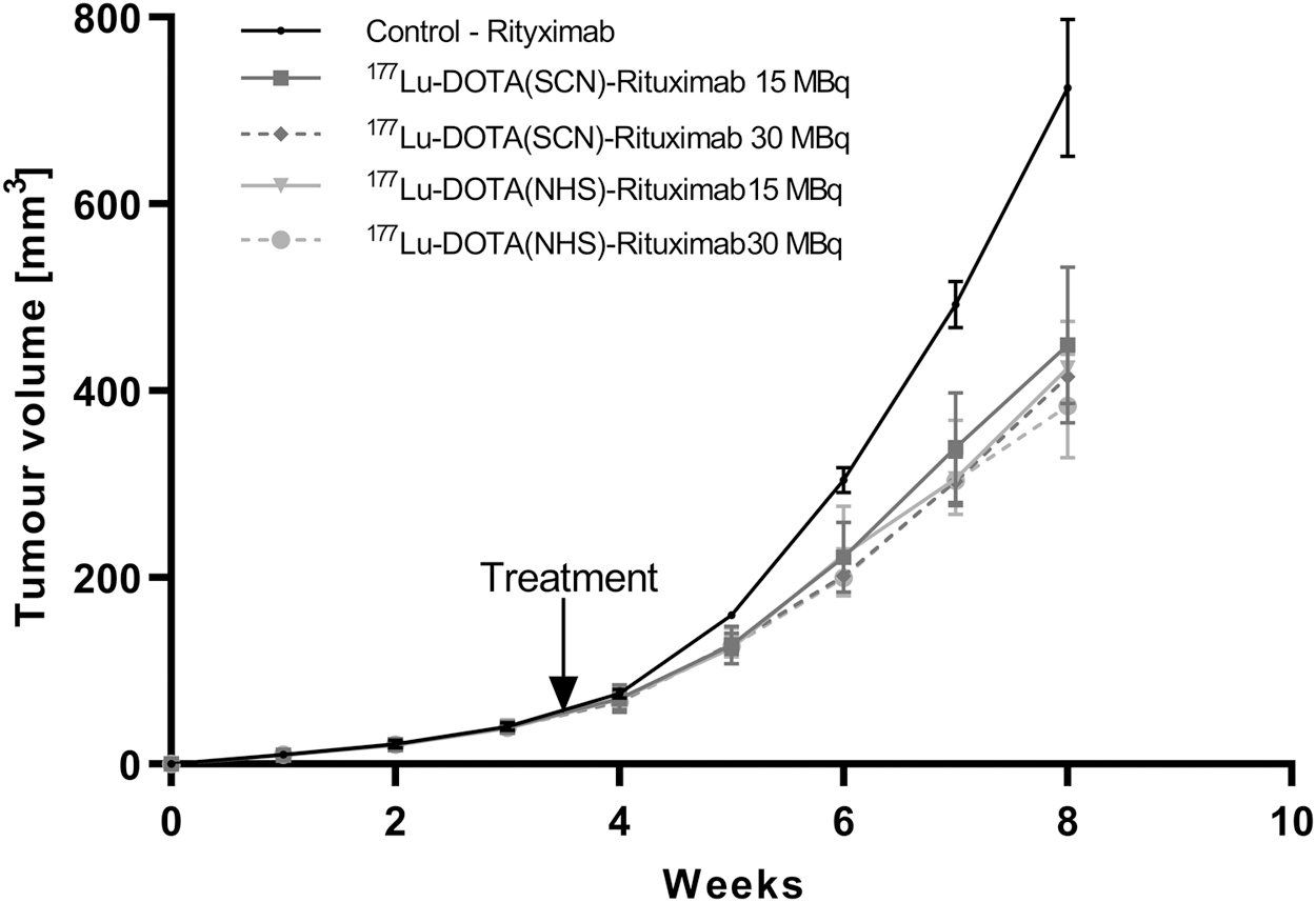
Changes of tumor growths after 177Lu-DOTA-Rituximab treatment in mice grafted Raji cells.
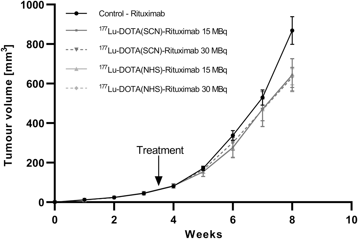
Changes of tumor growths after 177Lu-DOTA-Rituximab treatment in mice grafted Jurkat cells.
Summary of the Antitumor Activity of the Tested DOTA-Rituximab Complexes Observed in Mice Xenografted with Raji Cell
CI, confidence interval; GDF, growth delay factor; T/C, ratio of the relative tumor volumes of treated mice (RTVtr) to that of control mice (RTVcon); TD, tumor-doubling time.
However, the rate of tumor growths in the group treated with 177Lu-DOTA(NHS)-Rituximab was slightly lower in comparison to 177Lu-DOTA(SCN)-Rituximab, but the difference was not statistically significant. The most intensive antitumor activity was observed for the 177Lu-DOTA(NHS)-Rituximab, showing an activity of 30 MBq/kg. The tumor GDF was 0.22, whereas for the group treated with 177Lu-DOTA(SCN)-Rituximab it was 0.16.
In the group with the Jurkat tumor-bearing mice, no statistically significant inhibition of tumor growth was observed in the treated groups in comparison with the control group. Negative control (Jurkat cells) showed a higher T/C value 0.73–0.74 (at week 8 of the experiment) and a lower TD, which means faster tumor growth comparing to tumors growth induced by Raji cells in the animals treated with the same preparation (Table 2). Minor differences may be due to the presence of the radioisotope (Fig. 2). It is also possible that the observed low decrease of tumor growth was caused by appearing uncontrolled focal of fragile tumor blood vessels, leading to passive transport of 177Lu-DOTA-Rituximab.
Summary of the Antitumor Activity of the Tested DOTA-Rituximab Complexes Observed in Mice Xenografted with Jurkat Cell
CI, confidence interval; GDF, growth delay factor; T/C, ratio of the relative tumor volumes of treated mice (RTVtr) to that of control mice (RTVcon); TD, tumor-doubling time.
Consistent with our previous results, 18 we found that the DOTA chelators coupled to mAb molecule influenced the stability of radiolabeled complexes. It has been shown that DOTA-NHS-ester demonstrated slightly improved in vivo stability and caused a lower aggregation of mAb compared to p-SCN-Bn-DOTA. In the present study we observed a significant difference in the increase of the GDF for the DOTA(NHS) in comparison to DOTA(SCN) Rituximab conjugates [the difference between the overall GDFs is ca. 20% higher for DOTA(NHS).
The hematological and biochemical parameters remained within the normal range in all animals. Only small, nonsignificant, alterations were observed in WBC, RBC, and platelet counts after radioimmunoconjugate administration.
Dosimetry
Radiation absorbed dose to critical organs or tissue usually limits the amount of radioactivity that can be safely administered in radiotherapy. Most often the organs critically affected by radiolabeled antibodies are the liver, spleen, bone, and/or red marrow.
In this study, unsurprisingly it was noted (Table 3) that the organs/tissues receiving the highest absorbed dose from 177Lu-DOTA-Rituximab were the spleen [1.22 mGy/MBq for 177Lu-DOTA(SCN)-Rituximab and 1.40 mGy/MBq for 177Lu-DOTA(NHS)-Rituximab] and the liver [0.64 mGy/MBq for 177Lu-DOTA(SCN)-Rituximab and 0.57 mGy/MBq for 177Lu-DOTA(NHS)-Rituximab]. We observed very low absorbed radiation doses in osteogenic cells [0.21 mGy/MBq for 177Lu-DOTA(SCN)-Rituximab and 0.28 mGy/MBq for 177Lu-DOTA(NHS)-Rituximab] and in red marrow [0.01 mGy/MBq for 177Lu-DOTA(SCN)-Rituximab and 0.02 mGy/MBq for 177Lu-DOTA(NHS)-Rituximab].
Extrapolated Absorbed Dose in Adult Man (mGy/MBq)
Bold values indicate statistical significance.
Conclusions
The evaluation of the therapeutic efficacy of 177Lu-DOTA-Rituximab in murine lymphoma xenograft models confirmed that both immunoconjugates 177Lu-DOTA(NHS)-Rituximab and 177Lu-DOTA(SCN)-Rituximab demonstrate in vivo anticancer activity inhibiting tumor growth. This antitumor efficacy was dependent on the type of conjugated bifunctional chelators. The study pointed out that 177Lu-DOTA(NHS)-Rituximab was slightly more effective in suppressing the growth of tumor in athymic nude BALB/c mice.
Footnotes
Acknowledgments
This work was performed and partly financially supported within the framework of the International Atomic Energy Agency Co-ordinated Research Project No. 16639 “Development and Preclinical Evaluation of Therapeutic Radiopharmaceuticals Based on 177Lu and 90Y-Labeled Monoclonal Antibodies and Peptides.”
Disclosure Statement
There are no existing financial conflicts.
Funding Information
No funding was received for this article.