
Abstract
Objective:
Nanocapsules play a role in the targeted delivery of chemotherapy drugs. However, the traditional technology for preparation of nanocapsules is relatively complex with poor controllability, leading to large differences batch to batch. This study aimed to evaluate the quality of drugs-loaded nanocapsules (Drugs-NCs) fabricated by coaxial capillary microfluidic device, and inhibitory effect on malignant tumors.
Materials and Methods:
In this study, oxaliplatin, irinotecan, and 5-fluorouracil were selected as chemotherapy drugs, and Drugs-NCs were prepared by coaxial glass capillary microfluidic device. Next, transmission electron microscope was utilized to characterize surface morphology and particle size distribution of Drugs-NCs. Then, high performance liquid chromatography was used to determine the drug loading and encapsulation efficiency. Dialysis method was performed to measure the drug release of Drugs-NCs in vitro. To study the effects of Drugs + NCs on tumor growth in vivo, BALB/c (nu/nu) nude mice were used in vivo experiments.
Results:
The Drugs-NCs were spherical and uniform in size (103.4 nm). Besides, the encapsulation efficiencies of oxaliplatin, irinotecan, and 5-fluorouracil were 97.0%, 95.7%, and 15.6%, respectively. Moreover, drugs encapsulated in the nanocapsules released less and was pH-dependent, with more rapid release being observed at pH 5.5 group compared with pH 7.4 group. MTT assay and in vivo experiments indicated the inhibitory effect of Drugs-NCs on malignant tumors.
Conclusion:
The prepared nanocapsules had potential tumor targeting. Furthermore, coaxial capillary microfluidic device could be used as a promising microfluidic technology to fabricate multiple Drug-NCs.
Introduction
Currently, a great number of nano-formulations such as Doxil, Daunoxome, Abraxane, SP1049C, and Genexol-PM have been adopted for treatment of malignant tumor. Nano-formulations have become a hot topic for researchers in recent decades, because it plays an important role in improving the distribution of drugs in the lesion site and reducing adverse reactions. 1 As a means of drug delivery, nano-formulations could enhance the water solubility, stability, bioavailability of drugs, and prolong their circulation in blood compartments. 2 –4 They have been widely used in research and development of pharmaceutical preparations, especially in the research and development of hydrophobic drugs. In particular, currently marketed nano-formulations contain a single active component. 5
However, multidrug combination is an important principle in clinical cancer treatment, because chemotherapy may lead to a subclone, that is, a product that is not responsive to therapeutic drugs, and may also induce treatment-related mutations and generate new drug resistance. 6 FOLFIRINOX (5-fluorouracil, leucovorin, irinotecan, and oxaliplatin) is a common chemotherapy for the treatment of pancreatic cancer, gastrointestinal cancer, and colorectal cancer. 7 –9 However, adverse reactions are subsequently increased after the combined medication treatment, which is difficult for most of patients to tolerate. 10,11 Therefore, it is urgent to explore an innovative nano-drug delivery system involving multiple drugs.
As innovative nano-drug delivery systems containing multiple drugs that can reduce toxicity, multidrug nanocarriers are frequently reported. 12,13 Nanocapsules have special chemical and biological properties due to their hierarchical composition and structure, which can support a variety of active ingredients and have graded release effects. 14,15 Moreover, the delivery system in the form of nanoparticles makes use of the permeability enhancement and EPR effect to selectively accumulate anticancer drugs in the tumor and play a significant antitumor effect while reducing the drug toxicity. 16 The structures and surface properties to the inner and outer wall of nanocapsules vary in different preparation methods. The traditional technology for preparation of nanocapsules are emulsion interfacial self-assembly, emulsion polymerization, and emulsion template method. 17 Among them, emulsion interfacial self-assembly could better control the structure of polymer hollow spheres and the particle morphology of nanocapsules, but its preparation process is relative complex, the stability of batch to batch is poor, and the particle size of nanocapsules is not easy to control. 18
With the development of microfluidic technology, the coaxial glass capillary microfluidic device has attracted researchers' attention. 19 In a coaxial capillary microfluidic device, capillary tubes of different sizes are assembled and two or more solutions are injected into them, and then through nesting between capillary ports of different micron scale, three-dimensional (3D) shear fluid is formed to form dispersed droplets. 20 The initial research and application direction of the device is used as an emulsion generator to further treat the emulsion to prepare microcapsule. 21 In 2015 Othman et al. 22 first prepared PCL and PLA nanoparticles using a coaxial capillary glass tube microfluidic device. The particle sizes of the prepared nanoparticles were between 190 and 650 nm by adjusting the flow ratio of the internal phase (oil phase) to the external phase (aqueous phase). Subsequently, Liu et al. 23 also prepared paclitaxel-coated poly (lactic-co-glycolic acid) (PLGA), HPCS, and AcDX nanoparticles. Additionally, they have indicated that compared with traditional emulsion polymerization method, the paclitaxel-coated nanoparticles prepared by coaxial glass capillary microfluidic device have better encapsulation efficiency (EE) and uniformity of particle size.
This study focuses on quality evaluations of nanocapsules containing multiple drugs (oxaliplatin, irinotecan, and 5-fluorouracil) prepared by one-step method with coaxial capillary microfluidic device aiming their use in malignant tumor treatment. We also further reveal the inhibitory effect of prepared nanocapsules on tumors and tumor targeting of new dosage forms of nanodrugs.
Materials and Methods
Materials
Oxaliplatin (Oxa, API, purity >98.6%) and irinotecan (CPT-11, API, purity >99%) were gifts from Hisun Co., TaiZhou, Zhejiang, China. 5-fluorouracil (5-Fu, API, F100149) was purchased from Aladdin Coal, China. Polyvinylalcohol (PVA), Tween 20, and Tween 80 were purchased from Sigma-Aldrich (Dorset, UK). All chemicals were of analytical grade. The pure water was produced by reverse osmosis (Milli-Qs, Millipore). Human pancreatic cancer SW1990 cells and Panc-1 cells were purchased from the Shanghai Culture Collection. Healthy BALB/c (nu/nu) nude mice (male, 4–6 weeks, 20–30 g) were obtained from the experimental animal center of Wenzhou Medical University, and the laboratory animal number was SYXK2015-0009. A two-component epoxy glue and Sutter P-97 Flaming/Brown micropipette puller were purchased from ITW Devcon, Rushden and Linton Instrumentation, Norfolk, respectively (UK). Injectors were obtained from SGE Analytical Science Co. (1, 2.5, 5, and 10 mL; Australia).
One end of the cylindrical capillary (inner and outer diameter of 580 and 1000 μm; World Precision Instruments, Inc.) was tapered to a diameter of 20 μm using a micropipette puller (P-97; Sutter Instrument Co.), and then further enlarged to 100 μm. The tapered capillary was inserted into another bigger cylindrical capillary with an inner dimension of around 1100 μm (Vitrocom) and coaxial alignment. A transparent epoxy resin (5 Minute® Epoxi, Devcon) was used to seal the capillaries when required. Two miscible liquids were injected separately into the microfluidic device in the same direction through polyethylene tubes attached to syringes at constant flow rates. The flow rates of different liquids were controlled by pumps (PHD 2000; Harvard Apparatus).
Fabrication of the coaxial glass capillary microfluidic device
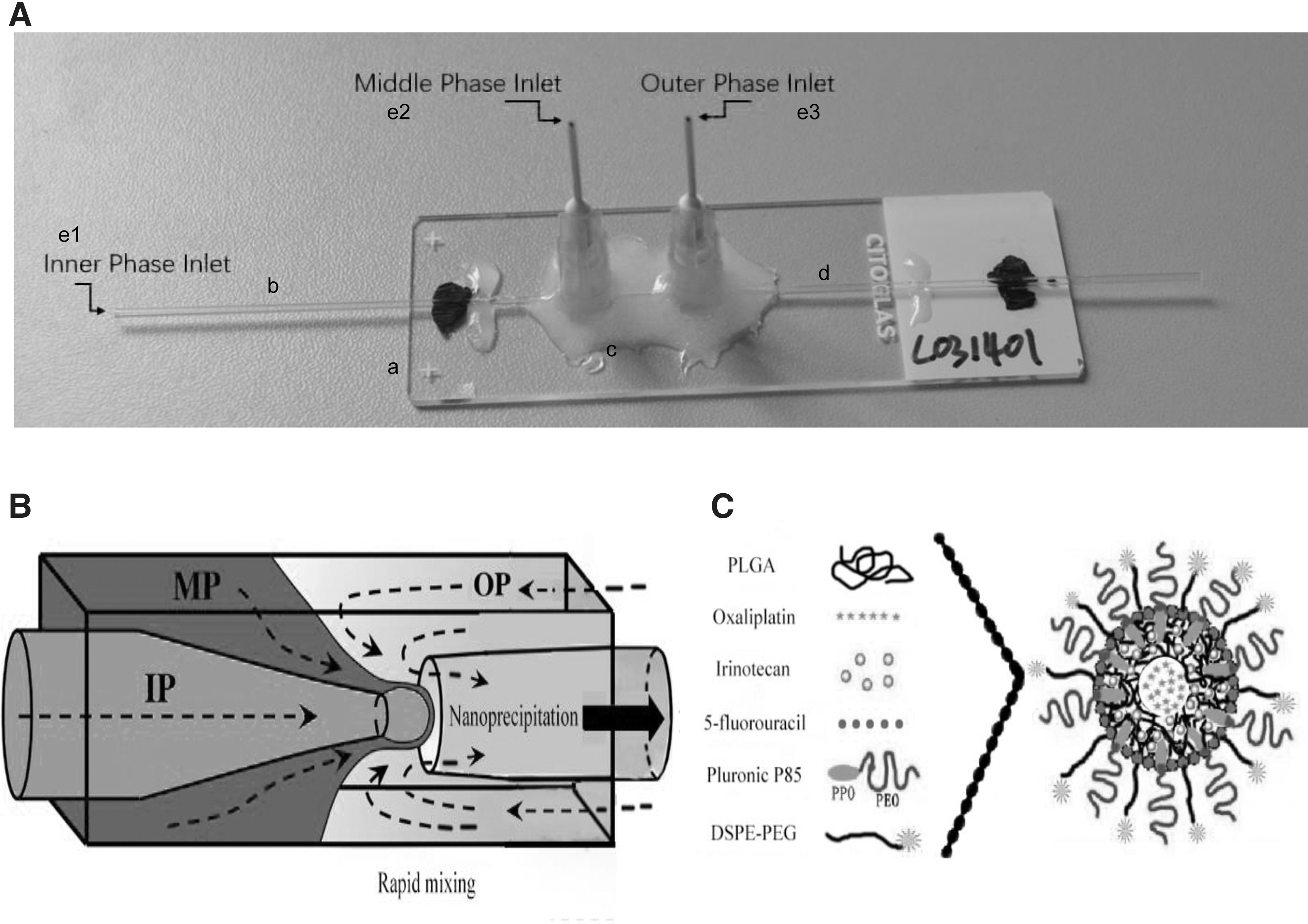
A 3D coaxial microfluidic nanoprecipitation device was fabricated by assembling borosilicate glass capillaries on a glass slide. The capillary glass microfluidic chip was composed of the following parts (Fig. 1A): (1) the middle part of glass slide (No. 7101; Chinese sailing brand) was used to support the glass microtubules and fixed needles of the chip, and the peripheral part was used to fix the chip while operation; (2) one end of inner phase glass capillary tube extended 5 mm out of the slide to connect the sample inlet pipe, and the other end could be extended into the middle phase glass capillary tube, namely outlet of the inner phase solution; (3) middle and outer phase glass capillary tube were nested with the inner phase glass capillary tube, and connected point needles at both ends; (4) the outlet of flow channel after formation of nanoparticles was a constricted part embedded in the stable phase channel; (5) several point needles (e1, e2, and e3) can be respectively used for the entry of inner, middle, and outer phases).
Preparation of Drug-NCs
Microfluidic device
Because colloidal particles could self-assemble into ordered structure in glass capillary channel, the channel must be kept clean, otherwise the impurity ion would seriously affect the self-assembly process of colloidal particles. In the fabrication of glass capillary microfluidic chips, high purity borosilicate glass capillary tubes (World Precision Instruments Co.) were used and cleaned by ethanol, in addition to syringe, sample probe and PE tubes (1.32 mm O.D., 0.86 mm I.D., #BB31695-PE/5; Scientific Commodities Company). In this study, seven apertures were set up at the end of narrowing orifice of the inner phase channel, including 80, 100, 150, 200, 250, 300, and 350 μm. The length of the narrowing part was set to 1 cm. The glass capillary microfluidic chips prepared by the method mentioned in 2.1 were used. The sample inlet was connected to Teflon microtubule and syringe, which pushed the solution into the microfluidic chip under the impetus of peristaltic pump (LSP01-1A; Baoding Lange Constant Current Pump Co., China). First, the water phase or the oil phase solution was discharged according to the experimental conditions, followed by inner phase, and then out of the middle phase.
Phase solution
The solvents that can dissolve PLGA included dichloromethane (density 1.32 g/mL), chloroform (density 1.48 g/mL), acetonitrile (density 0.98 g/mL), two methyl sulfoxides (density 1.10 g/mL), and N-dimethyl formamide (density 0.94 g/mL). Considering the density compatibility of the three-phase solution to stabilize emulsion, we chose acetonitrile dissolved PLGA as middle phase.
Each phase solutions were as follows: Inner phase: 5% glucose solution of Oxa; Middle phase: acetonitrile solution of CPT-11, DOTAP and PLGA; Outer phase: alcohol aqueous solution of 5-Fu, Pluronic P85, and DSPE-PEG; Stable phase: alcohol aqueous solution of Pluronic P85 and DSPE-PEG; Receiving solution: PBS buffer containing 0.5% PVA.
Flow rate control
The prepared solution was loaded into a syringe, removing excess bubbles to increase the stability of fluid. Then the parts of the microfluidic device were connected under the condition of sealing. During the experiment, glass capillary tube channels should be kept as horizontal as possible and decreased vibration to reduce fluid instability. Besides, peristatic pump parameters including syringe model, diameter, and flow rate were set. The operator switch was turned on. The inner, middle, and outer flow rates were set at 0.5, 5, and 50 mL/h-1, respectively. First, the excess air in the channel of microfluidic device was eliminated, and then the three-phase flow rate was adjusted, which played an important part in the production of nano-emulsion. The nano-particle size measurement instrument monitor was performed to detect the preparation of nano-emulsion.
Notably, uniform and stable nano-emulsion can only be produced within a certain range of flow rate. When solution viscosity and channel size are fixed, the size of nanocapsule is controlled by three-phase flow rate. The nano-emulsion was produced stably when the flow rate reached a stable range. The collected nano-emulsion was then freeze-dried in the dialysis bag to obtain the nanocapsule freeze-dried powder, which was used for further characterization and follow-up study.
The quality evaluations of Drug-NCs
Morphology and particle size distribution
The physical properties of the nanocapsules affect the distribution of the nanocapsules in the body after intravenous administration. In this study, transmission electron microscope was adopted to characterize its surface morphology and particle size distribution surface potential.
Determination of drug loading and EE
Sephadex microcolumn centrifugation was performed to separate the drugs in nanocapsule. High performance liquid chromatography (HPLC; Agilent LC1260H) was used to determine the amount of three free drugs and the conditions of drug contents. Then, the EE formula was adopted to calculate the EE of three drugs in the nanocapsule.

Drug release rate in vitro
The drug release rate in vitro was detected according to the third method of Chinese pharmacopoeia (2010 edition). Considering water-insoluble drugs and water-soluble drugs exist simultaneously, 800 mL solution containing 0.25% sodium lauryl sulfate was selected as the releasing medium based on experiment. In addition, the pharmacopoeia method was used to observe whether the nanocapsule had burst effect and leakage.
Long-term stability test and batch repeatability test
Long-term stability test was performed every month for 6 months. The particle size changes of nanocapsules were measured using dynamic light scattering (DLS) and transmission electron microscopy (TEM) methods; ζ potential charge was detected by potentiometer. Drug release was detected to see whether there is any burst effect.
In repeatability test, the preparation of Drug-NCs under the same conditions was in batches. Changes of DL and EE were tested according to HPLC conditions for determination of drug content; the particle size changes of nanocapsules were measured using DLS and TEM methods; Zeta potential charge was detected by potentiometer.
Tumor cell viability assay
After digesting with protease for a period, the cultured SW1990 cells and Panc-1 cells were washed twice with 1640 culture solution and concentrated to 1 × 10⁵/mL. Next, the cells were inoculated into 24-well culture plates, with 3 × 10⁴ cells in each well, and the cell culture plates were placed in a CO2 incubator for 48 h to reserve. The cultured SW1990 cells and Panc-1 cells were respectively divided into four groups with different treatments, that is, control group (saline), Free Drugs group (three free drugs: Oxa, CPT-11 and 5-Fu), Free Drugs + NCs group (three free drugs with blank nanocapsules), and drugs-loaded nanocapsules (Drugs-NCs) group (nanocapsules containing Oxa, CPT-11 and 5-Fu). After treatment, cells were incubated for 12, 24, and 48 h. Cell viability was then assessed by adding 5 mg/mL of MTT reagent (100 μL) to each well for 4 h. The reagent was stopped by adding dimethyl sulfoxide, the cell culture plate was shaken at room temperature for 10 min. Then, the absorbance value of each group was read at 570 nm by a microplate reader to calculate the cell inhibition rate (CI).

In vivo antitumor study
All mouse experiments were approved by the Ethics Committee of First Affiliated Hospital of Wenzhou Medical University. Healthy BALB/c (nu/nu) nude mice (male, 4–6 weeks, 20–30 g) were raised in a laminar flow clean fume hood with regular ultraviolet disinfection. Cell suspension was prepared by digesting human pancreatic cancer SW1990 cells and Panc-1 cells at logarithmic growth stage with 0.25% trypsin, then it was mixed with Gel Matrix in a 1:1 ratio to make the cell mixture. Next, each nude mouse was subcutaneously inoculated with 2 × 10⁶ tumor cells in the right shoulder. About 1 week after inoculation, the volume of tumors was close to 100 mm3. There was no difference between the skin of tumors and that of normal parts. Subcutaneous tumors showed prominent nodules, hard and clear margins. The tumors were in the axillary position, which was helpful to avoid skin abrasion in the prominent position of tumors caused by prolonged feeding, and had little effect on the daily activities of model mice.
After the establishment of cell tumor animal models, nude mice were divided into five groups, that is, control group (saline), NCs group (blank nanocapsules), Free Drugs group (three free drugs: Oxa, CPT-11, and 5-Fu), Free Drugs + NCs group (three free drugs with blank nanocapsules), and Drugs-NCs group (nanocapsules containing Oxa, CPT-11, and 5-Fu), with five mice per group. The drug was administered daily by tail vein (i.v.) on day 1. Tumor volume was calculated by measuring the length and width of the tumor with a vernier caliper at days 7, 9, 11, 13, and 15, and the tumor growth curve was plotted [V (mm3) = 0.52 × length (mm) × width (mm)2]. Meanwhile, tumor weight and survival time of mice were used as indicators to analyze the influence of each experimental group on the survival status of mice, and to determine the application technology scheme for the delivery of chemotherapy drugs by nanocapsules. All animals were handled according to the guidelines of the Institutional Animal Care and Use Committee (IACUC).
Statistical analysis
SPSS15.0 software was used for statistical analysis. The t-test for two independent samples was used for inter-group comparison; the paired t-test was used for intra-group comparison. Student–Newman–Keuls test (SNK-test) and two-way ANOVA were employed for comparisons among multiple groups. p value <0.05 was considered statistically significant.
Results
The preparation of nanocapsules
Figures 1A and B show a photograph and schematic of microchannel used to synthesize the Drugs-NCs. An aqueous solution composed of DSPE-PEG, Pluronic85, and PLGA (DSPE-PEG/Pluronic85/PLGA = 0.1/0.1/1 by volume ratio) were mixed. At the junction of the streams, the organic stream was hydrodynamically focused and enhanced mixing occurs through the Tesla structures as the focused streams flow along the channel. The Drugs-NCs had core-shell structure and properties of DSPE-PEG/Pluronic85/PLGA (Fig. 1C).
Particle size and distribution of nanocapsules
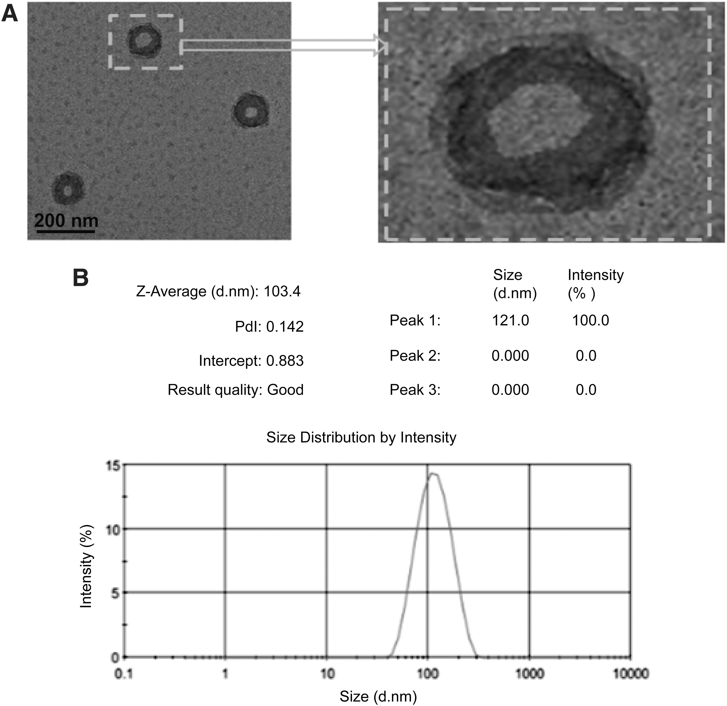
To investigate whether the particle size of nanocapsules prepared by coaxial glass capillary microfluidic device was uniform, TEM was used to observe the particle size of nanocapsules. Figure 2A showed the cavity and layered structure of the nanocapsules. In addition, Figure 2B indicated that the average particle size of the nanocapsule was 103.4 nm by DLS. These results demonstrated that the particle size of nanocapsules prepared by coaxial glass capillary microfluidic device was uniform.
Effect of organic solvent removal
The particle size of nanocapsules varied in different orifice diameter microfluidic device. As shown in Figure 3A, the larger the orifice diameter, the larger the diameter of the nanocapsules. In this study, an orifice diameter of 80 μm was chosen because previous results showed an average particle size of 103.4 nm. Additionally, after the solvent evaporation, the orifice diameter of nanocapsules shortened from 135 to 103 nm (Table 1).

The Average Size of Nanocapsules Before and After Solvent Removal
Determination of DL and EE
DL and EE are important indicators of nanoparticles, because they affect bioavailability, toxicity, and pharmacodynamics. 24 As shown in Figure 3B, the co-encapsulation of hydrophilic and hydrophobic drugs was effective. When the orifice diameter was 100 μm, the encapsulation efficiencies of CPT-11 and Oxa reached 97.0% (DL: 3.45%, w/w) and 95.7% (DL: 3.32%, w/w), respectively. Besides, the highest EE of 5-Fu was 15.6% (DL: 1.73%, w/w) (Table 2). These results confirmed the successful encapsulation of drugs in NCs, because the drug-loaded NCs had characteristic absorption bands (5-Fu, CPT-11, and Oxa).
The Drug Encapsulation Efficiency of Nanocapsules Prepared by Different Inner Phase End Pore Sizes
Drug release rate of CPT-11, 5-Fu, and Oxa encapsulated in nanocapsules in vitro
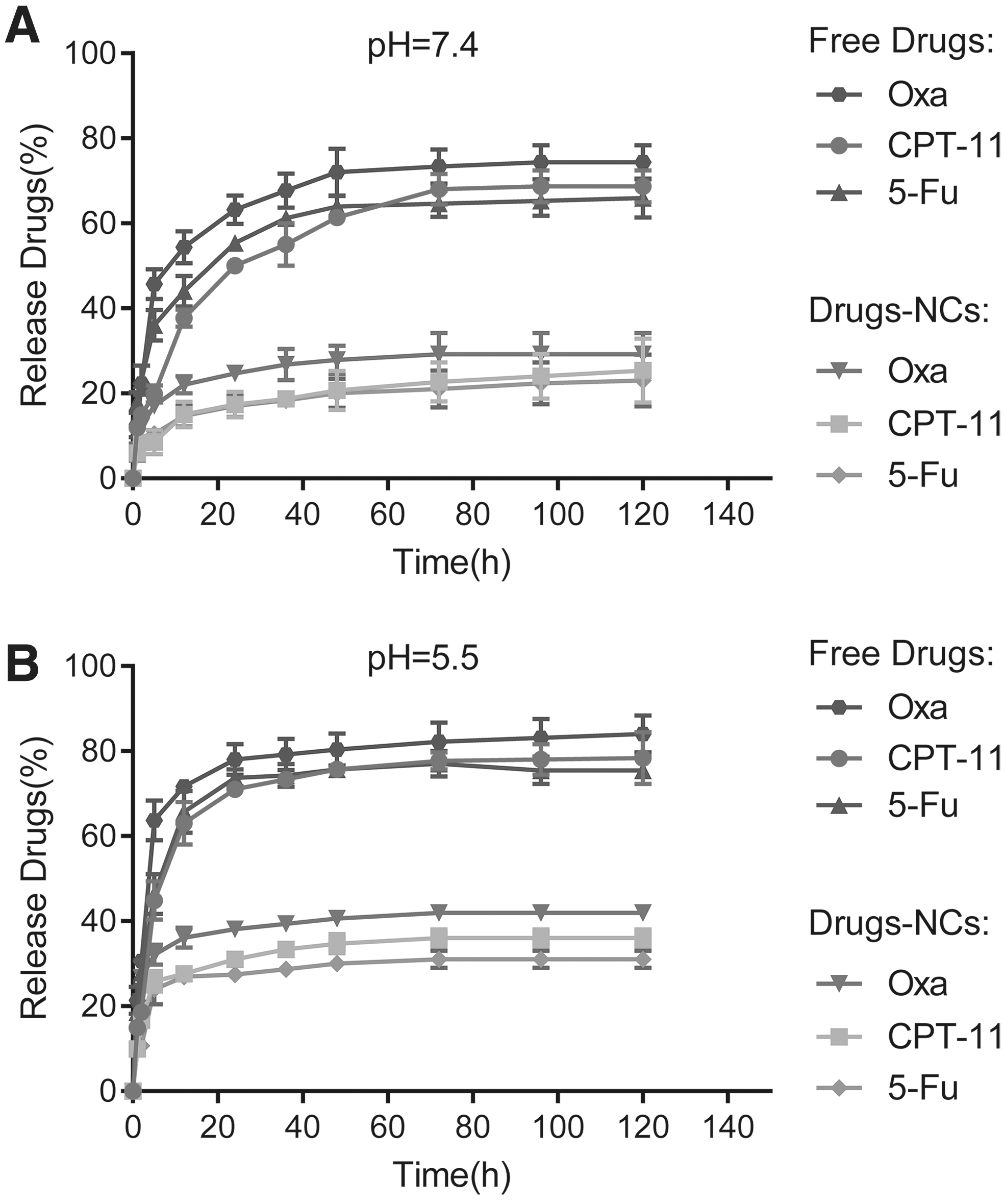
Dialysis method was performed to measure the release of three drugs in Free Drugs and Drugs-NCs at pH 5.5 and pH 7.4 at room temperature. As shown in Figure 4, the drug release curves of Free Drugs (CPT-11, 5-Fu, and Oxa) were pH-dependent, and more rapid release was noticed at pH 5.5 rather than at pH 7.4. On the contrary, the three drugs in Drugs-NCs group were released at low levels both at pH 5.5 and pH 7.4, and the release amount of Oxa with the highest release rate reached about 40% at pH 5.5. These results suggested that drugs encapsulated in the nanocapsules released less. Furthermore, Drugs-NCs released more quickly in acidic conditions.
The stability of drugs encapsulated in the nanocapsules
The variation of the size of Drugs-NCs (at 80 μm of orifice diameter) with time was a good indicator of particle stability. In this study, the variations in mean particle size were measured over a storage period of 30 and 60 d at 4°C and 60°C. Figure 5A–C showed that the particle size of the nanocapsules remained at about 100 nm within 60 d at 4°C, indicating good stability. However, the particle size distribution of nanoparticles appeared in different intervals within 30 d at 60°C, indicating that the nanocapsules had aggregation or destruction (Fig. 5D).

Long-term stability test and batch repeatability test.
Drugs-NCs inhibited SW1990 cell and Panc-1 cell proliferation
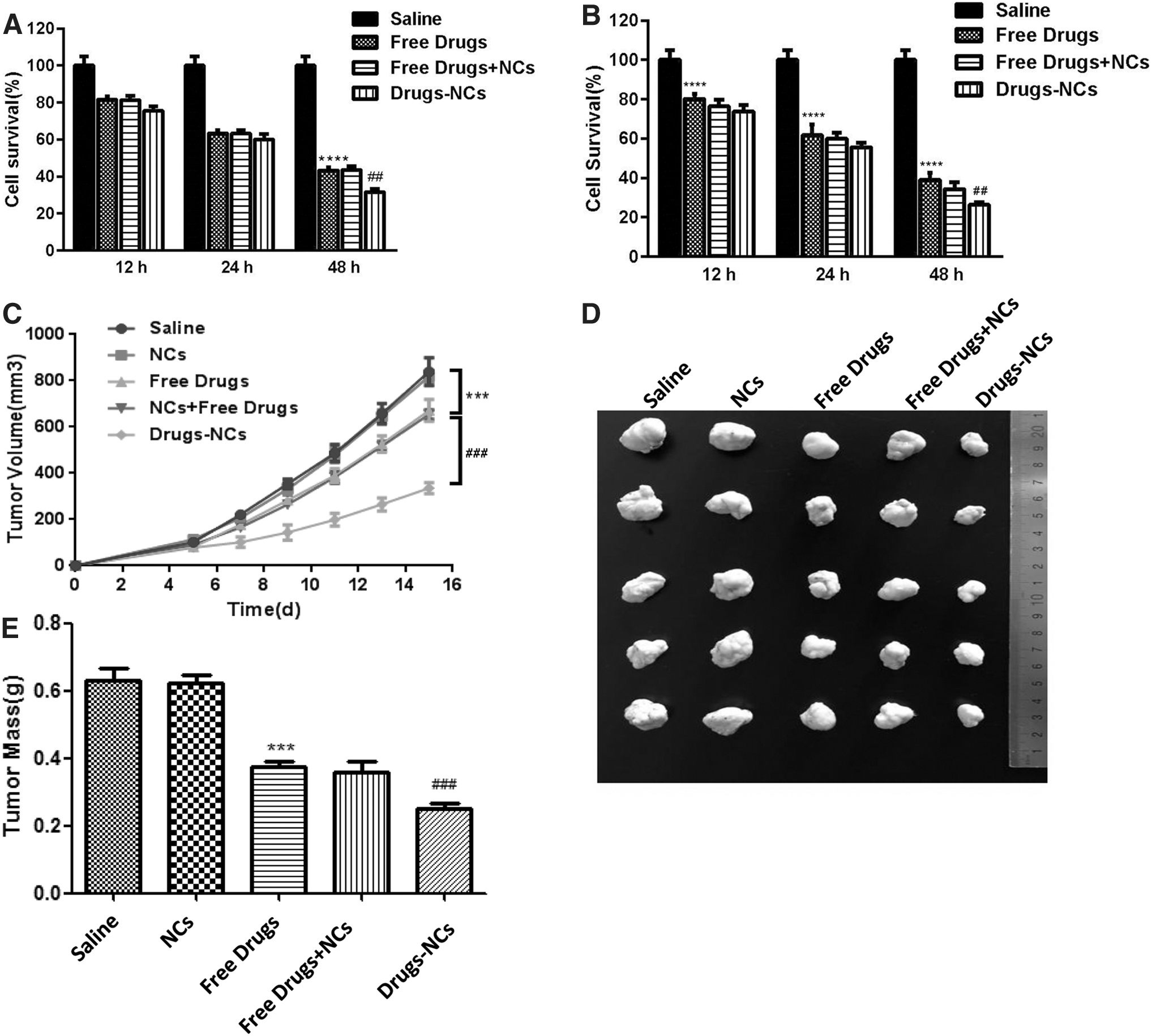
To find out the inhibitory effects of saline, Free Drugs, Free Drugs + NCs, and Drugs-NCs on human pancreatic cancer SW1990 cells and Panc-1 cells, MTT assay was used to measure SW1990 cell and Panc-1 cell survivals after different treatments. The total drug concentration (12.005 μM 5-Fu, 8.357 μM Oxa, and 5.095 μM CPT-11) was used for each treatment. As shown in Figure 6A and B, the survival rate of SW1990 cells and Panc-1 cells decreased with the extension of time, and there were significant differences among groups (p < 0.0001). There was no significant difference in the survival rate of SW1990 cells and Panc-1 cells between the three drug groups at 12 and 24 h. However, at 48 h, the cell survival rate of the Drugs-NCs group was significantly lower than that of the Free Drugs group or the Free Drugs + NCs group (p < 0.01). All these findings indicated that Drugs-NCs could inhibit SW1990 and Panc-1 cell survival.
MTT was used to measure the survival rate of
Antitumor effect of drugs-NCs in vivo
To study the effects of Drugs + NCs on tumor growth in vivo, BALB/c (nu/nu) nude mice were used in vivo experiments (n = 5, 6.0 μmol/kg 5-Fu, 4.2 μmol/kg Oxa, and 2.5 μmol/kg CPT-11). As shown in Figure 6C and D, there was no significant difference in tumor volume between the five groups within 5 d after administration. However, by day 15, the tumor volume of the Drugs-NCs group was significantly smaller than that of the Free Drugs and NCs groups (p < 0.001). As for the tumor weight, there was no significant change in tumor weight when treated with saline and NCs. However, mice treated with Drugs-NCs weighed significantly less than those treated with Free Drugs + NCs or Free Drugs (p < 0.001) (Fig. 6E).
Discussion
The most striking finding of this study was successful preparation of nanocapsules containing Oxa, CPT-11, and 5-Fu (Drugs-NCs) via one-step method with coaxial capillary microfluidization technique. In the design and fabrication process, the one-step method was mainly reflected in the structure and distribution of microfluidic chip. Briefly, this method mixed three-phase liquid containing drugs that occur in the same region at microscopic level, and can obtain nanocapsules with good uniformity and containing three kinds of chemotherapeutic drugs by adjusting the flow rate, fluid material composition, and ratio. 25
There are several reference values for evaluating the quality of nanocapsules, the first are their particle size and distribution. It has been reported that the collapsed blood vessels in pancreatic tumors lead to increased interstitial hydraulic pressure, which exceeds the hydrostatic pressure of collecting vessels and the osmotic pressure of the blood vessel wall, making it more difficult for large particle size nano-drugs to penetrate. 26 In 2017, the average size of hybrid liposomal nanospheres fabricated by Ji et al. was 200 nm. 27 In this study, TEM results revealed that the Drugs-NCs were spherical and uniform in size (103.4 nm), which was within the range of previous studies. 23,28
Second, DL and EE is of great importance to nanocapsules. Additionally, some researches have suggested that EE is closely related with nanocarrier materials. 29 In the present study, the encapsulation efficiencies of CPT-11, Oxa, and 5-Fu reached 97.0%, 95.7%, and 15.6%, respectively. Besides, CPT-11 was encapsulated by DOTAP and PLGA in the hydrophobic middle layer, and 5-Fu was encapsulated by Pluronic P85 and DSPE-PEG in the hydrophilic outer layer. As a Food and Drug Administration-approved excipient for injection, PLGA has good slow and controlled release performance for hydrophobic drugs 30 ; as a mixed emulsifier, Pluronic P85 and DSPE-PEG are used to regulate the physical and chemical properties of nanoparticles. 31 Third, the property of the drug release rate is also a vital indicator of nanocapsules. Dialysis method result suggested that compared with Free Drugs group, the drug release rate in Drugs-NCs group was longer, so that the drug release concentration could be maintained at the treatment level for a long time.
As a normal means in the treatment of malignant tumor, chemotherapy can kill tumor cells, but a lot of normal cells are killed by mistake with side effects. 32 To improve the effect of chemotherapy, target therapy of chemical-loaded nanocapsules on tumor tissues is a novel approach. In recent years, studies conducted by Kemp et al. 6 and Hu et al. 33 have reported that the successful preparation of nanoparticles containing multiple drugs has synergistic effect, which can improve the half-life of drugs in vivo circulation. Moreover, they have also confirmed that the passive targeting enrichment at tumor sites can improve the targeting of tumor therapy and reduce the damage to normal tissues. It should be noticed that the passive targeted enrichment of tumor sites is closely related to the sustained slow release of nanodrugs. 34
As mentioned before, the drug release of nanocapsules containing three chemotherapeutic drugs in the present study was long and sustained. This is because nanocarrier materials could optimize the biocompatibility of nanoparticles, reduce the agglomeration of particles, thereby prolonging the circulation time, so that the release concentration of three drugs in nanocapsules could be maintained at therapeutic level for a long time. 35,36 Moreover, nanocapsules can decrease the distribution of drugs in normal organs, playing a role in enhancing efficiency and reducing toxicity. 37,38 Additionally, physicians can reduce the frequency of drug administration, increase the patient's medication compliance, and greatly facilitate the medication. 39 Numerous studies have indicated that the release rate of drugs is significantly regulated by the pH value, more acidic the pH value, faster the nanodrug is released. 34,40,41 The lower pH value was chosen because the nanoparticles were primarily absorbed by the cells, eventually transferred to lysosomes, and then degraded. 42 In this study, the drug release rate of Drugs-NCs at pH = 5.5 was faster than that at pH = 7.4. The above findings indicated that the nanocapsules prepared in this study had potential tumor targeting.
Since survival rate is the main indicator for clinical evaluation of tumor, MTT viability assay showed that Drugs-NCs could significantly inhibit SW1990 and Panc-1 cell survival. Furthermore, in vivo experiments also proved the inhibitory effect of three Drug-NCs on malignant tumor. The tumor volume of in the Drugs-NCs group was significantly smaller than that of the Free Drugs and NCs groups (p < 0.001). As for the tumor weight, mice treated with Drugs-NCs weighed significantly less than those treated with Free Drugs + NCs or Free Drugs. There are still several limitations of this study. The study on the stability of nanocapsules is a long-term process, which requires not only the stability of preparation, but also the stability in vivo and in vitro. Meanwhile, the cytotoxicity test of Drugs-NCs and drug metabolism differences in different animals need further study.
Conclusions
This study successfully fabricates nanocapsules containing Oxa, CPT-11, and 5-Fu (Drugs-NCs) via coaxial capillary microfluidic device. The average particle size of the nanocapsules is 103.4 nm, with relative narrow size distribution. In addition, the encapsulation efficiencies of Oxa and CPT-11 are close to 100% and 5-Fu was 15.6%. MTT assay and anti-tumor experiment indicate the inhibitory effect of Drugs-NCs on malignant tumors. Finally, the sustain-release of Drugs-NCs can promote the local release of the three chemotherapy drugs at the target site, reducing the dose that enters the systemic circulation, thereby reducing the toxic effect on normal tissues. Therefore, coaxial capillary microfluidic device can be used as a promising microfluidic technology to fabricate multiple Drug-NCs.
Footnotes
Authors' Contributions
Y.X. designed the study. Y.X., B.H., J.X., J.W., and B.Y. performed the experiments. All coauthors have reviewed and approved the article before submission.
Disclosure Statement
No competing financial interests exist.
Funding Information
This research was supported by the Wenzhou Science and Technology Bureau of China (Y20170156).