
Abstract
Background:
Long non-coding RNA Zinc finger E-box binding homeobox 2 (ZEB2) antisense RNA 1 (ZEB2-AS1) has been shown to promote tumor progression. However, the clinical significance and fundamental function role of ZEB2-AS1 in osteosarcoma (OS) has been poorly understood.
Methods:
The expression of ZEB2-AS1 was determined in tumor tissues and matched normal tissues from 67 OS patients using quantitative reverse transcriptase PCR analysis. Clinical value of ZEB2-AS1 was evaluated by χ2 test and Kaplan-Meier method. Cell proliferation was analyzed using CCK-8 assay, colony formation. Cell apoptosis status was determined by caspase-3 activity assay. Cell migration, invasion and epithelial-mesenchymal transition (EMT) were investigated by scratch wound healing, transwell invasion assays and Western blotting.
Results:
Clinical association analysis revealed that high ZEB2-AS1 expression correlated with tumor size, distant metastasis and poor prognosis of OS patients. Moreover, ZEB2-AS1 expression was identified as an independent prognostic factor for OS patients. Loss-of-function assays demonstrated that ZEB2-AS1 knockdown suppressed the proliferation and induced apoptosis in OS cells. In addition, ZEB2-AS1 knockdown inhibited cell migration, invasion, EMT of OS cells in vitro.
Conclusions:
Taken together, our data demonstrate that ZEB2-AS1 serves a putative oncogenic role and associates with unfavorable prognosis in OS.
Introduction
As a type of skeletal system-associated tumor, osteosarcoma (OS) is most prevalent in a population with highest incidence reached 8–11 million cases each year within a 15–19 years old age group, commonly characterized as bone pain and swelling. 1 –3 Despite high efficacy found in traditional clinical treatment options, including surgery, chemotherapy, and radiation, 4,5 the prognosis is still unfavorable due to distant metastasis in OS patients in advanced stage. 6 Accordingly, it is of great importance to understand the molecular mechanisms underlying OS metastasis for clinically improving the prognosis of OS patients.
Long noncoding RNAs (lncRNAs), as a class of noncoding RNA longer than 200 nucleotides (>200 nt), recently have gained mounting attention for their key regulatory functions in different diseases, especially in cancers. 7 –9 A growing body of studies has revealed that lncRNAs are aberrantly expressed in human tumors and play critically important roles in a series of biological processes, such as proliferation, apoptosis, migration, and invasion. 10,11 A newly discovered lncRNA Zinc finger E-box-binding homeobox 2 (ZEB2) antisense RNA 1 (ZEB2-AS1) has been reported to serve important functions in several tumors. For example, Li et al. 11 demonstrated that ZEB2-AS1 was significantly upregulated and functioned as a prognostic factor for hepatocellular carcinoma (HCC) pathogenesis. Lan et al. 12 further elucidated that downregulation of ZEB2-AS1 was associated with decreased tumor growth and metastasis in HCC by the regulation of the expression levels of epithelial-mesenchymal transition (EMT)-induced markers. As shown in the study of Wang et al. 13 and Wu et al., 14 knockdown of ZEB2-AS1 reduced the proliferation, migration, invasion, and EMT, but increased the apoptosis of gastric carcinoma cells. In addition, ZEB2-AS1 has been demonstrated to be associated with poor clinical outcomes in gastric cancer 15 and acute myeloid leukemia. 16 Although previous studies have pointed many lncRNAs in OS are recognized as oncogenes, including urothelial carcinoma-associated 1 (UCA1) 17 and small nucleolar RNA host gene 17 (SNHG17), 18 as well as tumor suppressors, likely steroid receptor RNA activator 1 (SRA1) 19 and NKILA, 20 the clinical significance and regulatory functions of ZEB2-AS1 in OS still remain poorly understood.
In this study, the authors determined the expression profile of ZEB2-AS1 in clinical patients and cultured OS cell lines. Next, they evaluated the association between ZEB2-AS1 expression and survival prognosis in OS patients. By performing a series of functional experiments, they next investigated the effects of ZEB2-AS1 on cell proliferation, apoptosis, migration, and invasion of OS cells, as well as the underlying molecular mechanism. Their data may provide further intervention target to help improve the diagnosis and clinical treatment of OS.
Materials and Methods
Clinical tissue samples
A total of 67 paired tumor tissues and matched adjacent tissues derived from OS patients were obtained from the Affiliated Traditional Chinese Medicine Hospital, Southwest Medical University (Sichuan, China). All participants signed the written informed consent and received neither radiotherapy nor chemotherapy before surgical resection. Each clinical tissue sample was confirmed by pathologists who were blind to this study and immediately stored at −80°C for further analysis. The clinical information of all patients, including gender, age, and distant metastasis, was organized in Table 1. This study was conducted in accordance to the Declaration of Helsinki, which was approved by the Ethics Review Board of Affiliated Traditional Chinese Medicine Hospital, Southwest Medical University (approval number: CM34A-83).
Association Between ZEB2-AS1 Expression and Clinicopathological Characteristics of Osteosarcoma Patients
Statistical analyses were performed by the χ2 test.
p < 0.05 was considered significant.
OS, osteosarcoma; TNM, tumor-node-metastasis; ZEB2-AS1, Zinc finger E-box-binding homeobox 2 (ZEB2) antisense RNA 1.
Cell lines and siRNA transfection
American Type Culture Collection (ATCC) provided the following cell lines: human OS cell lines (Saos-2, MG-63 and U2OS) and one normal osteoblast cell line (hFOB1.19). All cell lines were cultured in Dulbecco's modified Eagle's medium (DMEM) mixed with 10% fetal bovine serum (FBS; Gibco, Grand Island, NY) at 37°C under humidified atmosphere containing 5% CO2. For ZEB2-AS1 knockdown, the specific small interring RNA targeting ZEB2-AS1 (si-ZEB2-AS1: 5′-CCUUUGGGAAAUUACCUCUTT-3′) and corresponding negative control siRNA (si-NC: 5′-AUACCAAAUCAGGUAGGUGTT-3′ were synthesized by RiboBio Co., Ltd (Guangzhou, China). Saos-2 and U2OS cells were seeded into six-well plates at a density of 1 × 10⁵ cells per well and transfected with 50 nM of si-ZEB2-AS1 or si-NC for 48 h based on the manufacturer's instructions provided by Lipofectamine 3000 reagent (Invitrogen, Carlsbad, CA).
Quantitative reverse transcriptase PCR
Total RNA was isolated from tissues or cells according to the protocols from TRIzol reagent (Invitrogen). Then, RNA was reverse transcribed to cDNA using PrimeScript RT Reagent Kit (Takara Biomedical Technology, Beijing, China). PCR was conducted using SYBR Green PCR Master Mix (Takara Biomedical Technology) with the following primer sequences: ZEB2-AS1 forward, 5′-ATGAAGAAGCCGCGAAGTGT-3′ and reverse, 5′-CACACCCTAATACACATGCCCT-3′; GAPDH forward, 5′-ATTTGATGGGTGAGGAATGGGTT-3′ and reverse, 5′TTCACACCCATCACAAAC-3′. The expression levels of ZEB2-AS1 were calculated by the 2−ΔΔCt method with GAPDH as the endogenous control.
Cell counting kit-8 assay
Transfected Saos-2 or U2OS cells in triplicate were seeded into 96-well plate at a density of 3000 cells per well and incubated form 0, 24, 48, and 72 h, respectively. Then, each well was treated with 10 μL of Cell Counting Kit-8 (CCK-8) reagent (Dojindo Molecular Technologies, Kumamoto, Japan) for 2 h at 37°C. The optical density (OD) was determined by a microplate reader at a wavelength of 450 nm.
Colony formation assay
Transfected Saos-2 or U2OS cells at 500 cells per well in six-well plates and cultured for 2 weeks, during which the medium was changed every 48 h. At the end-point, the cells were washed with PBS, fixed in methanol and stained with 0.1% crystal violet. The number of visible colonies (>50 cells per colony) in five random view fields was, respectively, counted and averaged as the final counts under a microscope.
Caspase-3 activity assay
Caspase-3 activity was analyzed in OS cells using Caspase-3 Assay Kit (Beyotime Institute of Biotechnology, Shanghai, China) according to the manufacturer's instructions. In brief, Saos-2 or U2OS cells were harvested and resuspended in chilled cell lysis buffer. After centrifugation at 4°C for 10 min, the supernatant was collected and treated with reactive buffer for 1.5 h at 37°C. The relative caspase-3 was estimated based on the OD value read by a microplate reader at 405 nm.
Scratch wound healing assay
In brief, transfected Saos-2 or U2OS cells were cultured in six-well plates until ∼80% confluency. Using a 200 μL aseptic pipette tip, the wounds were artificially created in a standard way through the monolayer. After replaced with a fresh complete culture medium, the cells were incubated for 24 h at 37°C. The wound area was photographed immediately at 0 (W0) and 24 h (W24) and relative migration distance in each group was calculated according to the following formula: (W0 − W24)/W0 × 100%.
Transwell invasion assay
The capacity of OS cell invasion was evaluated using a transwell chamber (8 μm pore size; Costar, Corning, NY) precoated with Matrigel (BD Biosciences, Franklin Lakes, NJ). In brief, appropriately 5000 transfected Saos-2 or U2OS cells were suspended in 100 μL serum-free medium and seeded into the upper transwell chambers. Meanwhile, 600 μL of DMEM medium supplemented with 10% FBS was added into the lower chamber as a chemoattractant. After incubation for 24 h at 37°C, the invasive cells on the lower chamber were fixed with 4% paraformaldehyde, stained with crystal violet (Sigma-Aldrich, China), and further counted in five random fields under a light microscope (Olympus Corporation) at 100 × magnification.
Western blotting
Transfected Saos-2 or U2OS cells were collected and lysed in RIPA buffer (Thermo Fisher Scientific, Inc.) following the manufacture's protocols. The lysed samples were centrifuged at 12,000 g for 20 min at 4°C and quantified using a BCA assay kit (Beyotime Institute of Biotechnology). After that, equal amounts of protein samples were separated by 12% SDS-PAGE electrophoresis and transferred to PVDF membranes (Millipore). The membrane was then blocked in 5% (w/v) nonfat dry milk for 2 h and incubated with primary antibodies against proliferating cell nuclear antigen (PCNA), E-cadherin, N-cadherin, Vimentin, and GAPDH against overnight at 4°C. After incubating with horseradish peroxidase-conjugated secondary antibody for 2 h at room temperature, the protein bands were visualized by an electrochemiluminescence Western Blotting Substrate (Thermo Fisher Scientific).
Statistical analysis
Data were analyzed using Statistical Package for Social Science software, version 22.0 (Chicago, IL) and expressed as mean ± standard deviation of at least three independent experiments. Wilcoxon signed-rank test was used to assess the differences of ZEB2-AS1 expression levels between OS tissues and adjacent tissues. The association between ZEB2-AS1 expression and clinicopathological features was determined by χ2 test. The curve of correlation between ZEB2-AS1 expression and overall survival was plotted by Kaplan–Meier method and evaluated by the log-rank test. The potential independent prognostic factors and 95% confidence interval of hazard ratios were assessed by univariate analysis and multivariate analysis based on Cox proportional hazard regression model. For the in vitro experiments, all p-values were calculated by unpaired Student's t test or ANOVA test with Tukey's post hoc test and considered to be statistically significant when it was <0.05.
Results
ZEB2-AS1 expression was correlated with clinicopathological features and poor prognosis in OS patients
To study the potential clinical significance of ZEB2-AS1 in OS patients, the expression of ZEB2-AS1 was determined in tumor tissues and adjacent tissues from 67 cases of OS patients using quantitative reverse transcriptase PCR (qRT-PCR). The results showed that ZEB2-AS1 expression levels were significantly upregulated in OS tissues, compared with adjacent normal tissues (Fig. 1A). Based on the expression of ZEB2-AS1 in OS tissues, the authors divided all OS patients into high ZEB2-AS1 expression group (n = 34) and low ZEB2-AS1 expression group (n = 33), with a cutoff point at the median ZEB2-AS1 level. The analysis of clinical data displayed in Table 1 manifested that ZEB2-AS1 expression was highly correlated with tumor size (p = 0.020) and distant metastasis (p = 0.038), while there was no significant correlation with gender, age, and location. Meanwhile, Kaplan–Meier survival analyses revealed that OS patients with high ZEB2-AS1 expression had a significantly worse overall survival than those with low ZEB2-AS1 expression (Fig. 1B). Moreover, univariate and multivariate Cox regression analysis identified distant metastasis and ZEB2-AS1 expression as independent prognostic factors for OS patients (Table 2). In sum, ZEB2-AS1 expression levels were markedly upregulated and strongly associated with poor prognosis.

ZEB2-AS1 expression was notably enhanced and closely associated with poor prognosis in OS.
Univariate and Multivariate Analysis for Overall Survival in Patients with Osteosarcoma
p < 0.05.
CI, confidence interval; HR, hazard ratio; NA, not analyzed; OS, osteosarcoma; TNM, tumor-node-metastasis.
ZEB2-AS1 knockdown suppressed cell proliferation and facilitated apoptosis in OS
In line with the above results, it was observed that the expression levels of ZEB2-AS1 in three OS cell lines (Saos-2, MG-63 and U2OS) were notably increased compared with that in one normal osteoblast cell line hFOB1.19 (Fig. 2A). Among them, Saos-2 and U2OS cells exhibited relative higher ZEB2-AS1 expression and were thus selected for loss-of-function assays to explore the biological function of ZEB2-AS1 in OS cells. At first, Saos-2 and U2OS cells were transfected with si-ZEB2-AS1 to downregulate the expression of ZEB2-AS1, as confirmed by qRT-PCR (Fig. 2B). Functionally, CCK-8 assay showed that silenced ZEB2-AS1 notably repressed the proliferation of Saos-2 (Fig. 2C) and U2OS (Fig. 2D) cells compared with proliferation of the corresponding control cells. The colony formation assay further demonstrated that the proliferation ability was impaired in both Saos-2 and U2OS cells after ZEB2-AS1 knockdown, as reflected by decreased colonies in si-ZEB2-AS1 group, in comparison with si-NC group (Fig. 2E). Concordant with decreased proliferation, it was discovered that ZEB2-AS1 silence significantly elevated cell apoptosis of Saos-2 and U2OS cells, as manifested by increased caspase-3 activity in si-ZEB2-AS1 group, in comparison with si-NC group (Fig. 2F). These results suggested that ZEB2-AS1 knockdown inhibited cell proliferation, whereas promoted apoptosis of OS cells.

ZEB2-AS1 knockdown suppressed cell proliferation and facilitated apoptosis in OS cells.
ZEB2-AS1 knockdown abrogated cell migration and invasion of OS
Next, Scratch wound healing and Transwell assay were performed to assess the migratory and invasive capacities, respectively, in OS cells. As shown in Figure 3A, si-ZEB2-AS1 transfection significantly retarded the wound closure processing compared with si-NC transfection in both Saos-2 and U2OS cells, which indicated that the migration ability of these two OS cell lines was remarkably decreased at the edge of the scratch. Likewise, ZEB2-AS1 knockdown obviously suppressed the invasive capacity as indicated by decreased invasive cells in Saos-2 (si-ZEB2-AS1 vs. si-NC: 23.3 ± 4.5 vs. 91.7 ± 3.8, p < 0.001) and U2OS (si-ZEB2-AS1 vs. si-NC: 16.0 ± 3.0 vs. 66.7 ± 4.2, p < 0.001) cells (Fig. 3B). Therefore, the authors' data clearly demonstrated that ZEB2-AS1 contributed to migration and invasion in OS.
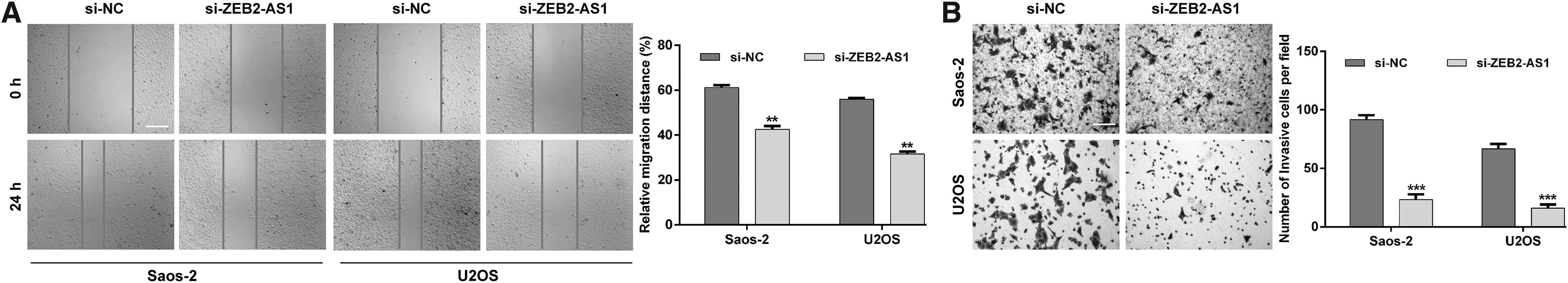
ZEB2-AS1 knockdown abrogated cell migration and invasion of OS.
ZEB2-AS1 knockdown inhibited PCNA expression and EMT process in OS cells
To further confirm the suppressive effects of ZEB2-AS1 on OS cell proliferation, migration, and invasion, the authors analyzed the protein expression associated with proliferation and EMT markers using Western blotting. As shown in Figure 4A and B, PCNA associated with proliferation was downregulated and EMT process was suppressed, as reflected by upregulation of E-cadherin and downregulation of N-cadherin and Vimentin in both Saos-2 and U2OS cells after ZEB2-AS1 knockdown. These results further supported that ZEB2-AS1 positively regulated OS cell proliferation, migration, and invasion.

ZEB2-AS1 knockdown inhibited PCNA expression and EMT process in OS cells.
Discussion
It has been widely accepted that genetic and epigenetic alterations are the driving factors of cancer. Currently, the 5-year survival rate remains relatively low worldwide, even though great effects have been made to improve the diagnosis and treatment of OS patients. 21,22 Abnormal expression of lncRNAs has recently been identified to contribute to the progression of OS. 23 ZEB2-AS1 is a newly discovered lncRNA, whose clinical meaning, biological roles, and explicit mechanism are poorly understood in OS. In this study, the authors first confirmed that ZEB2-AS1 was abnormally overexpressed in OS tissues and cell lines, compared with normal bone tissues and normal osteoblast cell line, respectively. By analyzing the clinical significance of ZEB2-AS1 expression, they observed that high expression of ZEB2-AS1 was correlated with tumor size, distant metastasis, and poor prognosis in OS patients. Consistently, the prognostic value of ZEB2-AS1 was previously reported in gastric cancer 15 and acute myeloid leukemia. 16 It was also demonstrated that overexpressed ZEB2-AS1 expression was significantly associated with large tumor size, cervical node metastasis and reduced overall and disease-free survival in head neck squamous cell carcinoma (HNSCC). 24 In addition, ZEB2-AS1 has been confirmed to be associated with poor prognosis in HCC 11 and laryngeal squamous cell carcinoma. 25 These facts suggested that ZEB2-AS1 might be an unfavorable prognostic biomarker in OS patients.
The biological function tests showed that ZEB2-AS1 knockdown significantly inhibited the proliferation and promoted the apoptosis of OS cells. ZEB2-AS1 is a noncoding antisense transcript from the promoters of ZEB2 as protein of the ZEB family and ZEB2 has been reported to promote tumor progression in a number of distinct types of cancer. 26 –29 ZEB2 protein could control cell cycle progression and cell differentiation in melanoma progression. 30 It is not hard to understand the downregulation of PCNA and impaired cell proliferation by ZEB2-AS1 knockdown according to the report by Wang et al., who pointed that ZEB2 could inhibit the apoptosis of vascular endothelial cells induced by high glucose. 31 In agreement with their data, ZEB2-AS1 silencing inhibited cell proliferation and promoted apoptosis in bladder cancer 32 and colorectal cancer. 33
In addition to uncontrolled proliferation, the ability of migrating and invading healthy tissues is another import biological feature of cancer cells. EMT is a key mechanism for tumor metastasis and recurrence, 34 which is characterized by loss of cell adhesion and acquisition of migration and invasive ability. 35,36 Related study shows that E-cadherin was decreased, while N-cadherin and Vimentin were increased, leading to EMT process. 37 ZEB2 protein could be upregulated by overexpression of ZEB2-AS1 in epithelial cells, which was also a transcriptional repressor of E-cadherin. 37 Here, the authors further validated that ZEB2-AS1 knockdown decreased the migratory and invasive capacities of OS cells by suppressing EMT process. Similarly, it was found that ZEB2-AS1 promoted HCC metastasis by regulating ZEB2 and some EMT markers. 12 Furthermore, Zhang et al. demonstrated that ZEB2-AS1 promotes the proliferation, metastasis, and EMT in triple-negative breast cancer. 38 The positive regulation of ZEB2-AS1 on EMT was also validated by Diao et al. in HNSCC. 24
In conclusion, the authors confirmed that ZEB2-AS1 was highly expressed in OS and its overexpression was significantly associated with unfavorable prognosis. Importantly, the present study is the first to demonstrate that ZEB2-AS1 promoted the proliferation migration, invasion, and EMT process in OS in vitro. Of course, more experimental studies will be needed to be performed to further reveal the oncogenic roles of ZEB2-AS1 in OS.
Ethics Approval and Consent to Participate
This study was conducted in accordance with the Declaration of Helsinki and the approval from the Ethics Review Board of Affiliated Traditional Chinese Medicine Hospital, Southwest Medical University (approval number: CM34A-83).
Footnotes
Authors' Contributions
Y.J.X. and Z.Y.G. performed the experimental study, data collection and analysis, and article writing. W.J., C.B., and L.C. carried out the most experiments. C.X.J. and S.H.R. performed statistical analyses. L.L. conceived and supervised the whole project. All authors read and approved the final article.
Disclosure Statement
No competing financial interests exist.
Funding Information
No funding was received for this article.