
Abstract
Purpose:
The authors explore the role of DNASE1L2 in breast cancer (BC) and its affect on the cell phenotype.
Methods:
Breast invasive ductal carcinoma RNA-Seq data set was downloaded from The Cancer Genome Atlas database for analyzing DNASE1L2 levels. Overall survival curve was plotted by Kaplan–Meier methods. The correlations between DNASE1L2 expression and clinical characteristics were analyzed by chi-square tests. Cox regression models were implemented for analyzing the potential prognosticators of BC. Small interference RNA-DNASE1L2 and pcDNA3.1-DNASE1L2 were transfected into BC cells to silence and overexpress DNASE1L2, respectively. Relative mRNA and protein levels were determined by quantitative real-time PCR (qRT-PCR) and Western blot, respectively. Cell counting Kit-8, clone formation, and Transwell assays were employed to measure the proliferative, invasive, and migratory abilities.
Results:
Bioinformatics analysis showed that the levels of DNASE1L2 were found to be elevated in BC tissues, which was further proved by qRT-PCR tests. Besides, high expression of DNASE1L2 was dramatically led to a poor overall survival. Furthermore, DNASE1L2 expression was remarkably associated with age and pathologic-stage. Silencing DNASE1L2 showed an inhibitory effect on the proliferation, invasion, and migration of MCF7 cells, whereas overexpression of DNASE1L2 in BT549 cells presented the opposite results. Mechanistically, downregulation of DNASE1L2 could significantly enhance the levels of E-cadherin, as well as suppress the levels of Vimentin, N-cadherin and Snail, whereas upregulation of DNASE1L2 showed the reverse outcomes.
Conclusion:
This study for the first time demonstrated that DNASE1L2 was upregulated in BC cells, and acted as an oncogene to affect the phenotype of BC cells by modulating the epithelial–mesenchymal transition process, which suggested that DNASE1L2 might be considered as a useful biomarker for BC therapeutics.
Introduction
Breast cancer (BC), a malignant tumor arising from the ductal epithelium of the breast, 1 is the most common cancer influencing women worldwide, and its mortality rates are expected to increase notably in the following years. 2 Despite great advances in treatment, BC remains a huge health burden, and the 5 year survival rate is only 26%. 3 This is largely due to its easy metastasis and poor prognosis. 4 Recently, lots of investigations have reported that detection of tumor-related biomarkers plays a key role in the preoperative diagnosis and therapy of BC. Such as human epidermal growth factor receptor-2 (HER-2) could be regarded as a prognostic marker, and its overexpression led to a stronger ability of infiltrating of breast tumor. 5,6 NM23, as a suppressor, could predict the overall survival and was associated with cancer cells growth and metastasis in BC. 7,8 Hence, the investigation of useful biomarkers is of great significance for predicting prognosis, controlling metastasis, understanding the deterioration, and finally achieving satisfactory control of BC through reasonable treatment.
Deoxyribonuclease 1-like 2 (DNASE1L2), a member of the DNase 1-like endonuclease family, 9 is considered to be a keratin cell-specific endonuclease that plays an important role in DNA degradation. 10 It has reported that DNASE1L2 was expressed specifically in cornifying keratinocytes, 11 preferentially expressed in the epidermis 12 . With the existing knowledge, studies on the expression of DNASE1L2 in tumor cells are rare, especially in BC cells. It was however reported that alkaline DNase could be used as a marker to assess the response of BC patients to anticancer therapy. 13 D'Antonio et al. identified 10 DNase 1 hypersensitive sites and found that these sites are significantly mutated and related to the abnormal expression of neighboring genes in BC. 14 These findings raise a question that what role of DNASE1L2 plays in BC, which is worthy for further investigation.
In view of this, with the purpose of investigating the function of DNASE1L2 expression in BC, the authors first downloaded RNA-Seq data set from The Cancer Genome Atlas (TCGA) database for analyzing DNASE1L2 levels, and assessed its prognostic values for BC patients. Beyond this, the effects of DNASE1L2 on the phenotype of BC were detected in MCF7 and BT549 cells. Furthermore, the correlation between DNASE1L2 expression and epithelial–mesenchymal transition (EMT)-related proteins was investigated. The results illustrated that DNASE1L2 might be a carcinogenic biomarker to enhance tumor cells viability and metastasis ability by regulating the levels of EMT-related markers in the progression of BC.
Materials and Methods
Bioinformatics analysis
First, the authors downloaded breast invasive ductal carcinoma RNA-Seq data set, which includes 1109 breast tumor samples and 113 normal samples from TCGA (
Cell culture and transfection
Human BC cell lines MDA-MB-231, MCF7 and BT549, and breast epithelial cell MCF-10A were obtained from Cell Resource Center, Shanghai Institute of Life Sciences, Chinese Academy of Sciences (Shanghai, China) and maintained in Roswell Park Memorial Institute-1640 (RPMI-1640) (Procell, Wuhan, China) medium with 0.1 mg/mL streptomycin and 100 U/mL penicillin under the humidified 5% CO2 atmosphere at 37°C.
Two small interfering RNAs targeting DNASE1L2 (si-DNASE1L2#1: 5′-CACGTGATGTGCTGCTCTGTA-3′; si-DNASE1L2#2: 5′- GAGATGTACCTGTTCGTGTAC-3′) and its control (si-con: 5′- CGAACTCACTGGTCTGACC-3′), as well as plasmids pcDNA3.1-DNASE1L2 and pcDNA3.1 were designed and synthesized by the GenePharma Co., Ltd (Shanghai, China). Transfecting process was performed using Lipofectamine 2000 kit (Invitrogen, CA) according to the manufactures' instructions.
Quantitative real-time PCR
Total RNA was extracted from cells using TRIzol reagent (Invitrogen) following the manufactures' instructions. The cDNA was reverse transcribed by PrimeScript RT Reagent Kit (TaKaRa, Dalian, China). mRNA level was detected by a SYBR Premix Ex Taq system (TaKaRa). The primers are listed as follows:
DNASE1L2 forward, 5′- GTGTACCTGGACGTGATCGACA -3′;
DNASE1L2 reverse, 5′- GAGCCACTTGAAGACCTCACTG -3′;
GAPDH forward, 5′- TGTGTCCGTCGTGGATCTGA -3′;
GAPDH reverse, 5′- CCTGCTTCACCACCTTCTTGA -3′.
Glyceraldehyde-3-phosphate dehydrogenase (GAPDH) was served as an internal reference. Relative levels of individual gene mRNA transcripts were calculated with the comparative quantification cycle (Cq) method (2−ΔΔCq).
Western blot
After transfection for 48 h, cells were lysed in the Radio Immunoprecipitation Assay (RIPA) lysis buffer (Beyotime, Nantong, China) on ice. Equal amounts of protein (20 μg protein each hole) was separated by sodium dodecyl sulfate-polyacrylamide gel electrophoresis and transferred onto polyvinylidene fluoride membrane. The membrane was blocked for an hour with 5% skimmed milk powder, then incubated with primary antibodies anti-DNASE1L2 (ab231232; Abcam, Cambridge, United Kingdom,
Subsequently, samples were washed with Tween-20 (TBST) thrice for 5 min, and incubated with horseradish peroxidase conjugated secondary antibodies for 1 h at room temperature. Positive bands were visualized by an enhanced Electro-Chemi-Luminescence kit (ECL; Beyotime, Nantong, China) on a ChemiDoc MP imaging system (Bio-Rad, Hercules, CA). The ImageJ software (National Institutes of Health, Bethesda, MD) was used to quantify the bands.
Cell proliferation assay
After transfection for 48 h, breast cells were seeded in 96-well culture plates at a density of 1000 cells per well, and then incubated for 24, 48, and 72 h. Cell counting Kit-8 (CCK-8) reagent (10 μL) was supplied to each well at the designated time point. Subsequently, the cells were maintained at 37°C for another 1.5 h. The optical density (OD) value was recorded at 450 nm under a microplate reader.
Plate clone formation assay
Equal number of cells (400 cells per plate) were plated onto 60 mm plate and cultured at 37°C in cell incubator for 1–2 weeks. When colonies were appeared, the culture process was terminated. Then the colonies were carefully washed with phosphate buffered saline (PBS) twice, and immobilized with 5 mL 4% paraformaldehyde for 30 min, and stained with 0.1% crystal violet for 30 min. Finally, total number of colonies in each culture dish was evaluated.
Transwell migration and invasion assays
After transfection for 48 h, cells were suspended in serum-free medium. The suspension (invasion assay 1 × 10⁴ cells per well, migration assay 5 × 10³ cells per well) was added into the upper chambers that were coated (invasion assay) or uncoated (migration assay) with matrigel (1:6 dilution of serum-free medium, 100 μL). The lower chambers were supplied with 500 μL complete medium. After incubating for 24 h, cells on the upper chambers were carefully wiped out with cotton swabs, whereas cells that had migrated or invaded to the lower chambers were washed with PBS, immobilized with 4% paraformaldehyde for 30 min, and stained with 0.1% crystal violet for 20 min. Finally, migrated or invaded cells were counted and photographed under an optical microscope (Olympus Corporation, Tokyo, Japan) at a magnification of 100 in five random fields.
Statistical analysis
Student's t-test was used to compare the difference of two groups. Kaplan–Meier, together with log-rank test, was used to perform survival analysis in SPSS version 22.0 (Armonk). Chi-square tests were applied to analyze the relationship between clinical features and DNASE1L2 expression. Cox regression models were utilized to assess whether DNASE1L2 can be used as an independent predictor for BC patients. Statistical significance was performed in GraphPad Prism version 5.0 (San Diegl) in this study. Data were expressed as mean ± standard deviation and p < 0.05 was considered to be statistically significant. All tests were repeated three times independently.
Results
The expression of DNASE1L2 in BC cells
First, the authors assessed DNASE1L2 expression in 1109 tumor tissues and 113 normal tissues based on TCGA database by bioinformatics analysis. Results in Figure 1A showed that DNASE1L2 expression was significantly upregulated in BC tissues in comparison with normal tissues (p < 0.05). To verify levels of DNASE1L2, three human BC cell lines MDA-MB-231, MCF7, and BT549, as well as normal cell line MCF-10A were selected to detect mRNA levels using quantitative real-time PCR (qRT-PCR). As shown in Figure 1B, DNASE1L2 mRNA levels were notably higher in all BC cell lines by contrasts with MCF-10A (p < 0.05), suggesting that DNASE1L2 might participate in the development of BC. Besides, among tumor cell lines, DNASE1L2 mRNA levels were the highest in MCF7 and the lowest in BT549. To make the results more persuasive, MCF7 cells were used to do the knockdown tests and BT549 cells were served to perform the overexpression experiments in the following tests.
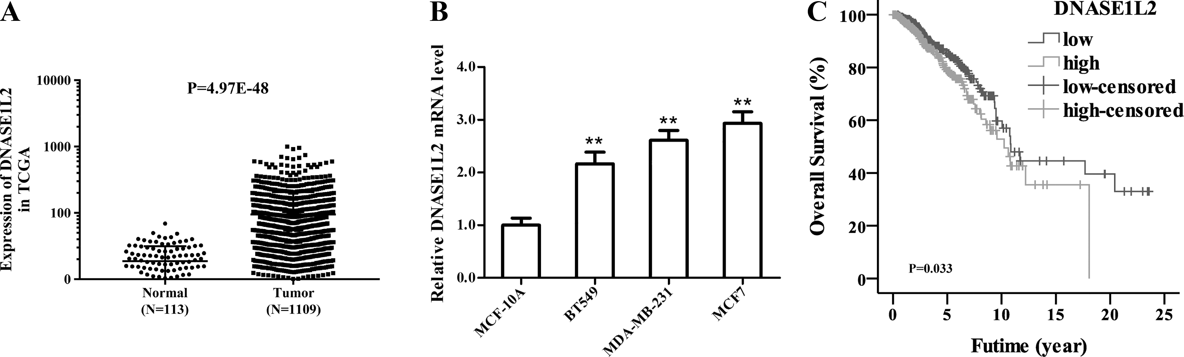
Levels of DNASE1L2 and survival curves.
The prognostic values of DNASE1L2
To explore DNASE1L2 prognostic values, the authors first evaluated the relevance between DNASE1L2 levels and the overall survival of BC patients. Kaplan–Meier plot indicated that patients with high DNASE1L2 levels appeared a cruel survival status in contrast with DNASE1L2 low levels group (p < 0.05, Fig. 1C). Next, using chi-square test, the authors assessed the relationship between DNASE1L2 levels and clinical symptoms of BC patients based on TCGA database. The data showed that DNASE1L2 expression was associated with age (p < 0.05) and pathologic-stage (p < 0.05, Table 1). Furthermore, to assess whether DNASE1L2 could be seen as an independent prognosticator, Cox regression analysis was employed. Univariate analysis demonstrated that DNASE1L2 expression, pathologic-stage, pathologic-tumor (T), -metastasis (M), and -node (N), and age were associated with the overall survival of BC patients (p < 0.05, Table 2). Then these candidate parameters with p-value <0.05 were further applied in multivariate analysis to identify independent prognostic values. Interestingly, no independent prognostic value of DNASE1L2 was showed (p > 0.05). In addition, pathologic-stage, -M, -N, and age were shown to be independent prognostic factors (p < 0.05). Although DNASE1L2 could not be regarded as an independent prognosticator, its abnormal expression in BC and its association with the overall survival suggested that DNASE1L2 may play a role in the development of BC.
Correlation Between Clinical Characteristics and DNASE1L2 Expression in Breast Cancer Patients
p < 0.05
Univariate and Multivariate Analysis for Prognosticators of Overall Survival in Breast Cancer Patients Based on The Cancer Genome Atlas Database
p < 0.05.
HR, hazard ratio.
Detection of DNASE1L2 silencing and overexpression transfection efficiency
The authors conducted two siRNAs and overexpression vector to down- and upregulate DNASE1L2 expression. To detect the transfection efficiency, qRT-PCR and Western blot were used to determine mRNA and protein levels of DNASE1L2, respectively. As shown in Figure 2A, B, after transfection with si-DNASE1L2#1 and si-DNASE1L2#2 in MCF7 cells, mRNA and protein levels of DNASE1L2 were notably reduced as contrasts with si-con group (p < 0.05). Considering that the silence efficiency of si-DNASE1L2#2 was better than that of si-DNASE1L2#1, si-DNASE1L2#2 was selected to perform the knockdown experiments in the following assays, and referred as si-DNASE1L2. In another aspect, mRNA and protein levels of DNASE1L2 were significantly enhanced after transfected with pcDNA3.1-DNASE1L2 by a contrast with pcDNA3.1 vector group in BT549 cells (p < 0.05, Fig. 2C, D). These data indicated that MCF7 cells with silencing DNASE1L2 and BT549 cells with overexpressed DNASE1L2 were successfully constructed.

Detection of DNASE1L2 silencing or overexpression efficacy qRT-PCR
Detection of proliferative capacity of BC cells with different expression of DNASE1L2
To detect the function of DNASE1L2 on BC cell viability, CCK-8 and plate clone formation assays were performed. The CCK-8 curve showed that the OD values in si-DNASE1L2 group were significantly reduced at 48 and 72 h compared with si-con group in MCF7 cells (p < 0.05, Fig. 3A). And clone formation assays indicated that silencing DNASE1L2 expression led to a poorer colony efficiency in comparison with si-con group (p < 0.05, Fig. 3C). Reversely, by enhancing DNASE1L2 expression, the OD values were increased compared with vector group (p < 0.05, Fig. 3B). And an increase in the number of colonies was notably observed after transfection with pcDNA3.1-DNASE1L2 compared with vector group in BT549 cells (p < 0.05, Fig. 3D). Above all, the authors deduced that the level of DNASE1L2 may have an important connection with BC cells proliferation.

Alteration expression of DNASE1L2 had effects on the cell viability. CCK-8 assay
Detection of invasive and migratory abilities of BC cells with different expression of DNASE1L2
To determine the effect of DNASE1L2 on the migratory and invasive abilities in BC cells, Transwell assay was implemented. Results of invasion assays revealed that, compared with si-con group (159.33 ± 32.65), the invasive MCF7 cell numbers of si-DNASE1L2 group (53.67 ± 28.1) showed an about threefold decrease (p < 0.05, Fig. 4A, B). Homoplastically, the migratory MCF7 cell numbers of si-DNASE1L2 group (162.67 ± 21.13) were fewer than that in si-con group (297.33 ± 37.5, p < 0.05, Fig. 4A, B). Yet, raising DNASE1L2 expression in BT549 cells, the number of invaded cells in pcDNA3.1-DNASE1L2 group (193.33 ± 30.57) was more than twice of that in vector group (79.33 ± 40.87, p < 0.05, Fig. 4C, D). Similarly, enhanced DNASE1L2 expression showed the same trend in migration assays. The migrated BT549 cell number in pcDNA3.1-DNASE1L2 group was 370.33 ± 56.19, and in vector group was 183.67 ± 42.52 (p < 0.05, Fig. 4C, D). These data illustrated that DNASE1L2 might serve a considerably promoting role on the metastasis capacity of BC cells.
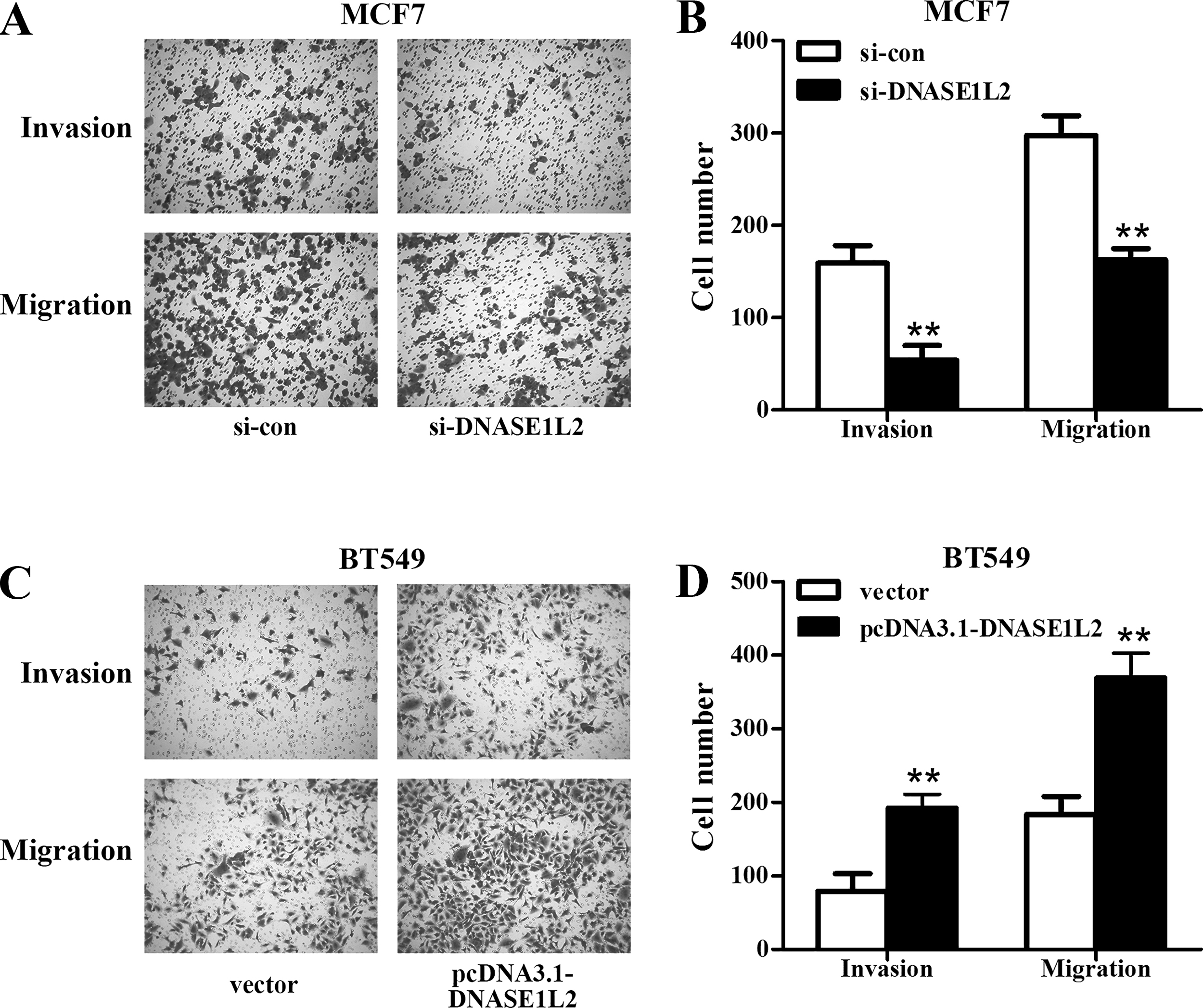
Transwell assays were adopted to detect influences of up- and downregulation of DNASE1L2 on the invasion and migration.
Association of DNASE1L2 levels and EMT process
For identifying the effects of DNASE1L2 levels on the EMT process in BC cells, qRT-PCR and Western blot were perform to evaluate the mRNA and protein levels of EMT-related markers. Results showed in Figure 4A and C indicated that knockdown of DNASE1L2 dramatically upregulated the levels of E-cadherin in MCF7 cells compared with si-con-transfected cells, whereas the levels of mesenchymal markers N-cadherin and Vimentin and transcription factor Snail were all significantly downregulated (p < 0.05, Fig. 5A, C). For upregulation of DNASE1L2, the authors observed that the EMT relative markers in BT549 cells showed the opposite effects, as with reduced E-cadherin levels and enhanced N-cadherin, Vimentin, and Snail levels (p < 0.05, Fig. 5B, D). To sum up, all these findings implied that DNASE1L2 expression plays roles on cells viability and movement partly by regulating EMT process in BC cells.
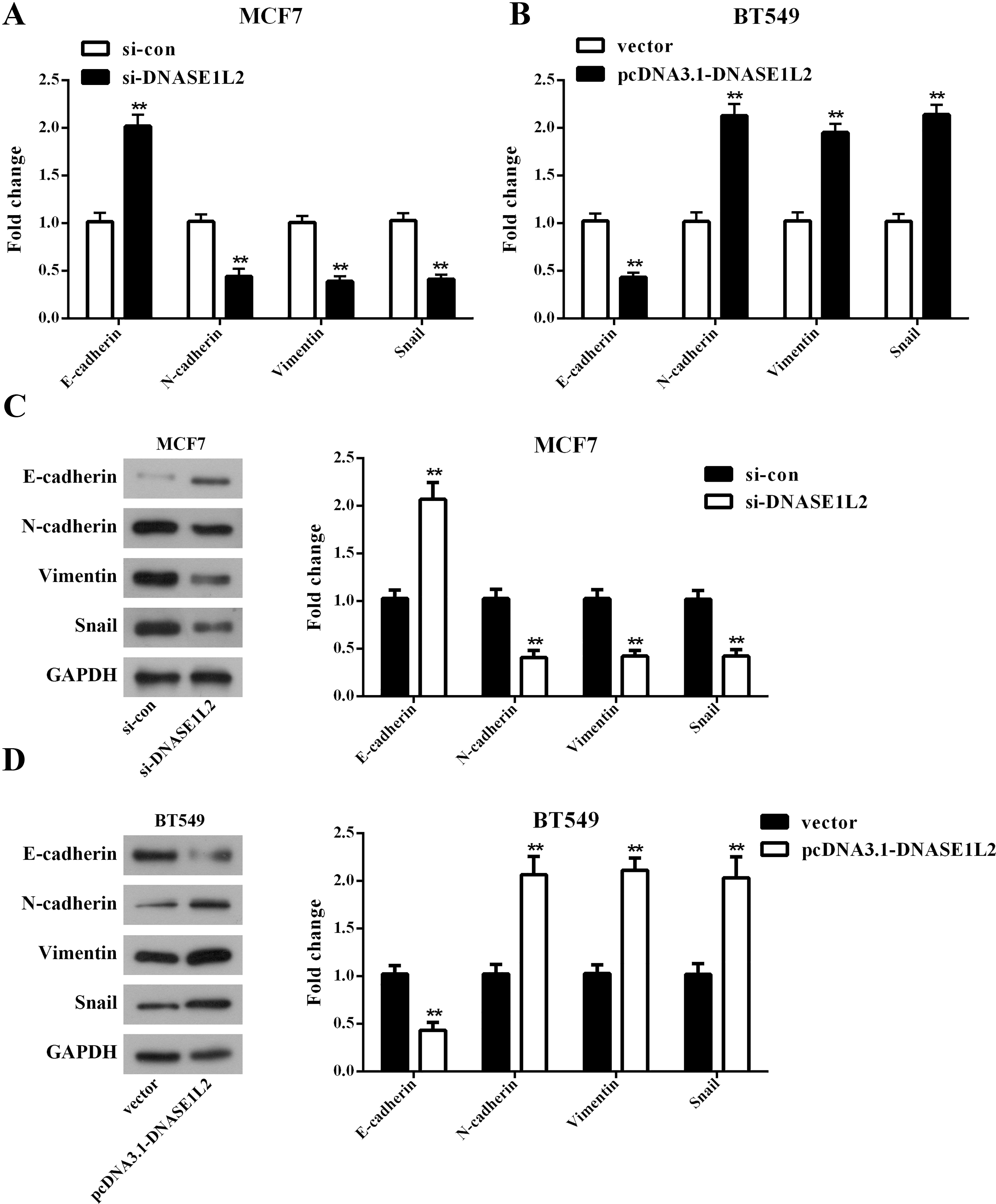
DNASE1L2 modulated the EMT process.
Discussion
Biomarkers can help clinicians make more accurate judgment on the prevention and therapy of BC, and choose more effective treatment measures. 15 At present, the application of many biomarkers in preoperative diagnosis of BC is still in the research stage. 16 Thus, the exploration of effective biomarkers is the current and future trend for BC therapeutics. In this report, it is found that DNASE1L2 was highly expressed in BC cells, and could be seen as a carcinogenic biomarker to affect cell proliferative, invasive, and migratory capacities by regulating EMT process in BC.
DNASE1L2 is a member of DNase family, which is involved in the degradation of chromatin in apoptotic cells. 17 It was reported that DNase 1 family members are usually highly expressed in a limited number of tissues or cell types, and poorly expressed in a few others. 18 As currently reported, most members of DNase 1 family are not characterized, but their elimination can lead to a variety of diseases, such as keratosis and systemic lupus erythematosus. 19,20 Owing to the enzymatic nature of the members, the lower expression level could be biologically relevant. 18 Therefore, the level of DNASE1L2 expression may be crucial to the healthy organism development and survival.
By a literature review, the authors found that DNASE1L2 is mainly expressed in keratinocytes and tissues containing keratinocytes, such as skin. 21 In addition, its expression was also detected in brain. 22 It has been shown that DNASE1L2 is a necessary and specific regulator of programmed cell death in skin appendages. 21 At present, the role of DNASE1L2 in tumors has been rarely reported. But it is worth mentioning that DNASE1L3, also a member of DNase 1 family, was reported to be downregulated in the late stage of clear cell renal cell carcinoma. 22,23 Furthermore, Malecki et al. inserted human DNASE1L3 gene into a plasmid regulated by EGFR promoter, and then introduced the plasmid into ovarian cancer cell line, resulting in destruction of cell genome and apoptosis. 24 All of these stimulate everyone to wonder whether DNASE1L2 expression plays a role in cancer.
Thus, in this study, the authors initially revealed that DNASE1L2 was upregulated in BC tissues based on TCGA database, and the next in vitro tests confirmed its upregulation in three BC cell lines. Furthermore, they assessed the prognostic values of DNASE1L2 in BC, and observed that high expression of DNASE1L2 led to a short overall survival. And the following univariate Cox regression analysis demonstrated that it has prognostic values for BC patients. But the multivariate analysis did not demonstrate that it is an independent prognosticator. The authors conjectured that clinical specificity and tumor heterogeneity may be appropriate explanations for these results. Nonetheless, the results suggest that DNASE1L2 might play a role on the progression of BC, which might provide a new point of view for the role of DNASE1L2 in BC.
Despite significant advances in the therapy of BC, metastatic BC remains a deadly disease. 25 Therefore, a thorough understanding of the systemic process of cancer cell proliferation is essential for the development of new therapies. EMT is a cell biological program that transforms epithelial cells into mesenchymal cells, confers cancer cells metastasis diffusion ability. 26 And it is frequently used as a popular explanation for how tumor cells gain migratory and invasive properties to leave the original tumor site and spread throughout the body, eventually forming distant metastases. 27,28
At present, studies on molecular markers related to EMT have been very mature. E-cadherin, an epithelial marker, can maintain tight connections between cells and prevent invasion, metastasis, and spread of cell activities. The conversion of cadherin between E-cadherin and N-cadherin is a classic example of cancer-related EMT. 29 Vimentin maintains cytoskeletal integrity, 30 and it was reported that inhibition of 70% Vimentin expression would effectively reduce the invasion and migration of tumors. 31 Furthermore, Snail, as a transcription factor of EMT-related marker, has been proved to participate in the progression and metastasis of BC cells. 32,33 In view of this, to gain insight into this underlying mechanism of action, the authors detected mRNA and protein-level changes of E-cadherin, N-cadherin, Vimentin, and Snail by up- and downregulation of DNASE1L2 expression.
The data showed that reducing DNASE1L2 levels in MCF7 cells could significantly restrain the EMT process, which raised the levels of E-cadherin and lowered the levels of N-cadherin, Vimentin, and Snail. Increasing DNASE1L2 expression reversed these results. These outcomes implied that DNASE1L2, as a cancer-promoting gene, facilitates the cell proliferative, invasive, and migratory abilities partly by regulating the EMT relative markers in BC cells. Evidence has demonstrated that EMT plays a key role in the progression and metastasis of BC, and participates in antagonizing chemotherapy. 34 Thus, it is of great clinical significance to elucidate the specific molecular mechanism by which EMT regulates metastasis, and more in-depth studies are needed.
In summary, this study is the first to explore the role of DNASE1L2 in BC. The data indicated that DNASE1L2 was significantly upregulated in BC cells, and had a correlation with the overall survival, and illustrated that DNASE1L2 affects the cells phenotype through modulating EMT-related markers. All these outcomes suggested that DNASE1L2 might serve as a carcinogenic biomarker in the progression and metastasis of BC cells, which provide the basis for further investigation of underlying mechanism and exploitation of novel target options for BC therapeutic.
Footnotes
Authors' Contribution
Chang-Rui Liu and Fan-Hua Meng designed this study. Chang-Rui Liu performed the experiments and wrote the article. Fan-Hua Meng explained the data and revised the article. All the authors approved the final version of this article.
Disclosure Statement
No competing financial interests exist.
Funding Information
No funding was received for this article.