
Abstract
LncRNA ELF3-AS1 has been characterized as an oncogenic lncRNA in bladder cancer and oral cancer, whereas its role in non-small cell lung cancer (NSCLC) is unknown. In this study, the authors observed that ELF3-AS1 was upregulated in NSCLC tissues in comparison with that in paired nontumor tissues collected from 68 NSCLC patients. High expression levels of ELF3-AS1 predicted the poor survival of NSCLC patients. Expression levels of miR-212 were inversely and significantly correlated with the expression levels of ELF3-AS1 across NSCLC tissue samples. In NSCLC cells, overexpression of ELF3-AS1 led to downregulated miR-212 and increased methylation of miR-212 gene. In addition, overexpression of ELF3-AS1 inhibited the role of miR-212 in suppressing cancer cell invasion and migration. Therefore, ELF3-AS1 is upregulated in NSCLC and promotes cancer cell invasion and migration by downregulating miR-212 through methylation.
Introduction
For decades, lung cancer is the most common type of malignancy in clinical practice for both incidence and mortality rates. 1 In 2018 alone, lung cancer affected a total number of 2,093,876 new cases (11.6% of all new cancer cases) and caused 1,761,007 deaths (18.4% of all cancer deaths). 2 Non-small cell lung cancer (NSCLC) is the major subtype of lung cancer and accounts for 85% of all cases. 3 Smoking is the major risk factor for NSCLC, whereas ∼10–15% NSCLC occurs in never-smokers, 4 indicating the complex pathogenesis of this disease. Even with advances in anticancer therapies, prognosis of NSCLC is still poor. It is estimated that only <15% of patients can live >5 years after initial diagnosis. 5,6 Therefore, novel cancer therapies are still needed.
Occurrence, development, and progression of NSCLC involve dysregulation of molecular pathways. 7,8 Functional analysis of genes involved in NSCLC provides novel targets for the development of targeted therapies. 9 Noncoding RNAs (ncRNAs), such as long (>200 nt) ncRNAs (lncRNAs) and miRNAs, encode no protein but regulate cancer-related gene expression at multiple levels to participate in cancer biology. 10 In effect, regulation of the expression of certain key ncRNA players in cancer provides novel insights into the development of anticancer approaches. 11 However, functions of most lncRNAs in cancer biology remain unknown. ELF3-AS1 is a recently identified oncogenic lncRNA in bladder cancer and oral cancer, 12,13 whereas its role in NSCLC is unknown. The authors' preliminary RNA-seq analysis revealed altered expression of ELF3-AS1 and its inverse correlation with miR-212, which is a tumor suppressor in NSCLC. 14 This study was, therefore, carried out to investigate the interactions between ELF3-AS1 and miR-212 in NSCLC.
Materials and Methods
NSCLC patients and specimens
This study was approved by the Ethics Committee of Fudan University Shanghai Cancer Center. This study enrolled a total number of 68 NSCLC patients (43 men and 25 women, age range 47–69 years, mean age 57.2 ± 8.1 years) who were admitted to the cancer center between January 2011 and January 2014. No other severe clinical disorders were observed among patients. The 68 patients included 37 cases of lung squamous cell carcinoma and 31 cases of lung adenocarcinoma. All patients signed the informed consent. Paired NSCLC and nontumor tissues were collected from each patient during surgical resections. All specimens were stored in liquid nitrogen before total RNA extractions.
Treatment and a 5-year follow-up
The 68 NSCLC patients included 12, 18, 20, and 18 cases at American Joint Committee on Cancer (AJCC) stages I, II, III, and IV, respectively. Based on patients' health conditions and AJCC stages, therapies such as radiotherapy, chemotherapy, surgical resection, targeted therapy, or combined therapies were applied. From the day of admission, the 68 patients were followed up for 5 years to record their survival. All 68 patients completed this 5-year follow-up.
NSCLC cell lines and cell culture
Human NSCLC cell lines H522, H23, H460, A549, and adjacent tumor cell line (16HBE) were used. Cells of both cell lines were obtained from ATCC. Cell culture conditions were 37°C, 95% humidity, and 5% CO2. Cell culture medium was composed of 10% FBS and 90% RPMI-1640 medium. Cells were harvested at ∼85% confluence to perform subsequent transfections.
Cell transfections
ELF3-AS1 expression vector was constructed with pcDNA 3.1 vector (Invitrogen) as backbone. MiR-212 mimic and negative control (NC) miRNA were purchased from Sigma-Aldrich. H522 and H23 cells were transfected with either 12 nM vector or 40 nM siRNA using lipofectamine 2000 (Invitrogen). All operations were completed following the instructions from Invitrogen. Control (C) cells were untransfected cells in all cases. NC cells were cells transfected with either empty pcDNA 3.1 vector or NC miRNA. At 48 h post-transfection, cells were harvested to perform subsequent experiments.
RNA preparations
Total RNAs were isolated from both tissues and in vitro cultivated cells using RNAzol (Sigma-Aldrich). Precipitation of miRNAs was performed using 85% ethanol. NanoDrop™ 2000 Spectrophotometer (Thermo Fisher) was used to measure RNA concentrations. To remove genomic DNAs, all RNA samples were digested with DNase I at 37°C for 1 h.
Reverse transcriptase-quantitative polymerase chain reaction
RNA samples with satisfactory quality were reverse transcribed into cDNA using MMLV Reverse Transcriptase 1st-Strand cDNA Synthesis Kit (Lucigen). With cDNA as template, quantitative polymerase chain reaction (qPCR) was performed using QuantiTect SYBR Green PCR Kit (Qiagen). The expression levels of ELF3-AS1 were measured with glyceraldehyde-3-phosphate dehydrogenase as endogenous control. Measurement of the expression of mature miR-212 was performed using All-in-One™ miRNA qRT-PCR Reagent Kit (Genecopoeia). All PCRs were repeated three times and 2−ΔΔCT method was used to normalize gene expression levels.
Methylation-specific PCR
Isolation of genomic DNA from H522 and H23 cells was performed using Monarch® Genomic DNA Purification Kit (NEB). DNA concentrations were measured using NanoDrop 2000 Spectrophotometer. To perform methylation-specific PCR (MSP), DNA was converted using EZ DNA Methylation-Gold™ kit (ZYMO RESEARCH) and PCRs were performed using Taq DNA polymerase kit (NEB).
Transwell assays
Cell invasion and migration were analyzed by performing Transwell assays using Transwell filters (8 μm; BD Biosciences). Matrigel-coated membranes and uncoated membranes were used in invasion and migration assays, respectively. The upper compartment was filled with 10,000 cells in 0.5 mL cell culture medium, and the lower compartment was filled with medium containing 20% FBS. Cells were cultivated under aforementioned conditions for 24 h, followed by staining of the lower surface of membranes by crystal violet (0.1%). Cells were counted under a light microscope.
Assay of colony formation
H522 and H23 cells with the same density were seeded into six-well plates after transfection with different vectors. Two weeks later, the cells were fixed with 4% paraformaldehyde (Sigma-Aldrich), stained with 0.5% crystal violet (Sigma-Aldrich), and then washed with phosphate-buffered saline (Sigma-Aldrich). The total number of colonies from three separate transfections were counted. The average value was used to evaluate colony formation ability.
Statistical analysis
Experiments were performed in triplicate and data were expressed as mean ± SEM. Paired t-test was used to compare differences between nontumor and NSCLC tissues. One-way ANOVA and Tukey test were used to compare differences among multiple groups. Spearman's correlation coefficient was used to perform correlation analysis. The 68 patients were divided into high and low ELF3-AS1 level groups (n = 34 for each) with median expression level of ELF3-AS1 in NSCLC as cutoff value. Chi-squared test was performed to analyze the associations between patients' clinical data and expression levels of ELF3-AS1 in NSCLC tissues. Survival curves were compared using log-rank test. p < 0.05 was statistically significant.
Results
Upregulation of ELF3-AS1 predicted the poor survival of NSCLC
Expression levels of ELF3-AS1 in paired nontumor and NSCLC tissue samples collected from 68 NSCLC patients were measured using reverse transcriptase-qPCR (RT-qPCR). Compared with nontumor tissues, NSCLC tissues showed significantly increased expression levels of ELF3-AS1 (Fig. 1A, p < 0.05). Survival curves were plotted for both high and low ELF3-AS1 level groups. Log-rank test analysis showed that overall survival rate or patients in high ELF3-AS1 level group was significantly lower than that of the patients in low ELF3-AS1 level group (Fig. 1B). Chi-squared test showed that the expression levels of ELF3-AS1 in NSCLC tissues were significantly associated with patients' clinical stage, but not with other clinical factors (Table 1).
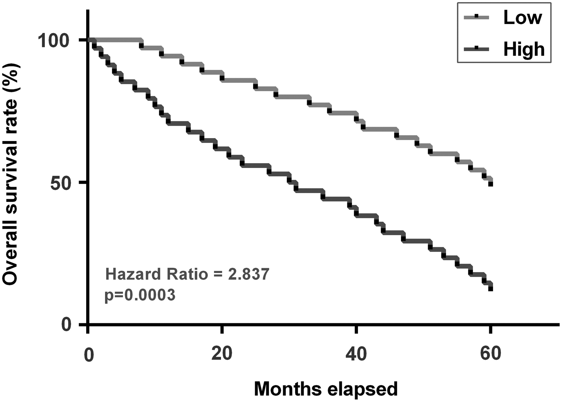
Downregulation of ELF3-AS1 predicted the poor survival of NSCLC. Expression levels of ELF3-AS1 in paired nontumor and NSCLC tissue samples collected from 68 NSCLC patients were measured using RT-qPCR. PCRs were repeated three times and mean values were presented
Associations Between Patients' Clinical Data and Expression Levels of ELF3-AS1 in Non-small Cell Lung Cancer Tissues
LUSC, lung squamous cell carcinoma; LUAD, lung adenocarcinoma.
Expression levels of miR-212 inversely correlated with ELF3-AS1
Expression levels of miR-212 in paired nontumor and NSCLC tissue samples collected from 68 NSCLC patients were also measured using RT-qPCR. Compared with nontumor tissues, NSCLC tissues showed significantly decreased expression levels of miR-212 (Fig. 2A, p < 0.05). Correlation analysis showed that the expression levels of miR-212 inversely correlated with ELF3-AS1 across both NSCLC tissues (Fig. 2B) and nontumor tissues (Fig. 2C).
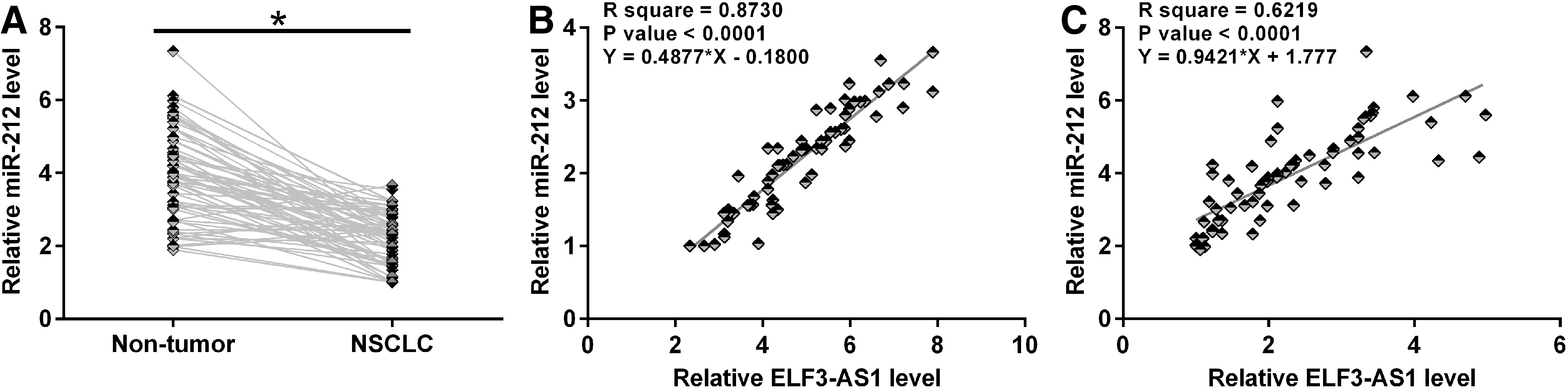
Expression levels of miR-212 correlated inversely with ELF3-AS1. Expression levels of miR-212 in paired nontumor and NSCLC tissue samples collected from 68 NSCLC patients were also measured using RT-qPCR. PCRs were repeated three times and mean values were presented
Overexpression of ELF3-AS1 led to downregulated miR-212 and increased methylation of miR-212 gene
H522 and H23 cells were transfected with either ELF3-AS1 expression vector or miR-212 mimic. Overexpression of ELF3-AS1 and miR-212 was confirmed by RT-qPCR at 48 h post-transfection (Fig. 3A, p < 0.05). Compared with C and NC groups, overexpression of ELF3-AS1 led to downregulated miR-212 (Fig. 3B, p < 0.05). In contrast, overexpression of miR-212 did not affect the expression of ELF3-AS1 (Fig. 3C). The effects of overexpression of ELF3-AS1 on the methylation of miR-212 gene were analyzed by MSP. Compared with the cells transfected with empty pcDNA3.1 vector, cells transfected with ELF3-AS1 expression vector showed obviously increased methylation of miR-212 gene (Fig. 3D).
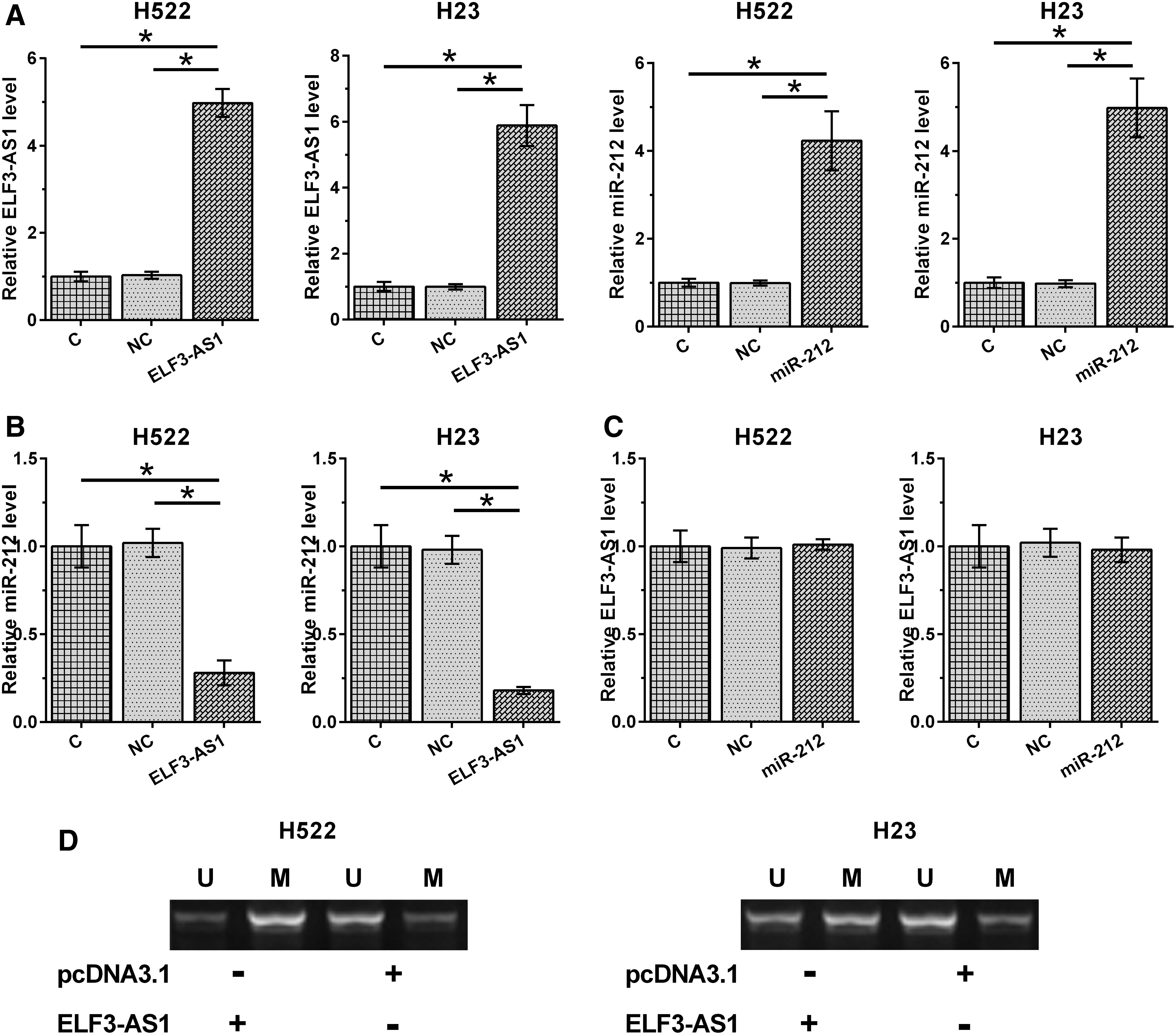
Overexpression of ELF3-AS1 led to downregulated miR-212 and increased methylation of miR-212 gene. H522 and H23 cells were transfected with either ELF3-AS1 expression vector or miR-212 mimic. Overexpression of ELF3-AS1 and miR-212 was confirmed by RT-qPCR at 48 h post-transfection
ELF3-AS1 promoted NSCLC cell invasion and migration through miR-212
The effects of overexpression of ELF3-AS1 and miR-212 on the invasion and migration of NSCLC cells were analyzed by performing Transwell assays. Compared with C group, overexpression of miR-212 led to decreased invasion (Fig. 4A) and migration (Fig. 4B) rates of NSCLC cells (p < 0.05). Overexpression of ELF3-AS1 played an opposite role and inhibited the role of miR-212 in suppressing cancer cell invasion and migration (p < 0.05). In the colony formation assay, overexpression of miR-212 led to partial recovery of ELF3-AS1 overexpression-induced promotion of cell proliferation for both H522 and H23 cells (Fig. 4C, p < 0.05).

ELF3-AS1 promoted NSCLC cell invasion and migration through miR-212. The effects of overexpression of ELF3-AS1 and miR-212 on the invasion
Discussion
This study investigated the role of ELF3-AS1 in NSCLC. The authors found that ELF3-AS1 was upregulated in NSCLC and may downregulate miR-212 through methylation to promote cancer cell invasion and migration.
The functionality of ELF3-AS1 has only been investigated in bladder cancer and oral squamous cell carcinoma (OSCC). 12,13 ELF3-AS1 is upregulated in bladder cancer and can regulate Krüppel-like factor 8 to promote cancer progression. 12 In OSCC, ELF3-AS1 reprograms glucose metabolism to promote cancer cell proliferation. 13 This study is the first to report the upregulation of ELF3-AS1 in NSCLC. In addition, overexpression of ELF3-AS1 led to increased invasion and migration rates of NSCLC cells. Therefore, ELF3-AS1 has oncogenic functions in NSCLC.
Accurate prognosis is critical for the selection of anticancer therapies and the development of postoperative care programs. 15 In this study, the authors showed that the high expression levels of ELF3-AS1 before treatment can be used to predict the poor survival of NSCLC patients. However, this study is limited by the small sample size. Future studies are needed to further confirm these conclusions with bigger sample size.
MiR-212 plays different roles in different cancers. For instance, the expression of miR-212 in pancreatic cancer can be induced by HIF-1α to promote cancer progression. 16 In contrast, miR-212 plays a role as tumor suppressor by regulating cancer cell behaviors, such as the inhibition of cancer cell invasion and migration in most other cancers, including NSCLC. 14,17 This study confirmed the downregulation of miR-212 in NSCLC and its inhibitory effects on cancer cell invasion and migration.
The key finding of this study is that ELF3-AS1 can promote the methylation of miR-212 gene to downregulate its expression. A recent study also reported that an lncRNA PVT1 can induce the methylation of miR-146a to participate in cancer biology. 18 However, the molecular mechanism is unknown. More studies are still needed.
In conclusion, ELF3-AS1 is upregulated in NSCLC and may downregulate miR-212 through methylation to promote cancer cell invasion and migration.
Footnotes
Disclosure Statement
The authors declare that they have no competing interests.
Funding Information
No funding was received for this article.