
Abstract
Cancer Biotherapy and Radiopharmaceuticals
is officially retracting the article entitled, A Novel CircRNA Circ_0095424 Regulates Proliferation, Metastasis, and Apoptosis of Osteosarcoma Cells Via the PI3K/AKT Signaling Pathway Through Targeting the miR-1238/HMGB1 Axis by Zhang et al., (Cancer Biother Radiopharm epub 19 Aug 2020; DOI: 10.1089/cbr.2020.3563), due to manipulated images appearing in the published paper.
The Editor of the journal received an email on August 31, 2020 from the corresponding author of the article, Dr. Chuan Qin, indicating that, ‘‘due to our negligence in organizing the pictures, the protein pictures are repeatedly placed in Figure 7G PI3K. For this, we express our sincerest apologies. We need to [issue] an [erratum] on this issue. We have replaced the protein picture of Figure 7G with the correct picture.’’ However, one of the attachments submitted with the request appeared to be the original version of Figure 7 from the accepted manuscript. A second attachment appeared to be the data from three replicates to be used (by the journal) to construct a revised version of Figure 7. The Editor, in turn, informed the authors that it was not at the journal’s discretion to create a new image for them, and asked the authors to create the revised figure and supply it to the publisher. Below is the response from Dr. Qin, dated September 2, 2020.
“In fact, our team’s Western blot experiment commissioned a third-party company for testing. At present, some peers have found that the company has forged experimental reports. We believe that the authenticity of the data provided by the company is problematic. After contacting the company, they were unable to provide the original images. In view of the problems in this manuscript, all the authors discussed and agreed to withdraw the manuscript.”
As the entirety of the situation is unacceptable, the Editor officially retracts the article based on the “forged experimental reports” and the questionable validity of the data provided.
The Editor and Publisher of Cancer Biotherapy and Radiopharmaceuticals is dedicated to preserving the integrity of the scientific literature and the community it serves.
Introduction
Among primary bone malignancies, osteosarcoma (OS) is a common solid tumor that occurs more frequently in children and adolescents. 1,2 According to statistics, the incidence of OS in adolescents under the age of 19 is 3%, and the 5-year survival rate is between 65% and 70%. 3 However, the occurrence of metastasis significantly reduces the survival rate of OS patients, and this is also the main cause of death in OS patients. 4,5 Similar to other tumors, the progression of OS is a multistep process and is regulated by multiple factors. 6 Therefore, it is essential to identify effective diagnostic markers and potentially key molecules that influence OS progression.
Circular RNAs (circRNAs) are a class of noncoding RNAs that have been studied more in recent years. Compared with traditional linear RNAs, circRNAs are characterized by covalently closed loop and have extremely high stability. 7,8 There is a lot of evidence that circRNAs have a major role in the development of cancers, and their great potential as biomarkers and therapeutic targets for cancers is being elucidated. 9 –11 Of course, a number of circRNAs have been implicated in regulating OS progression, such as circMMP9 and circ_0102049. 12,13 Liu et al. used microarray analysis to screen for circRNAs that were differentially expressed in OS tissues and normal tissues and uncovered that circ_0095424 was significantly upregulated in OS tumor tissues. 14 But the role of circ_0095424 in OS is not yet known.
At present, the biological function of circRNA as a microRNA (miRNA) sponge has been widely known, which has become an important way to elucidate the molecular mechanism of circRNA. 15,16 MiR-1238 is markedly underexpressed in many cancers and is involved in the regulation of cancer progression, including cell growth and metastasis. 17,18 Qi et al. report that miR-1238 is lowly expressed in OS and participates in the regulation of circ_0000502 on OS progression. 19 However, there are few studies on miR-1238 in OS.
High mobility group box-1 (HMGB1) is believed to be a vital regulator involved in a wide range of biological processes in cancer. 20,21 Studies have shown that the expression of HMGB1 can mediate the activity of phosphatidylinositol 3 kinase (PI3K)/serine-threonine kinase (AKT) signaling pathway, which plays an important role in various cellular processes, such as cell proliferation, apoptosis, and metastasis. 22,23 In OS, many studies have suggested that HMGB1 is highly expressed in OS and is related to the progression of OS. 24,25 Therefore, studies on HMGB1 can help everyone better understand the pathogenesis of OS.
In this study, the authors focused on exploring the role and mechanism of circ_0095424 in OS progression with a view to providing a potential target for OS treatment.
Materials and Methods
Clinical tissue samples
A total of 51 OS patients were recruited from Rizhao City Traditional Chinese Medicine Hospital, and OS tissues and adjacent normal tissues from OS patients were collected and stored at −80°C. All patients provided informed content. This study was allowed by the Ethics Committee of Rizhao City Traditional Chinese Medicine Hospital.
Cell culture and transfection
Human OS cell lines (U2OS and MG63) and the human osteoblastic cell line (hFOB1.19) were purchased from American Type Culture Collection (Manassas, VA) and cultured in Dulbecco's modified Eagle medium (Gibco, Gran Island, NY) containing 10% fetal bovine serum (Gibco), 100 U/mL penicillin (Invitrogen, Carlsbad, CA) and 100 mg/mL streptomycin (Invitrogen) at 37°C with 5% CO2 incubator. Circ_0095424 small interfering RNA (si-circ_0095424#1: 5′-GCCCCTCAGGTGTTTTGTCCA-3′; si-circ_0095424#2: 5′-ACAAATGCCCCTCAGGTGTTT-3′; si-circ_0095424#3: 5′-AATGCCCCTCAGGTGTTTTGT-3′) and lentiviral short hairpin RNA (sh-circ_0095424: 5′-TACAAATGCCCCTCAGGTGTT-3′) or their negative controls (si-NC: 5′-CAGUACUUUUGUGUAGUACAA-3′; and sh-NC: 5′-TACGTGCATCGATCGCACGT-3′), circ_0095424 and HMGB1 overexpression plasmids (circ_0095424 and HMGB1) or negative control (pcDNA) were bought from RiboBio (Guangzhou, China). MiR-1238 mimic and inhibitor (miR-1238 and in-miR-1238) or their negative controls (miR-NC and in-miR-NC) were obtained from Genepharm (Shanghai, China). U2OS and MG63 cells were seeded in 6-well plates, and these plasmids (100 ng) were transfected into cells using Lipofectamine 3000 (Invitrogen).
Quantitative real-time polymerase chain reaction
Total RNAs were extracted by Trizol reagent (Invitrogen), and cDNA was synthesized using BeyoRT II cDNA Synthesis Kit (with gDNA Eraser) (Beyotime, Shanghai, China). Then, quantitative real-time polymerase chain reaction (qRT-PCR) was applied using SYBR Kit (Takara, Dalian, China). The relative expression data were normalized by β-actin or U6 and analyzed by relative quantification (2−ΔΔCt). The primers were designed as follows: circ_0095424, F 5′-ATGGCTCTATACGTGGCAGC-3′, R 5′-AGACACTGGTCCATGGTTGG-3′; miR-1238, F 5′-GTCGTATCCAGTGCAGGG-3′, R 5′-CGACGCTTCCTCGTCTG-3′; β-actin, F 5′-CACAGAGCCTCGCCTTTGCC-3′, R 5′-ACCCATGCCCACCATCACG-3′; U6, F 5′-CTCGCTTCGGCAGCACATATACT-3′, R 5′-ACGCTTCACGAATTTGCGTGTC-3′.
Cell proliferation assay
Transfected U2OS and MG63 cells were collected and seeded in 96-well plates (2 × 10³). After incubation for 12 h, cell counting kit 8 (CCK8) solution (Yeasen, Shanghai, China) was added into cells at the indicated time points and incubated for another 4 h. The absorbance was analyzed at 450 nm on a microplate reader.
Transwell assay
Transfected U2OS and MG63 cells (5 × 10⁴) suspended in serum-free medium were seeded in the upper chamber (24-well; Corning, Inc., Corning, NY), which was coated with Matrigel (Corning, Inc.) to detect cell invasion and uncoated to detect cell migration. Then, the lower chamber was filled with 600 μL serum medium. After incubation for 24 h, the upper chamber cells were removed and the lower chamber cells were fixed and stained. Finally, the number of cells was counted under the microscope.
Flow cytometry
After transfection for 48 h, U2OS and MG63 cells were harvested and resuspended with binding buffer containing Annexin V/fluorescein isothiocyanate (FITC) and propidium iodide (Beyotime) for 15 min. Then, the fluorescence of cells was measured using a flow cytometer and the apoptosis rate was counted by FlowJo software (FlowJo, San Francisco, CA).
Western blot analysis
Tissues or cells were lysed with RIPA lysate (Beyotime). Next, protein products were fractionated by 10% sodium dodecyl sulfate-polyacrylamide gel electrophoresis gel and transferred to polyvinylidene fluoride membranes (Millipore, Billerica, MA). Next, the membrane was incubated with skimmed milk, primary antibodies (phosphorylated-AKT [p-AKT], AKT, p-PI3K, PI3K, HMGB1, β-actin), and secondary antibody in this order. The protein signals were visualized with enhanced chemiluminescence detection reagent (Yeasen). All antibodies were bought from Amyjet (Wuhan, China) with a dilution of 1:1000.
Tumor model
MG63 cells were stably transfected with sh-circ_0095424 or sh-NC and then subcutaneously injected in the flank of nude mice (BALB/c, male, 4-week old; Junke Bio, Nanjing, China). The length and width of the tumor were measured every 7 d and tumor volume was calculated by width 2 × length/2 until day 28. After the mice were euthanized, the tumor was removed for tumor weight, circ_0095424 expression and p-AKT/AKT and p-PI3K/PI3K protein levels detection. Animal experiments were approved by the Committee of Rizhao City Traditional Chinese Medicine Hospital.
Dual-luciferase reporter assay
Fragments of circ_0095424 containing predicted or mutant target sites of miR-1238 were amplified and subcloned into a psiCHECK-2 vector (Promega, Madison, WI) to create the circ_0095424 wild-type (WT) or circ_0095424 mutant-type (MUT) vector. At the same time, the HMGB1 3′UTR (3′ untranslated region) WT and HMGB1 3′UTR MUT vectors were built in the same form. Then, U2OS and MG63 cells were co-transfected with the aforementioned vectors and miR-1238 mimic or miR-NC. After 48 h, the luciferase activities were detected using the Dual-Lucy Assay Kit (Solarbio, Beijing, China). All experiments were repeated at least three times.
RNA pull-down assay
Magnetic beads (Thermo Fisher Scientific, Waltham, MA) were incubated with Biotin-labeled circ_0095424 WT probe (Bio-circ_0095424 WT; Viagene Bio, Jiangsu, China), Bio-circ_0095424 MUT probe (Bio-circ_0095424 MUT; Viagene Bio), or Bio-negative control probe (Bio-NC; Viagene Bio) at 4°C overnight. U2OS and MG63 cells were lysed, and then cell lysates were collected and incubated with magnetic beads. Then, magnetic beads were purified and the enrichment of miR-1238 in Bio-circ_0095424 WT, Bio-circ_0095424 MUT, or Bio-NC was detected using qRT-PCR.
RNA immunoprecipitation assay
According to the manufacturer's instructions of RNA immunoprecipitation (RIP) Kit (Millipore), RIP assay was performed. In brief, U2OS and MG63 cells were lysed, and cell lysates were collected. Afterward, the cell lysates were incubated with magnetic beads conjugated with antibodies against IgG (Anti-IgG) or Ago2 (Anti-Ago2). After extracted RNA using TRIzol reagent, the enrichments of circ_0095424 and miR-1238 were tested by qRT-PCR. The RNA extracted from lysate not incubated with magnetic beads was used as “Input.”
Statistical analysis
Data were expressed as the mean ± standard deviation on GraphPad Prism5.0 software (GraphPad, San Diego, CA). Statistical comparisons were performed using Student's t-test or one-way analysis of variance. Pearson correlation analysis was used for analyzing the correlation among circ_0095424, miR-1238, and HMGB1. P < 0.05 was presented as statistically significant.
Results
Expression of circ_0095424 was increased in OS
The authors first evaluated the clinical significance of circ_0095424 in OS by detecting its expression. As exhibited in Figure 1A, circ_0095424 expression was elevated in OS tissues compared with adjacent normal tissues. Furthermore, they also measured circ_0095424 expression in two OS cell lines (U2OS and MG63) and hFOB1.19 cells, and the results indicated that the expression of circ_0095424 was higher in OS cells than that in hFOB1.19 cells (Fig. 1B). These data suggested that circ_0095424 might have an important function in OS.
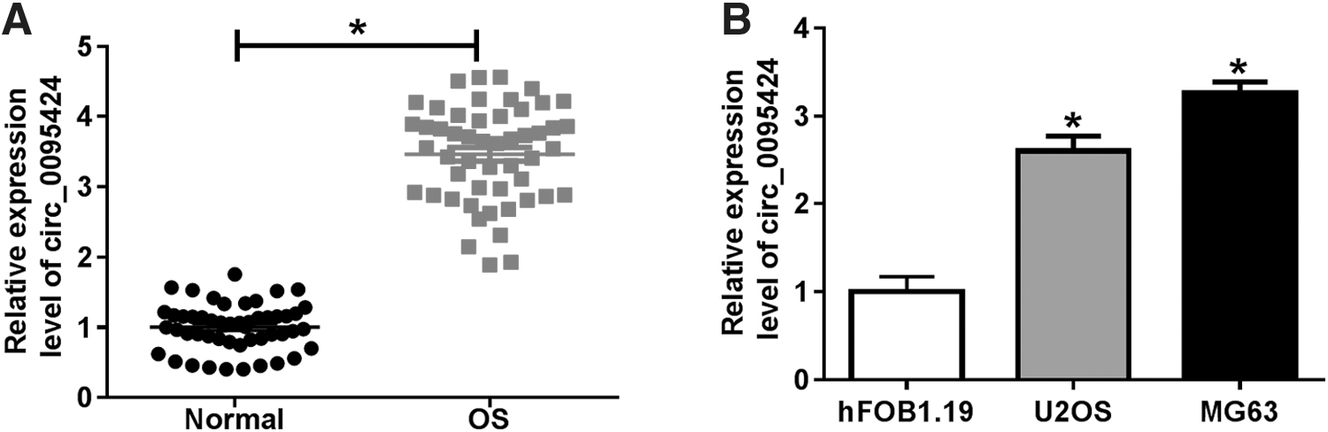
Expression of circ_0095424 was increased in OS.
Circ_0095424 silencing suppressed OS cell progression in vitro
For investigating the function of circ_0095424 in OS, the authors performed the loss-functional experiments using siRNA. By detecting the expression of circ_0095424, they found that si-circ_0095424#2 had a relatively good inhibitory effect on circ_0095424 expression and a relatively high transfection efficiency compared with si-circ_0095424#1 or si-circ_0095424#3 (Fig. 2A). Therefore, subsequent experiments were conducted using si-circ_0095424#2. CCK8 results suggested that circ_0095424 knockdown suppressed the proliferation of U2OS and MG63 cells (Fig. 2B, C). Besides, the results of measuring the migration and invasion of cells showed that silenced circ_0095424 inhibited the migration and invasion abilities of U2OS and MG63 cells (Fig. 2D, E). Moreover, through flow cytometry, the authors uncovered that the knockdown of circ_0095424 promoted the apoptosis rates of U2OS and MG63 cells (Fig. 2F). In addition, the detection of the protein levels of p-AKT/AKT and p-PI3K/PI3K showed that silencing of circ_0095424 reduced the relative expression of p-AKT/AKT and p-PI3K/PI3K in U2OS and MG63 cells, indicating that its knockdown repressed the activity of the PI3K/AKT signaling pathway (Fig. 2G). In view of this, the authors speculated that circ_0095424 might act as an oncogene in OS.

Circ_0095424 silencing regulated the progression of OS cells.
Knockdown of circ_0095424 restrained OS tumor growth and the activity of the PI3K/AKT signaling pathway in vivo
To further confirm the function of circ_0095424 in OS, the authors constructed the xenograft models of OS. As shown in Figure 3A, B, the growth rate of tumor volume derived from the sh-circ_0095424 group was markedly inhibited, and the tumor weight was also reduced. A significant decrease in circ_0095424 expression in the sh-circ_0095424 group indicated that its transfection was successful (Fig. 3C). Besides, the authors also discovered that the protein levels of p-AKT/AKT and p-PI3K/PI3K were decreased in the sh-circ_0095424 group, suggesting that the activity of the PI3K/AKT signaling pathway was remarkably suppressed (Fig. 3D). Therefore, the authors concluded that circ_0095424 expression was critical for OS tumor growth in vivo.

Circ_0095424 knockdown regulated the tumor growth of OS.
Circ_0095424 could bind with miR-1238 in OS
In view of the molecular mechanism of circRNA as a “miRNA sponge,” the authors explored the mechanism of circ_0095424 in OS. Using the CircInteractome tool, miR-1238 was predicted to have binding sites with circ_0095424 (Fig. 4A). The dual-luciferase reporter assay was performed to evaluate the relationship between circ_0095424 and miR-1238, and the results showed that miR-1238 markedly inhibited the luciferase activity of circ_0095424 WT; however, it had not any effect on the luciferase activity of circ_0095424 MUT (Fig. 4B, C). Also, RNA pull-down assay results revealed that miR-1238 expression was remarkably enriched in Bio-circ_0095424 WT, whereas its expression was not significantly changed in Bio-circ_0095424 MUT (Fig. 4D, E). Importantly, RIP assay results showed that circ_0095424 and miR-1238 was notably enriched in Anti-Ago2 compared with Anti-IgG (Supplementary Fig. S1A, B). The aforementioned evidence indicated that there had been an interaction between circ_0095424 and miR-1238 in OS. To further confirm this, the authors detected the miR-1238 expression in OS tissues and cells. The results were consistent with the expectations, and miR-1238 expression in OS tissues and both OS cells was lower than in the control groups (Fig. 4F, G), and miR-1238 expression was negatively correlated with circ_0095424 in OS (Fig. 4H). Furthermore, miR-1238 expression was suppressed by circ_0095424 overexpression in U2OS and MG63 cells (Fig. 4I), whereas circ_0095424 expression was not affected by miR-1238 mimic or inhibitor (Supplementary Fig. S1C, D). Hence, the data indicated that circ_0095424 served as a sponge of miR-1238 in OS.
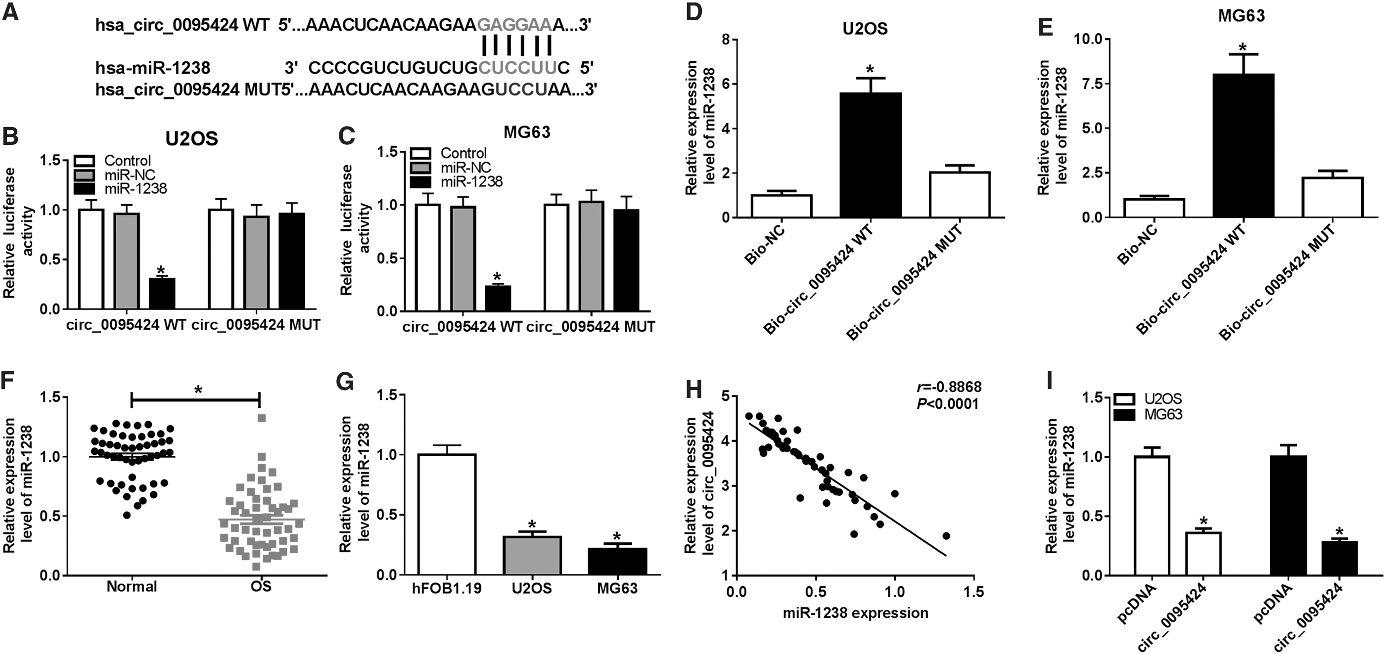
Circ_0095424 could bind with miR-1238 in OS.
Circ_0095424 regulated OS cell progression through targeting miR-1238
Subsequently, the authors further investigated whether miR-1238 participated in the oncogenic function of circ_0095424 in OS. MiR-1238 expression was inhibited using in-miR-1238, and the detection of the miR-1238 expression suggested that in-miR-1238 reversed the promotion effect of si-circ_0095424 on miR-1238 expression in U2OS and MG63 cells (Fig. 5A). The rescue experiments revealed that miR-1238 inhibitor could partially reverse the inhibition effect of circ_0095424 knockdown on the proliferation, migration, and invasion of U2OS and MG63 cells, as proved by CCK8 assay (Fig. 5B, C) and transwell assay (Fig. 5D, E), respectively. Also, the increasing effect of silenced circ_0095424 on the apoptosis of U2OS and MG63 cells also could be inverted by miR-1238 inhibitor (Fig. 5F). The detection of the protein levels of p-AKT/AKT and p-PI3K/PI3K also showed that the inhibitory effect of circ_0095424 silencing on the PI3K/AKT signaling pathway activity could also be restored by transfection of miR-1238 inhibitor (Fig. 5G). The aforementioned findings revealed that the oncogenic role of circ_0095424 in OS was partially dependent on the regulation of it on miR-1238 expression.

Circ_0095424 silencing and miR-1238 inhibitor regulated OS cells progression. U2OS and MG63 cells were transfected si-NC, si-circ_0095424#2, si-circ_0095424#2 + in-miR-NC or si-circ_0095424#2 + in-miR-1238.
HMGB1 was a downstream target of miR-1238 in OS
To find the downstream targets that could bind with miR-1238, the authors used the Targetscan tool for prediction and found that HMGB1 could bind to miR-1238 (Fig. 6A). Then, dual-luciferase reporter assay was carried out and the results revealed that the luciferase activity of HMGB1 3′UTR WT could be inhibited by miR-1238 overexpression in U2OS and MG63 cells, whereas the luciferase activity of HMGB1 3′UTR MUT had no change (Fig. 6B, C). Besides, the authors found that HMGB1 expression was markedly upregulated in OS tissues and both OS cells (Fig. 6D, E). Through analyzing the correlation between HMGB1 and miR-1238 or circ-0095424 expression, the authors concluded that HMGB1 expression was negatively correlated with miR-1238, whereas positively correlated with circ_0095424 in OS (Fig. 6F, G). Meanwhile, in miR-1238 mimic transfected U2OS and MG63 cells, the authors also discovered a significantly decreased expression in HMGB1 (Fig. 6H). At the same time, it could be seen from Figure 6I that overexpressed circ_0095424 had an obviously promoting effect on HMGB1 expression, which could also be reversed by miR-1238 overexpression.

HMGB1 was a downstream target of miR-1238 in OS.
The regulation of miR-1238 on OS cell progression was achieved by mediating HMGB1 expression
To perfect the hypothesis of the circ_0095424-miR-1238-HMGB1 regulation axis, the authors conducted a recovery experiment using miR-1238 mimic and HMGB1 overexpression plasmid. The results in Figure 7A showed that HMGB1 overexpression plasmid markedly restored the expression of HMGB1 inhibited by miR-1238 mimic, indicating that the transfection of both was successful. CCK8 assay and transwell assay results suggested that miR-1238 overexpression inhibited the proliferation, migration, and invasion of U2OS and MG63 cells, whereas overexpressed HMGB1 could invert this effect (Fig. 7B–E). Meanwhile, the promotion effect of miR-1238 overexpression on the apoptosis of U2OS and MG63 cells also could be reversed by HMGB1 overexpression (Fig. 7F). Besides, the detection results of the protein levels of p-AKT/AKT and p-PI3K/PI3K also revealed that the inhibitory effect of miR-1238 mimic on the PI3K/AKT signaling pathway activity could be eliminated by HMGB1 overexpression in U2OS and MG63 cells (Fig. 7G). Collectively, these data illustrated that miR-1238 regulating the progression of OS through targeting HMGB1.
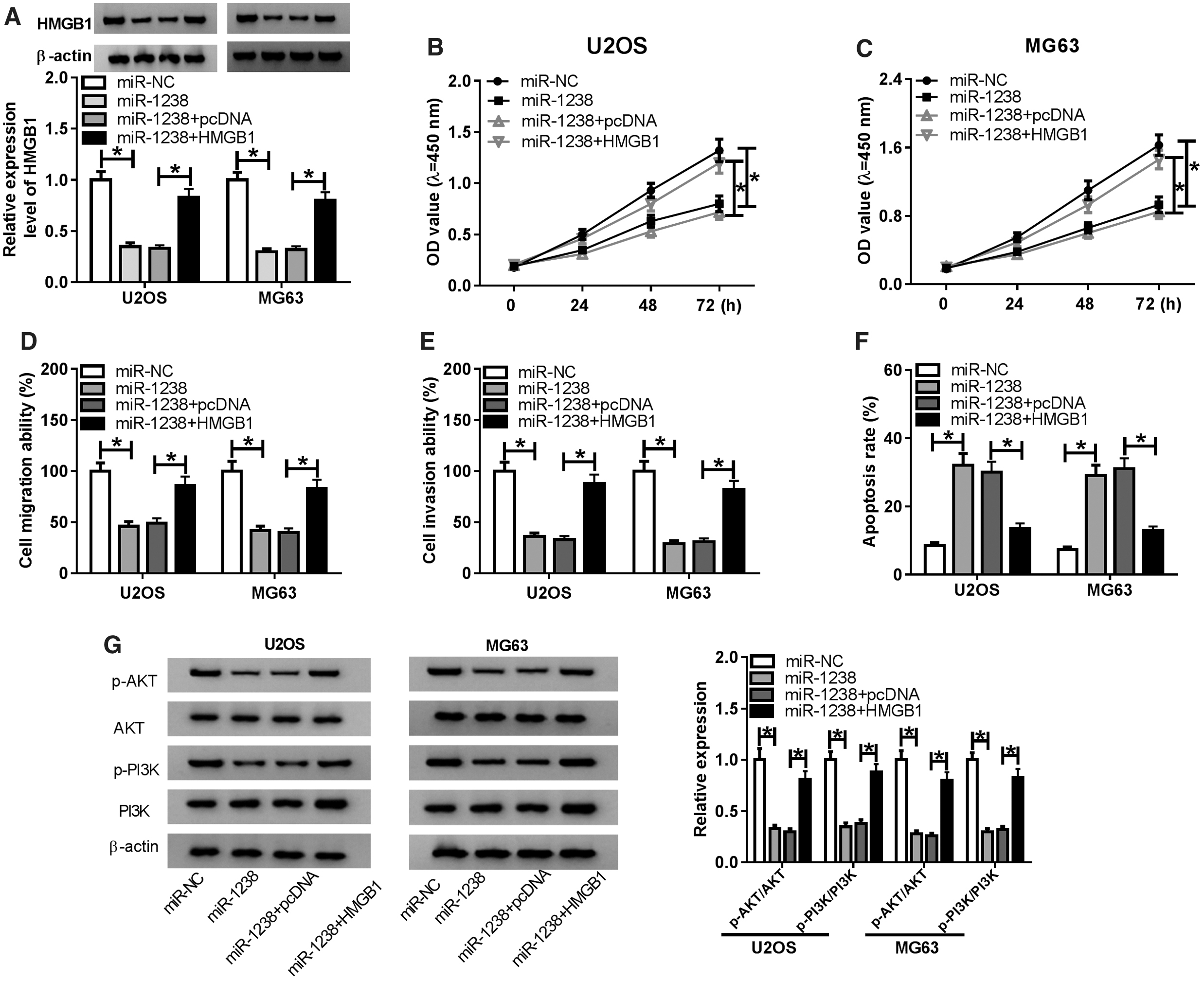
MiR-1238 mimic and HMGB1 overexpression regulated OS cells progression. U2OS and MG63 cells were transfected miR-NC, miR-1238, miR-1238 + pcDNA or miR-1238 + HMGB1.
Discussion
At present, the treatment of OS is mainly a combination of surgical treatment and chemotherapy. 26 However, incomplete surgical resection and side-effects of chemotherapy can bring secondary damage to OS patients. 27 Therefore, searching for new molecular targets may provide new clinical strategies for the treatment of OS. Wang et al. counted the circRNAs that were differentially expressed in OS in recent years and emphasized their potential as biomarkers for OS treatment and prognosis. 28 More recently, many new functions of circRNAs were constantly being explored, for example, they could be used as tumor suppressors or oncogenes in OS. 29,30 Circ_0095424 was a newly discovered circRNA that, to the authors' knowledge, had not been reported to regulate cancer progression. The authors found that circ_0095424 was upregulated in OS, which was agreed with the results of the microarray analysis by Liu et al. 14 Functional tests had confirmed that circ_0095424 silencing could inhibit the proliferation and metastasis, enhance the apoptosis of OS cells in vitro, and reduce the OS tumor growth in vivo, indicating that circ_0095424 knockdown might be an effective strategy for the treatment of OS and had important clinical significance.
PI3K/AKT is an important signaling pathway regulating various biological functions of cells. Many studies have found that the activity of the PI3K/AKT signaling pathway is closely related to the development of human cancers, including OS. 31,32 Therapeutic strategies that target key molecules of the PI3K/AKT signaling pathway have become a new direction for cancer treatment. 33,34 In this study, the authors measured the protein levels of p-AKT/AKT and p-PI3K/PI3K in OS cells and discovered that circ_0095424 knockdown could restrain the activity of the PI3K/AKT signaling pathway in vitro and in vivo. This confirms that the regulation of circ_0095424 on OS progression was achieved by regulating the activity of the PI3K/AKT signaling pathway.
There is increasing evidence that miRNAs are often maladjusted in OS and are related to OS tumorigenesis. 35 Wang et al. reported that miR-384 expression was decreased in OS and participated in the proliferation, migration, invasion, and apoptosis of OS. 36 Besides, miR-204-5p might result in the increase of apoptosis and decrease of migration and invasion in OS. 37 These all indicated the importance of miRNA expression for OS progression. Although there were few studies on miR-1238 in OS, the low expression of miR-1238 involved in cutaneous squamous cell carcinoma and cholangiocarcinoma development could also provide some references for this study. 17,18 In this study, miR-1238 was underexpressed in OS, which was agreed with the results of Qi et al. 19 Also, miR-1238 was believed to interact with circ_0095424, and the reversal effect of miR-1238 inhibitor on the inhibition effects of circ_0095424 silencing on OS progression and the PI3K/AKT signaling pathway activity also confirmed that circ_0095424 regulated OS progression through miR-1238.
HMGB1 was originally considered to be an important late-stage proinflammatory factor, but as research continues, it had been found that it could participate in tumor cell proliferation, differentiation, and migration, and also could affect the functions of nerve cells, vascular endothelial cells, and immune systems. 38,39 Lv et al. found that miR-1284 restrained the proliferation and migration of OS through targeting HMGB1, 24 and Liu et al. showed that miR-935 inhibited HMGB1 expression to suppress the proliferation and invasion of OS. 25 Consistent with this, the authors also discovered that HMGB1 was regulated by miR-1238. The reversal effect of overexpressed HMGB1 on miR-1238 mimic suppressed OS progression and the PI3K/AKT signaling pathway activity also revealed that HMGB1 was indeed a target of miR-1238 in OS. At the same time, this reversal effect was not complete, suggesting that there might be other target genes for miR-1238, which needed further exploration.
Conclusion
In summary, the authors concluded that circ_0095424 was highly expressed in OS, and knockdown of it could directly regulate the proliferation, metastasis, and apoptosis of OS by mediating the activity of the PI3K/AKT signaling pathway through the miR-1238/HMGB1 axis. Clarifying the specific role of circ_0095424 in OS might help all to better understand the pathogenesis of OS and develop new treatments for OS.
Footnotes
Authors' Contributions
Conception and design were by Z.Z. and Y.S.; development of methodology by L.L.; acquisition of data was by B.L.; analysis and interpretation of data were done by C.Q.; writing, review, and revision of article were taken care of by Z.Z., Y.S., and C.Q.; all coauthors have reviewed and approved of the article before submission.
Disclosure Statement
No competing financial interests exist.
Funding Information
No funding was received for this article.
Supplementary Material
Supplementary Figure S1