
Abstract
Background:
Prostate cancer is the most common type of cancer for men in many countries. One of the various prostate cancer therapy methods is hormone therapy, and explaining the association between androgen hormones and prostate cancer is a critical role for successful prostate cancer treatment.
Materials and Methods:
In the current study, the behavior of 3,4-divanillyltetrahydrofuran (DTH) was examined against prostate cancer cells, which have androgen sensitivity differences [LNCaP (+), PC3 (-)]. For this aim, DTH was obtained by extraction of Urtica dioica roots. The molecular structure of isolated compound was confirmed as DTH by liquid chromatography-mass spectrometry and nuclear magnetic resonance spectroscopy analyses. To evaluate the association of androgen sensitivity, DTH was radiolabeled with 131I, and cell uptake assay was performed by using 131I-radiolabeled DTH. Also, cytotoxicity (WST-1) assay of DTH was performed against LNCaP and PC3 cells to determinate the toxic effects of DTH on different androgen mechanisms.
Results:
The results of assays on cells have shown that DTH lignan behaves different like being more toxic to LNCaP cells than PC3 cells, depending on androgen sensitivity.
Conclusion:
The results may contribute both the research topics of phytolignan prostate cancer and androgen-sensitive prostate cancer.
Introduction
Prostate cancer is the most common male malignancy in many countries along with lung and colorectal cancer. 1 Prostate cancer risk factors are age, ethnicity, and genetic, and the strongest factor is age. African and American men have the highest incidences, and Chinese men have the lowest. Also, mutations of BRCA1 and BRCA2 genes contribute as a risk factor. Fatness and smoking cigarettes can be associated as other risk factors. 2 There are many therapy methods used for prostate cancer, and hormone therapy is one of the most important ones. The androgen hormones, androstenedione, testosterone, and dihydrotestosterone, control the development and maintenance of male characteristics and primarily synthesized from cholesterol and are produced by ovarian follicular cells. 3 Androgen hormones have a critical role about prostate physiology and male sexual development. Testosterone is produced by testicular Leydig cells and has a central role for male health such as diminished libido, decreased energy, and erectile dysfunction. Dihydrotestosterone is produced from testosterone in peripheral tissues by 5-α reductase. 4 The androgen receptor (AR) stimulates the expression of specific genes that cause prostate cells to grow by stimulating aerobic glycolysis and anabolism in prostate cancer cells. 5 Androgen deprivation therapy with medical or surgical castration is the main treatment of metastatic prostate cancer. AR activity and expression about causing the tumor growth during prostate cancer development and progression through genetic and epigenetic mechanisms are shown in many reports. 6 Studies about production of antiandrogens, especially for the treatment of castration-resistant prostate cancer, is very important. With this effort, explaining the complex regions of the biology of androgen-AR mechanism and various mechanisms of drug resistance of androgen-deprivation therapy may be easier. 7
The association between androgen sensitivity of prostate cancer cells and apoptosis of these cells as a result of inhibiting androgens was reported in many studies, 8 “the lowest testosterone level possible” was accepted as a hormone therapy's main goal in Expert Consensus Meeting. 9
Lignans and neolignans are diphenolic compounds, which consist of two propylbenzene (C6–C3) structures derived from the phenylpropanoid pathway. 10 Lignans have been identified in many plants 11 and known with health benefits for protection against cancer type and osteoporosis. 12 The lignans influence the estrogen metabolism by their antioxidant activity. This influence on estrogen metabolism cause the lignans are functionally classified as phytoestrogens. 13 One of the main lignans is a 3,4-divanillyltetrahydrofuran (DTH) compound, which can be obtained from stinging nettle roots. Schöttner et al. reported that they tested the affinity to human sex hormone binding globulin of polar extracts of the stinging nettle roots contain the lignans which were either isolated from Urtica roots or obtained semisynthetically. The affinity of DTH was outstandingly high (95.5%, n = 5), and this finding shows the potential beneficial effects of plant lignans. 14 131I radionuclide was used as a tracer for evaluating the differences of DTH against PC3 and LNCaP cells about affinities and effects. 131I was chosen because of the wide usage in nuclear medicine and the idea of easily radiolabeling on aromatic rings of DTH.
The aim of this study is evaluating the association between the DTH lignan, which was obtained from the root extract of stinging nettle, and different prostate cancer cells, which have different AR sensitivities [androgen sensitivity: LNCaP (+), PC3 (−)].
Materials and Methods
Chemicals and reagents
Iodine-131 (Na131I) (Ege University, Medical Faculty), Thin Layer Chromatography paper ITLC-cellulose, n-octanol, acetic acid, isopropyl alcohol, hexane, ethylacetate, methanol, and dichloromethane n-butanol were purchased from Merck, iodogen was purchased from Fluka. Dulbecco's Minimum Essential Medium Eagle, RPMI-1640 medium, min. essential amino acid,
Extraction of nettle plant
Procuring of nettle roots
Stinging nettles were gathered up from the northeast of Turkey in February. Plant systematic of nettle plants is approved by Ege University, Department of Pharmacognosy. Roots were separated and pulverized by a triturator machine.
Extraction procedure of nettle roots
A procedure of Meagher et al. was performed for extraction of nettle roots. 15 Ten grams of root powder were dissolved in of 70% aqueous methanol solution with continuous stirring for 4 h at 55°C. Methanol was evaporated and the aqueous solution was filtrated (Whatmann No. 1). The extraction procedure was repeated three times and the total extract (42.1 mg) was dried by the stove (30°C, a day). The extract was acid hydrolyzed with HCl (1 M, at 95°C, an hour). Acid hydrolysate was extracted with ethyl acetate/hexane, and DTH compound was obtained in the organic phase.
Analysis of whole extract and isolation of DTH by high-performance liquid chromatography
A procedure of Meagher et al. was used for high-performance liquid chromatography (HPLC) analysis of whole extract and isolation of DTH. 15 A low-pressure gradient HPLC system (SPD-M20A DAD detector, 5-μm C18-ODS column, LC-10ATvp quaternary pump, a syringe injector equipped with a 500 μL loop, CTO-10AS column oven, SIL 20A-HT auto sampler, FRC-10A fraction collector, and [250 × 4.6 mm I.D.; Gl sciences, Inc.]) was used for analysis and isolation experiments. The column was eluted with a two mobile phases, which were A (distilled water/glacial acetic acid, 99.8:0.2) and B acetonitrile, injection was started by 70A: 30B 0 min and continued by 50A:50B 55 min, the flow rate was 0.6 mL/min, wavelength was 280 nm, and oven temperature was 26°C.
Molecular structural analysis of DTH
Nuclear magnetic resonance spectroscopy
1 H-nuclear magnetic resonance spectroscopy (NMR) was performed for the structural analysis of DTH and all spectra were obtained by the Erzurum Atatürk University. ACD/LogP 6 Algorithm Computer Program (Version 6.0) was used for preparing the theoretical spectra.
Liquid chromatography–mass spectrometry
Liquid chromatography–mass spectrometry (LC-MS) (Thermo Scientific/TSQ Quantum Access Max) analysis was performed by Pharmaceutical Sciences Research Centre, Faculty of Pharmacy, Ege University. LC-MS conditions: C18 column, mobile phase: water +0.1% Formic acid/MeOH, and flow rate: 20 μL/min. LC-MS chromatogram and fragments were given as Supplementary materials in Figure S1 and Figure S2.
Radiolabeling of DTH (131I-DTH)
Radiolabeling procedure
Iodogen-coated tubes were prepared to oxidize the 131I for chemical reaction. For this step, 0.25 mg iodogen/0.25 mL dicholoromethane solutions were prepared and coated to reaction tubes. By evaporation of dichloromethane, 0.05 mg DTH/0.05 μL methanol was radiolabeled with 0.1 mCi 131I (30 min, at room temperature) in these iodogen-coated tubes.
Quality control by chromatographic methods
Thin layer radio chromatography
Thin layer radio chromatography (TLRC) was used for determination of the radiolabeling yield and R f values of each radioactive component (131I-DTH, 131I, and oxidized 131I). Cellulose TLC plates (three plates for each component) were used as stable phase, and solvent mixture was used as mobile phase. The content of solvent mixture was n-butanol/water/acetic acid (4:2:1). By the end of the incubation of radiolabeling, 2.0 μL of the mixture was dribbled to TLC plates and these plates were added mobile phase. Drop of reaction mixture was wafted by mobile phase and plates were counted by Bioscan AR2000 to determine R f values of radioactive components.
High-performance liquid radio chromatography
A same HPLC procedure with analysis of whole extract and isolated DTH was used for high-performance liquid radio chromatography. 15 A low-pressure gradient HPLC system was equipped with a radioactivity detector [NaI(Tl) radioactivity detector (Gabi Star; Raytest)] to determinate the retention times of radioactive components (131I and 131I-DTH).
Paper electrophoresis
Paper electrophoresis of 131I-DTH was performed with Thermo Fisher Scientific Horizontal Electrophoresis System supply using Whatman no. 1 electrophoresis strip. Cellulose strips were moistened by n-butanol/distillated water/acetic acid (4:2:1) solution and placed in electrophoresis chamber. A sample of 131I-DTH solution (2 μL) was dripped to the midpoints of the strips and power supply was set to 250 V (120 min). The dried strips were counted by Bioscan AR-2000, and R f values of radioactive components were calculated.
Stability of radiolabeling
The radiolabeling yield of 131I-DTH was determined at 0, 1, 2, 4, 8, 12, and 24 h by using TLRC method and the same conditions. The stability graph of 131I-DTH was drawn with these yields.
In vitro studies
Cell culture of PC3 and LNCaP
PC3 (human prostate adenocarcinoma cells) and LNCaP (androgen-sensitive human prostate adenocarcinoma cell line) cells were obtained from the American Type Culture Collection, Rockville, MD. PC3 cells were grown in Dulbecco's Minimum Essential Medium Eagle, supplemented with 10% FBS, 2.0 mM glutamine, 1.5 g/L sodium bicarbonate, 0.1 mM nonessential amino acids, and 1 mM sodium pyruvate. LNCaP cells were grown in RPMI-1640 Medium with 10% FBS, 2.0 mM glutamine, 1.5 g/L sodium bicarbonate, 0.1 mM nonessential amino acids, and 1 mM sodium pyruvate.
The incubation condition of both cells was 37°C with 5.0% CO2. Trypsin-EDTA was used for suspending cells by enzymatic method.
Cytotoxicity assay (WST-1)
Cells were suspended in own medium, and cell suspensions (104 per a well) were grown in 96-well plates. DTH solution was prepared in mediums, and five concentrations (5, 10, 25, 50, and 100 μg/mL) of DTH solution were diluted by DMEM and RPMI-1640, for each PC3 and LNCaP cells included wells, respectively. Solely, medium (without cell or any reagent) was used as a positive control, blank (empty well) was used as a negative control, and DTH solution dilutions (three replication) were prepared in plates and incubated at the 37°C in a humidified incubator equilibrated with 5% CO2. Three incubation times were tested as 24, 48, and 72 h. At the end of incubation time, 10 μL of WST solution [(2-(4-iodophenyl)-3-(4-nitrophenyl)-5-(2,4-disulfophenyl)-2H-tetrazolium, monosodium salt)] was added to each well and incubated 4 h more. Thermo Scientific Varioskan Flash multimode reader, Vantaa Finland microplate reader, was used for measurement of absorbance values (at 450/690 nm).
[Measured absorbance value/absorbance value of negative control) × 100] formula was used for calculation of cell viabilities.
Cell uptake of 131I-DTH
PC3 and LNCaP cells were preferred for cell uptake study to examine the uptake against the androgen sensitivity differences. Furthermore, six different time parameters (1, 2, 4, 8, 12, and 24 h) were tested to clarify the uptake mechanism depending on time. For this aim, both the cells (104 cells per well) were seeded in own medium in 24-well plates and incubated (37°C, 5.0% CO2, a day) in the medium to be adsorbed as a monolayer on the bottom of the wells. Each well was washed three times with phosphate-buffered saline (PBS, pH = 7). 131I-DTH solution was prepared as 1.0 μg/mL DTH and 1.0 μCi/mL 131I per well in medium and added to each well, which contents monolayer cells. The same procedure was repeated with solely 131I. The cells were incubated at 37°C, 5.0% CO2 during the time parameters. By the end of incubation time, monolayered cells were washed with PBS two times and suspended by treating with RIPA buffer (200 μL) solution. The protein quantity of 25 μL of lysed cell suspension was determinated by bicinchoninic acid (BCA) kit, and radioactivity of 100 μL of lysed cell suspension was counted by the liquid scintillation counter (Packard TriCARB-1200). Radioactivity per cell was calculated with an amount of protein and radioactivity values (n = 6).
Statistical analysis
According to one-way ANOVA (GraphPad [prism 8.1]) analysis (p) <0.05 was found for the cell uptake study.
Results and Discussion
The HPLC chromatogram of the whole extract of stinging nettle root is given in Figure 1. Two main peaks were obtained and the first one with 7.05 min retention time was chosen as DTH compound according to literature. 15 To perform the molecular structural analysis, first peak was isolated by HPLC with same analysis conditions. Isolated DTH was in (distilled water/glacial acetic acid, 99.8:0.2) and acetonitrile solution due to HPLC mobile phase so this solution was lyophilized. For identifying the molecular structure of the isolated compound (peak I), LC-MS analysis was performed, and theoretical and experimental m/z values of fragmentized components with chemical drafts are given in Table 1. The retention time of LC-MS analysis of DTH lignan was found as 9.12 min. When the theoretical and experimental m/z values were compared, it was seen that fragmentized components have similar values with theoretical ones. Furthermore, NMR analysis of the isolated sample was made, and theoretical and experimental σ (ppm) values are given in Table 2. According to NMR results, experimental σ (ppm) values of carbons were found similar. Both LC-MS and NMR analyses showed that DTH was correctly isolated from the extract. TLRC technique was preferred for quality control of 131I radiolabeling of DTH. According to TLRC results, R f values of 131I, oxidized, 131I and 131I-DTH measured were 0.45, 0.11, and 0.65, respectively (given in Fig. 2 and Table 3). This measurement showed that 131I-DTH has different R f value from 131I and oxidized 131I. According to paper electrophoresis results, R f values are 0.15 (−), 0.05–0.05 (neutral), and 0.26 (+) for 131I, oxidized 131I, and 131I-DTH, respectively. The electrical charge of 131I-DTH was found positive, although oxidized 131I was neutral. The time-dependent changes of radiolabeling yields after 1, 2, 4, 8, 12, and 24 h were calculated by TLRC and stability graphic of 131I-DTH is given in Figure 3. 131I-DTH has radiochemical stability for 12 h. Cytotoxic effects of five different concentrations (100, 50, 25, 10, and 5 μg/mL) of DTH were evaluated on LNCaP and PC3 cells and cell viabilities (%) at the end of 24, 48, and 72 h are given in Figure 4. Tested concentrations of DTH have no cytotoxic effect for PC3 although having against to LNCaP cells. This result showed them that DTH behaves different for tested cancer cells, even though both of them belong to prostate tissue. The reason of this behavior should be related to androgen sensitivity differences of LNCaP and PC3 cells. In addition, DTH starts to show cytotoxicity with effect from 25 μg/mL concentration for LNCaP cells and there was no significant changes depending on the time. Antiproliferative effect of methanolic extract of stinging nettle roots was evaluated against LNCaP and hPCPs cells in Konrad et al. study and significant antiproliferative effect of the extract was observed on LNCaP cells during 7 days. 16 Husein et al. studied with Urtica pilulifera plant, which belongs to the same family with Urtica dioica (Urticaceae), and the ethanolic extract of this plant showed a high cytotoxic effect against breast cancer cells (MCF7), 85% of the cells were dead in the concentration of 500 mg/mL. 17 Antiproliferative effect of the U. dioica roots' aqueous, chloroform, and ethyl acetate extracts on KG-1 cell line was performed by MTT assay. Various concentrations (250–1000 μg/mL) of root extracts showed cytotoxic effects against KG-1 cells, and IC50 values of U. dioica roots' aqueous, chloroform, and ethyl acetate extracts were found as 282.1, 219.3, and 251.2 μg/mL, respectively. 18
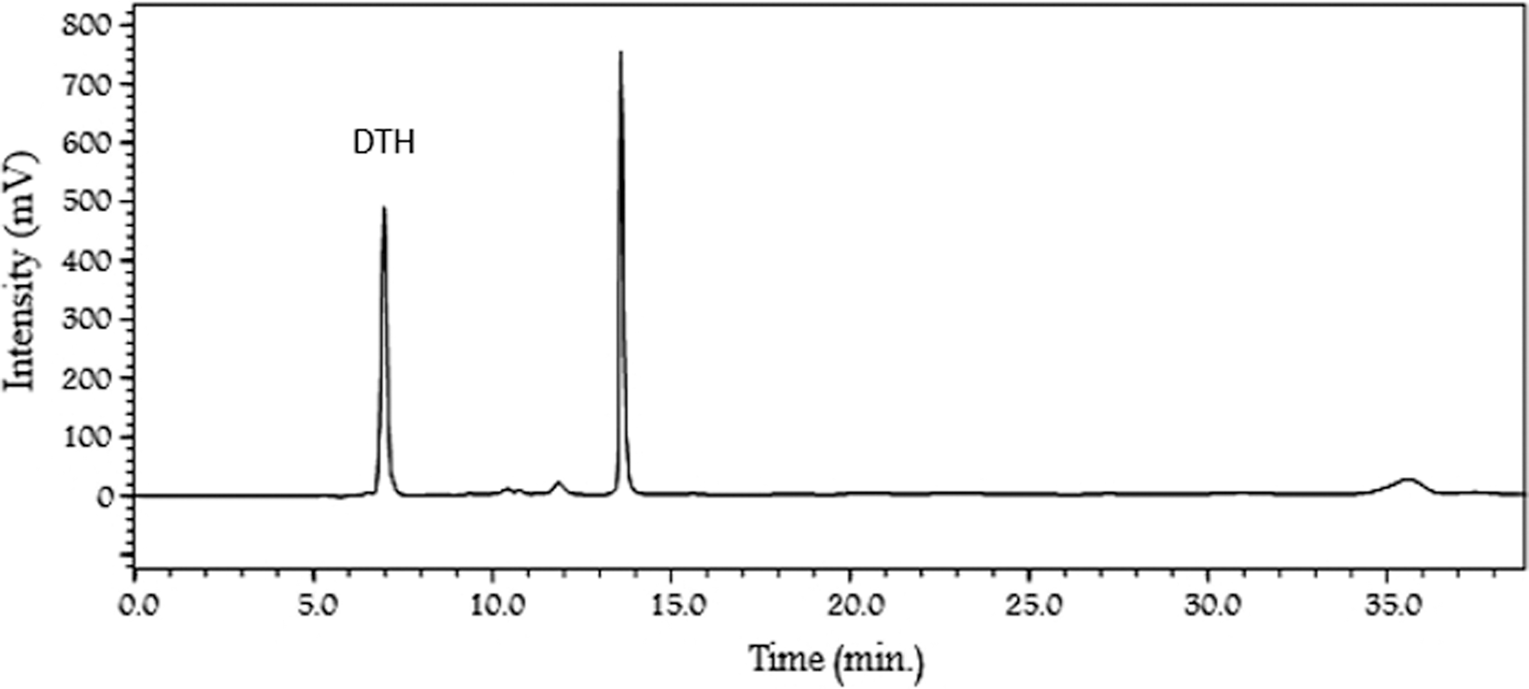
HPLC chromatogram of Urtica dioica extract. DTH, 3,4-divanillyltetrahydrofuran; HPLC, high-performance liquid chromatography.
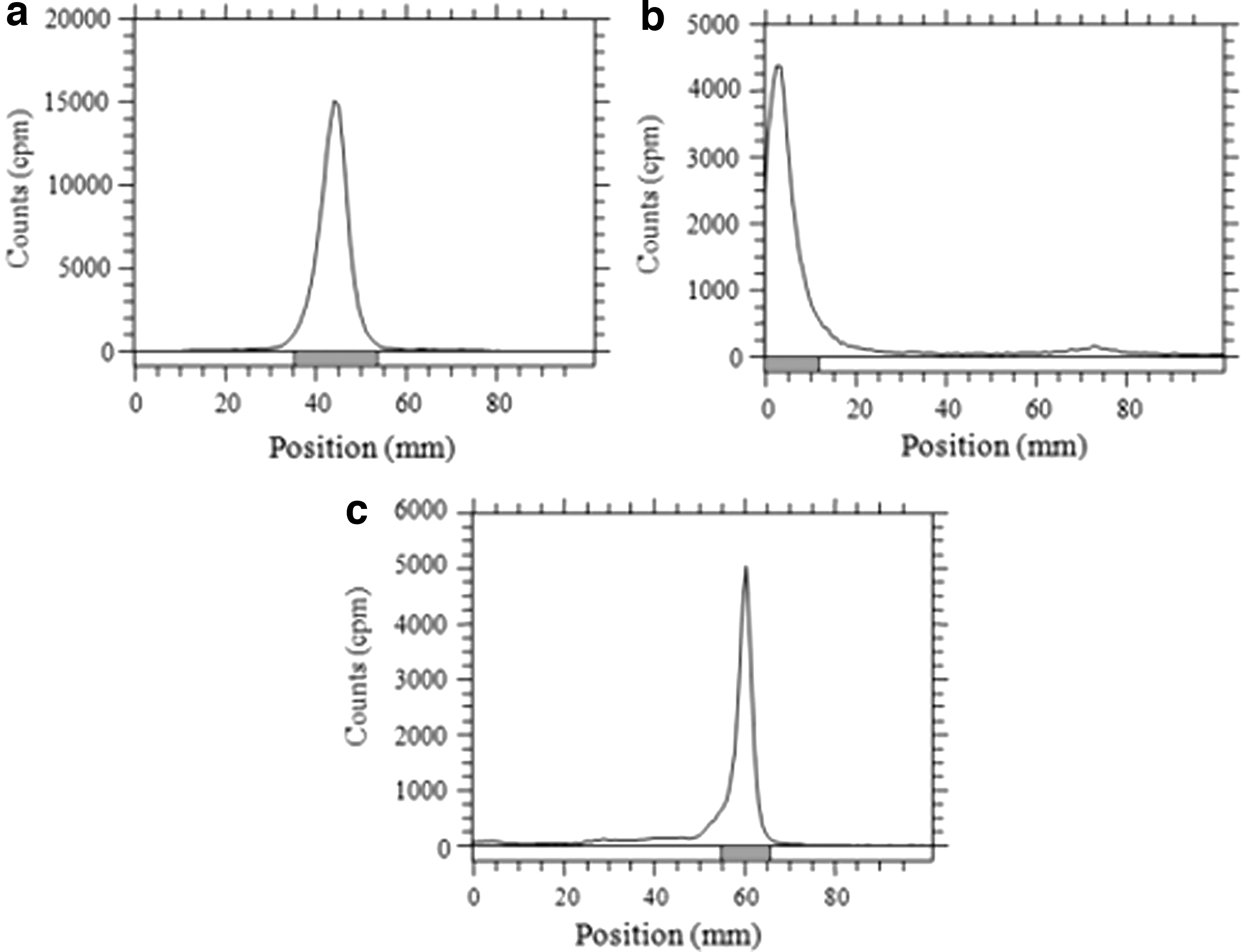
TLRC chromatograms of 131I
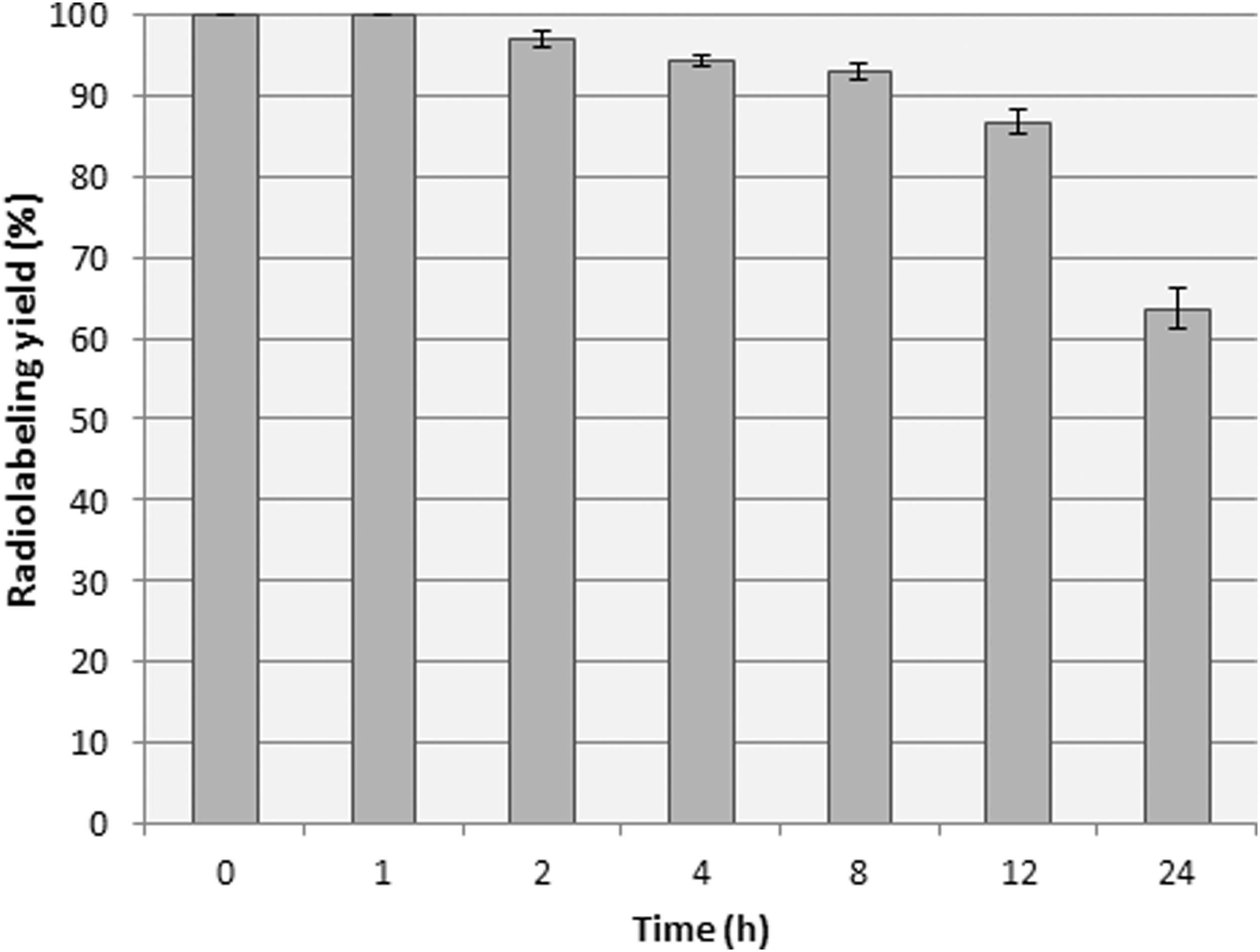
Stability graphic of 131I-DTH according to radiolabeling yields depending time. DTH, 3,4-divanillyltetrahydrofuran.
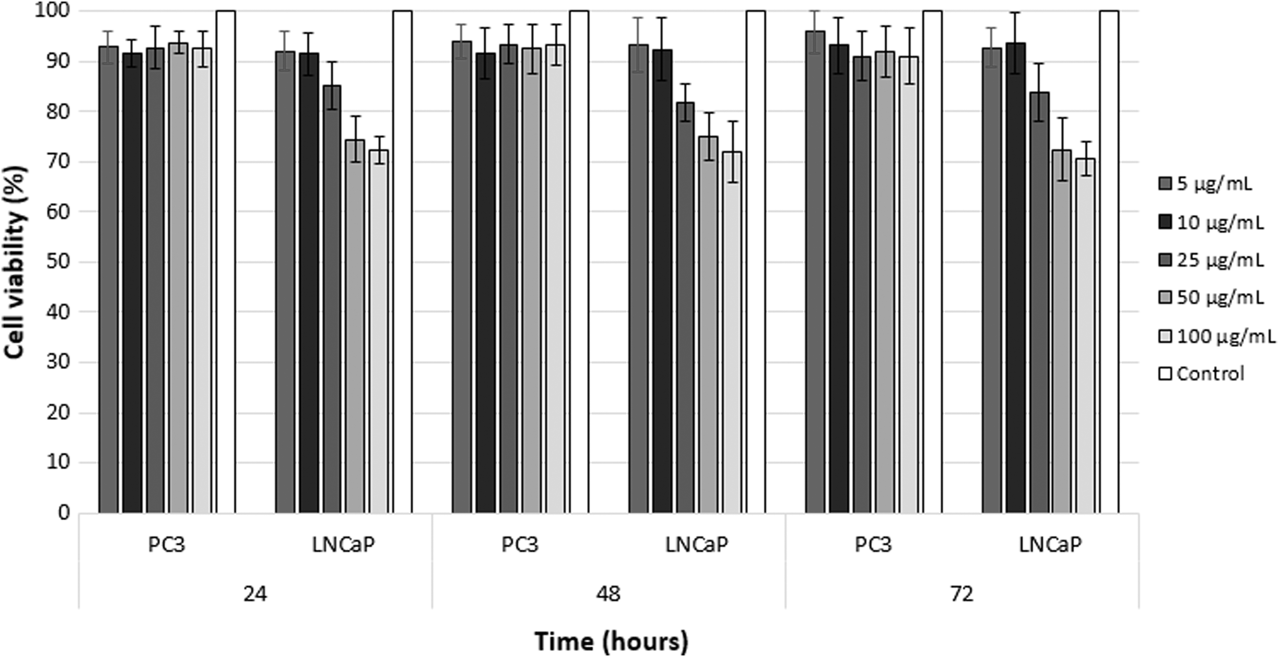
Cell viabilities of PC3 and LNCaP cells after 24, 48, and 72 h incubation with DTH. DTH, 3,4-divanillyltetrahydrofuran.
Theoretical and Experimental m/z values of 3,4-Divanillyltetrahydrofuran
Theoretical and Experimental σ (ppm) Values of 3,4-Divanillyltetrahydrofuran
R f Values of 131I, Oxidized 131I, and 131I-3,4-Divanillyltetrahydrofuran
DTH, 3,4-divanillyltetrahydrofuran.
IC50 values of DTH against LNCaP cells were calculated by using cell viability values and the results are given in Table 4. IC50 values were found as 187.68, 183.24, and 167.14 μg/mL for 24, 48, and 72 h, respectively. Mohammadi et al. performed MTT assay to determine cytotoxic effects of 0–60 μg/mL concentrations of U. dioica dichloromethane extract on PC3 cells treated with extract, in 24 and 48 h. They reported that IC50 values were found as 29.46 and 15.54 (mg/mL) for 24 and 48 h, respectively. 19
IC50 Values (μg/mL) of 3,4-Divanillyltetrahydrofuran Against LNCaP Cells
DTH, 3,4-divanillyltetrahydrofuran.
PC3 and LNCaP cell uptake % values were calculated and given in Table 5, and cell uptake graphic is given in Figure 5. Sole 131I was tested along with 131I-DTH, nevertheless, 131I-DTH was uptake by both of the cells. The uptake by LNCaP cells is more than PC cells, this result showed that DTH lignan is in interaction with LNCaP cells instead of PC3 cells. The possible uptake mechanism is entering into cell by simple diffusion according to During et al. study, which is performed to evaluate the interaction between lignan derivatives and cancer cells (Caco-2). 20 The difference of the cell uptake mechanism of DTH against PC3 and LNCaP cells showed that the association between DTH lignan and androgen sensitivity [LNCaP (+)] causes similar results in common with cytotoxicity assay. During et al. reported that lignan standards (secoisolariciresinol diglucoside [SDG], secoisolariciresinol [SECO], pinoresinol [PINO], lariciresinol, matairesinol [MAT], and hydroxymatairesinol) did not show any cytotoxic effect (<5% of LDH activity released in cell culture media, compared with the positive control) at the different concentrations tested (#275 mmol/L) to Caco-2 cells for 24 h. 19 Furthermore the uptake % value increased by the time until 8 h. After 8 h, the uptake value started to decrease and this result showed that the optimum time of LNCaP cell uptake should be between 8 and 12 h.

PC3 and LNCaP cell uptake values of 131I-DTH by comparing of 131I. DTH, 3,4-divanillyltetrahydrofuran.
Cell Uptake (%) Values of 3,4-Divanillyltetrahydrofuran on PC3 and LNCaP Cells
DTH, 3,4-divanillyltetrahydrofuran.
Conclusions
In the current study, with the aim of examining the association of DTH lignan and androgen sensitivity, DTH compound was isolated from U. dioica extract. Molecular structural identification of DTH was performed by both LC-MS and NMR. 131I radiolabeled to DTH to trace the DTH on cell uptake study. TLRC quality control results showed that radiolabeling was performed with high yield, chemical stability continues about 12 h. According to both cytotoxicity and cell uptake assays, DTH has specificity to androgen sensitivity. DTH has a cytotoxic effect against LNCaP cells, although it has not for PC3 cells. Furthermore, DTH was uptake by LNCaP cells, but there was not found a remarkable uptake % value for PC3 cells. The association between phytolignans such as DTH and androgen sensitivity differences of prostate cancer cells may contribute to the researches about hormone-prostate cancer therapy.
Footnotes
Acknowledgments
The authors are thankful to PhD student Talha Siddik Akkaya for supporting to obtain stinging nettle plant.
Disclosure Statement
No competing financial interests exist.
Funding Information
No funding was received for this study.
Supplementary Material
Supplementary Figure S1
Supplementary Figure S2
References
Supplementary Material
Please find the following supplemental material available below.
For Open Access articles published under a Creative Commons License, all supplemental material carries the same license as the article it is associated with.
For non-Open Access articles published, all supplemental material carries a non-exclusive license, and permission requests for re-use of supplemental material or any part of supplemental material shall be sent directly to the copyright owner as specified in the copyright notice associated with the article.