
Abstract
Background:
To explore the clinical significance of miR-125b-5p and its potential mechanisms in lung squamous cell carcinoma (LUSC).
Materials and Methods:
An integrated analysis of data from in-house quantitative real-time polymerase chain reaction (qRT-PCR), microRNA-sequencing, and microarray assays to appraise the expression level of miR-125b-5p in LUSC tissues compared to adjacent noncancerous controls. The authors identified the candidate targets of miR-125b-5p and conducted functional analysis using computational biology strategies from gene ontology, the Kyoto Encyclopedia of Genes and Genomes (KEGG) analysis, disease ontology (DO), and protein–protein interaction (PPI) network analyses to investigate the prospective mechanisms.
Results:
According to qRT-PCR results, the expression level of miR-125b-5p was markedly decreased in LUSC tissues compared to noncancerous control tissues. Receiver operating characteristic and summary receiver operating characteristic analyses showed that miR-125b-5p had good specificity and sensitivity for distinguishing LUSC tissue from noncancerous lung tissue. The standard mean difference revealed that men and women with lower expression levels of miR-125b-5p may have a higher risk for LUSC. KEGG analysis and DO analysis intimated that target genes were evidently enriched in pyrimidine metabolism and pancreatic carcinoma. The PPI network of the top assembled KEGG pathway indicated that RRM2, UMPS, UCK2, and CTPS1 were regarded as crucial target genes for miR-125b-5p, and RRM2 was eventually deemed a key target.
Conclusions:
The authors' findings implicate a low expression level of miR-125b-5p in LUSC. A tumor-suppressive role of miR-125b-5p is proposed, based on its effects on LUSC tumor growth, clinical stage progression, and lymph node metastasis.
Introduction
In 2018, 18.1 million people were newly diagnosed with cancer around the world, and it caused 9.6 million deaths. 1 Cancer has therefore become one of the most significant factors affecting human health and cannot be ignored. In 2020, cancer statistics for the United States and China ranks lung cancer as the top cause of cancer mortality for male patients and the second leading cause for female patients, 1,2 implicating lung cancer as a particular therapeutic challenge.
Lung cancer is categorized into two subtypes: non-small cell lung cancer (NSCLC) and small cell lung cancer. NSCLC has the highest morbidity and is further divided into lung adenocarcinoma (LUAD) and lung squamous cell carcinoma (LUSC), which is the main type of NSCLC. 3 –5 In the past 5 years, targeted therapy for NSCLC has begun to make remarkable progress, 6 but only a small proportion of patients with LUSC are currently receiving advanced treatment. In addition, no molecular target has yet been identified for the prediction of prognosis and effective treatment of LUSC, which has a survival rate of only about 18%. 7 Therefore, improvement of prognosis in patients with LUSC requires further research to identify therapeutic targets.
One potential area of research is the use of microRNAs (miRNAs), which are small (19–22 nucleotides) endogenous noncoding single-stranded RNAs. 8 Extensive research has already established a deep understanding of miRNA function, 9,10 especially regarding its involvement in transcriptional regulation of basic cellular processes. 11 Upon release into the blood system, various miRNAs can serve as either tumor suppressors or oncogenes. 12 One important miRNA family with known cancer involvement is the miR-125b family, which appears to occupy a vital position in several cancers. For instance, miR-125b can promote the malignant progression of skin tumors, 13 whereas, in esophageal squamous cell carcinoma, miR-125b-5p serves as a cancer-suppressor gene and inhibits cancer cell proliferation, invasion, and migration. 14 The level of miR-125b is reduced in numerous cancers, including head and neck verrucous carcinoma, hepatocellular carcinoma, laryngeal squamous cell carcinoma (LSCC), breast cancer, thyroid cancer, gallbladder cancer, and multiple myeloma. 15 –21
There are many significant researches about the mechanism of miRNAs in LUSC. Previous studies have used bioinformatics methods to examine the expression of miRNA family in LUSC and its impact on the prognosis of patients with LUSC, with the aim of studying its potential biomarkers and molecular mechanisms. 22 The latest research has shown that miR-195 could inhibit the metastasis and angiogenesis of LUSC by reducing the expression of vascular endothelial growth factor. 23 FAM13A, official symbol of family with sequence similarity 13 member A, is a gene relating to aging lung diseases. There was a study finding that rs9224 variants, located in the 3′untranslated region (UTR) of FAM13A, might increase the risk of LUSC by combining with miR-22-5p. 24
In the case of LUSC, hsa-miR-183 and hsa-miR-135b have been shown to be useful in distinguishing LUSC from normal clinical samples. 25 In addition, miR-200a and miR-331-3p have shown tumor-suppressor activity in LUSC. 26,27 A low expression level of miR-125b has been reported in NSCLC and has shown a relationship with poor prognosis 28 ; however, the clinical value and underlying effect of miR-125b-5p in LUSC remain unclear.
The objective of this study was therefore to examine the clinical significance of the expression level of miR-125b-5p and its potential involvement in LUSC. The authors evaluated data obtained from multiple methods, including RNA sequencing, microarray, and in-house quantitative real-time polymerase chain reaction (qRT-PCR), to appraise the expression level of miR-125b-5p in LUSC tissues in comparison to adjacent noncancerous controls. They also explored the correlations between expression levels and clinical features.
In addition, the authors identified the candidate targets of miR-125b-5p and conducted functional analysis with the computational biology strategies of gene ontology (GO), the Kyoto Encyclopedia of Genes and Genomes (KEGG), and disease ontology (DO), and protein–protein interaction (PPI) network analyses. The overall aims were to determine the clinical significance and prospective molecular machinery of miR-125b-5p in LUSC and to provide new ideas for future LUSC research.
Materials and Methods
Clinical samples and in-house qRT-PCR
From January 2012 to February 2014, 23 clinical formalin-fixed, paraffin-embedded (FFPE) samples were recruited from the pathology department at the First Affiliated Hospital of Guangxi Medical University. Independent pathology diagnosis and reports were made and affirmed by two senior pathologists (G.C. and W.-J.M.). All patients and their families signed informed consent in advance, and the hospital ethics committee authorized the application. The expression level of miR-125b-5p in LUSC was evaluated by in-house qRT-PCR using an Applied Biosystems PCR 7900 instrument. All RNAs were abstracted and standardized using the method previously reported by the authors' research team. 29,30 The expression level of miR-125b-5p was evaluated with a miRVana qRT-PCR miRNA test kit (Ambion, Inc., Austin, TX). The authors used RNU6B as an endogenous control. The in-house qRT-PCR used the TaqMan micro-RNA kit from Applied Biosystems. The sequences of RNU6B and miR-125b-5p were shown as follows: RNU6B, CGCAAGGAUGACACGCAAAUUCGUGAAGCGUUCCAUAUUUUU (cat. no. 4427975-000490); and miR-125b-5p, UCCCUGAGACCCUAACUUGUGA. 31 The expression level of miR-125b-5p in the FFPE tissues was calculated using the 2−Δcq formula.
The above results were statistically examined using IBM SPSS Statistics software, version 22.0. The miR-125b-5p expression levels in LUSC tissues and corresponding noncancerous lung tissues were compared by paired sample t-tests. The authors generated a receiver operating characteristic (ROC) curve to estimate the predictive accuracy of miR-125b-5p. p < 0.05 was set up to be statistically significant.
The Cancer Genome Atlas data acquisition
The Cancer Genome Atlas (TCGA) is a freely accessible and comprehensive archive that provides 33 cancer genomic datasets that researchers can screen, download, and analyze. 32 The authors downloaded the miRNA-sequencing (miRNA-seq) and clinical features of miR-125b-5p in LUSC. In total, they retrieved 478 LUSC samples, 45 adjacent noncancerous samples, and information about the clinical features. The level of miR-125b-5p in LUSC and its correlation with clinicopathological data were summarized using IBM SPSS Statistics software, version 22.0. The authors then used univariate Cox regression and multivariate Cox regression models to appraise the hazard ratio (HR) of the expression level of miR-125b-5p and several clinicopathological factors in LUSC; these analyses were then displayed as forest plots. For all these analyses, p < 0.05 was deemed statistically significant.
Collection of high-throughput datasets from Gene Expression Omnibus, ArrayExpress, Sequence Read Archive, Oncomine, and published literature
The authors obtained microarray datasets from the Gene Expression Omnibus (GEO) database, ArrayExpress, Sequence Read Archive (SRA), Oncomine, and published literature through the following search strategy: (lung OR pulmonary OR respiration OR respiratory OR bronchi OR bronchioles OR aspiration OR pneumocytes OR alveoli OR airway) AND (cancer OR carcinoma OR tumor OR tumour OR neoplas* OR malignanc*). The last dataset search was performed on November 1, 2019. Microarray or miRNA-seq data were excluded if they met the following exclusion criteria: (1) datasets failed to meet the inclusion criteria; (2) datasets did not have information about miR-125b-5p; (3) datasets only offered information on LUSC tissues, with no noncancerous tissue samples; (4) there were fewer than three cases in each series for analysis; or (5) datasets contained samples based on cell lines.
NCBI PubMed, Wiley Online Library, Google Scholar, Web of Science, Science Direct, Medline, EMBASE, LILACS, Ovid, Wan Fang, Chinese CNKI, and China Biology Medicine Disc were searched for supplemental datasets using the same retrieval methods. However, neither the mean nor the standard deviation (SD) of miR-125b-5p levels in LUSC and the controls was provided in the literature screening.
Integrated expression level of miR-125b-5p in LUSC
Integrated analysis was performed using Stata 14.0 software and MetaDisc software for the data from in-house qRT-PCR, miRNA-seq, and microarray assays. The mean and SD of each study were used in a fixed-effect model to evaluate the overall standard mean difference (SMD) and 95% confidence interval (CI). The authors implemented I-square (I 2 ) tests and a chi-squared-based Q-test to assess the heterogeneity across this research. Heterogeneity was inferred from studies when I 2 > 50% or p < 0.05. Due to the significant heterogeneity (I 2 > 50%), they ultimately chose a random-effects model, and performed a sensitivity analysis to trace the reason for the heterogeneity. Assessment of the data by Egger's and Begg's funnel plots indicated no notable publication bias in the integrated analysis when p > 0.05. 33 The authors also appraised the prospective predictive value of miR-125b-5p levels in LUSC by plotting the summary receiver operating characteristic (sROC) curve. Forest plots were also constructed to show the sensitivity, specificity, positive likelihood ratio (PLR), and negative likelihood ratio (NLR).
Prediction of putative miR-125b-5p targets
The putative miR-125b-5p targets were predicted using the following 12 online algorithms: miRNAMap, Microt4, miRWalk, miRDB, miRanda, miRbridge, miRMap, PITA, Pictar2, RNAhybrid, RNA22, and Targetscan. Genes that appeared in at least four or more algorithms were deemed candidate target genes of miR-125b-5p. Also, since miR-125b-5p was downregulated in LUSC, the overexpressed genes in LUSC were identified from high-throughput data, including RNA-seq and microarray, when |log2-fold changes (FC) | ≥ 1.0 and p < 0.05, to analyze their relationship to reverse control. The overlapping genes of online algorithms and upregulation differentially expressed genes (DEGs) from RNA-seq and microarray were identified by Venn diagrams and deemed candidate target genes. The pertinence analysis between these genes and miR-125b-5p was also described.
Functional analysis for promising target genes
The potential mechanism of miR-125b-5p in LUSC was investigated by functional analysis. The DEGs commonly involved in upregulation and the 12 algorithms were subjected to GO, KEGG pathway, and DO analysis using R software and the procedures from the “cluster profiler” function package. If p < 0.05 and q < 0.05, a gene was deemed statistically significant. The authors considered genes participating in the most enriched pathway as crucial target genes and then constructed a PPI network through the online STRING database 34 to display the relationship of these genes. 35,36 The selected candidate target genes were overexpressed DEGs, so these were expected to show an upregulation trend. They used these microarray data from high-throughput datasets and mRNA sequences to validate the expression level of these crucial genes. The Human Protein Atlas 37 (HPA) was also utilized to validate the protein expression by immunohistochemical staining with corresponding antibody.
Results
Expression level of miR-125b-5p in clinical LUSC samples by in-house qRT-PCR verification
The expression level of miR-125b-5p was markedly decreased in LUSC tissues compared to noncancerous control tissues (2.177 ± 1.147 vs. 6.039 ± 2.231, p < 0.001; Fig. 1A and Table 1). The ROC curve generated from the in-house qRT-PCR data for miR-125b-5p in LUSC gave an area under the curve (AUC) of 0.948 (p < 0.001; Fig. 1B). When the authors divided the 23 LUSC tumors into two different groups using a tumor threshold size of 3 cm, the in-house qRT-PCR showed a lower expression of miR-125b-5p in the group with a larger tumor size than that with a smaller tumor size (1.711 ± 0.839 vs. 3.243 ± 1.077, p = 0.004; Table 1). The ROC curve for miR-125b-5p versus tumor size showed an AUC of 0.879 (p = 0.005; Fig. 1C). The level of miR-125b-5p was lower in LUSC with lymph node metastasis (LNM) than without LNM (1.489 ± 0.435 vs. 2.927 ± 1.225, p = 0.010), and the ROC curve displayed an AUC of 0.814 (p = 0.011; Fig. 1D). No significant relationship was found between the level of miR-125b-5p and other clinicopathological parameters (p > 0.05; Table 1).
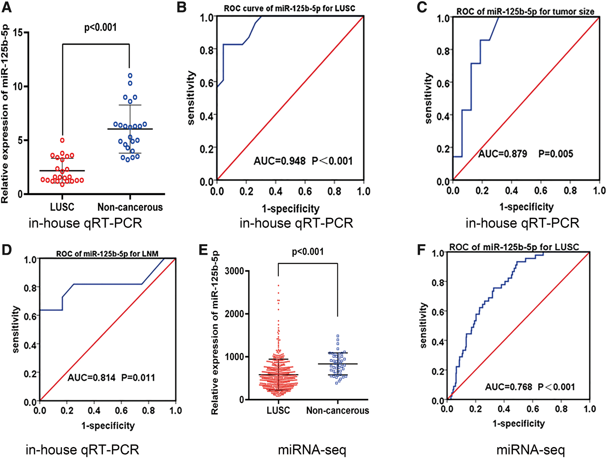
Clinical significance of miR-125b-5p in LUSC based on in-house qRT-PCR and miRNA-seq.
Associations Between miR-125b-5p Expression and Clinicopathological Features in LUSC Based on In-House qRT-PCR Data
p < 0.05 was considered statistically significant.
LUSC, lung squamous cell carcinoma; n, number; qRT-PCR, quantitative real-time polymerase chain reaction; SD, standard deviation.
Expression level of miR-125b-5p in LUSC based on other miRNA-seq and miRNA microarray datasets
Table 2 shows that, after removing samples with data deficiency, 478 anonymized LUSC and 45 adjacent noncancerous tissue samples were finally retrieved from the miRNA-seq dataset. The expression level of miR-125b-5p was remarkably reduced in LUSC compared to noncancerous control tissues (583.767 ± 360.765 vs. 835.672 ± 255.248, p < 0.001; Table 2; Fig. 1E). The ROC curve for differentiating LUSC from noncancerous tissues gave an AUC of 0.768 (p < 0.001; Fig. 1F).
Relationship Between miR-125b-5p Expression and Clinicopathological Parameters of LUSC from miRNA-Seq
p < 0.05 was considered statistically significant.
Kruskal-Wallis test was performed to assess the distribution difference of miR-125b-5p in three or more groups of clinicopathological parameters.
miRNA-seq, data form TCGA.
miRNA-seq, microRNA-sequencing; TCGA, The Cancer Genome Atlas.
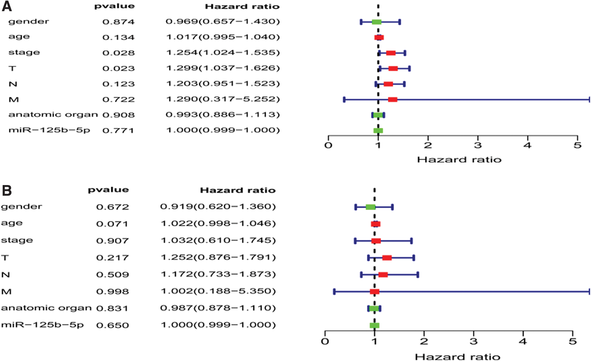
Table 2 summarizes the miR-125b-5p expression levels and clinicopathological features. No significant relationships were found between miR-125b-5p levels and any clinicopathological factor of LUSC, including gender, age, and TNM stage. Univariate and multivariate Cox regression models were also constructed to analyze the HR of miR-125b-5p expression levels and several clinicopathological factors in LUSC, respectively (Fig. 2). The forest plot for the univariate Cox regression model indicated that a poorer condition of the primary tumor (T: T3–T4; p = 0.023; HR = 1.299, 95% CI: 1.037–1.626) and a higher stage of LUSC (III–IV; p = 0.028, HR = 1.254, 95% CI: 1.024–1.535) might serve as risk factors for poorer prognosis (Fig. 2A).
Forest plots of HR of miR-125b-5p expression and other clinicopathological factors in LUSC.
In total, 9 microarrays were screened and downloaded from the GEO database; these contained 185 LUSC tissue samples and 230 noncancerous lung samples. A Student's t-test revealed that miR-125b-5p was significantly lower in LUSC than noncancerous lung controls in five microarrays (GSE56036, GSE51853, GSE47525, GSE40738, and GSE19945).
Integrated analysis of all possible miR-125b-5p expression data
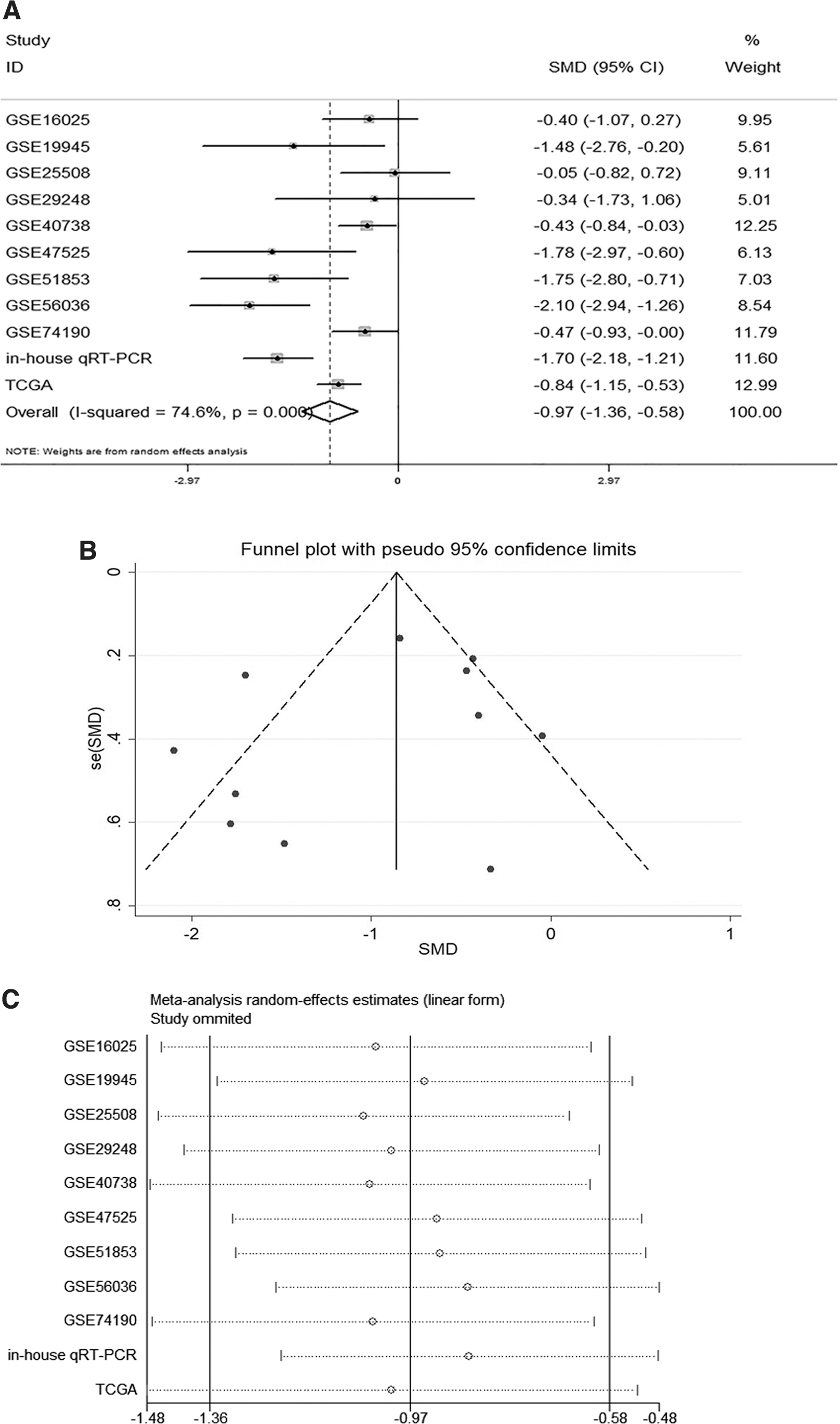
The miR-125b-5p data from in-house qRT-PCR and all available miRNA microarray datasets were combined for comprehensive analysis. This included, in total, 686 LUSC and 298 noncancerous tissue samples (Table 3). The pooled SMD and its 95% CI (Fig. 3A) revealed that men and women with lower expressions of miR-125b-5p may have a higher risk for LUSC (SMD = −0.97; 95% CI: −1.36 to −0.58). The authors pooled the effect variables showing heterogeneity by choosing a random-effects model (I 2 = 74.6%, p < 0.001). Publication bias was also appraised within a funnel plot (Fig. 3B). The computations from Egger's and Begg's tests were 0.414 and 0.640, respectively, so no publication bias was identified. A sensitivity analysis (Fig. 3C) showed no obvious difference across the microarrays. The predictive implication of miR-125b-5p in LUSC was addressed by constructing the sROC curve to obtain an AUC value. As shown in Figures 4 and 5, the AUC value was 0.88 (95% CI: 0.85–0.90). The values for sensitivity, specificity, diagnostic odds ratio, PLR, and NLR were 0.58 (95% CI: 0.54–0.62, p < 0.001), 0.78 (95% CI: 0.73–0.82, p < 0.001), 16.61 (95% CI: 6.37–43.29, p < 0.001), 3.97 (95% CI: 2.27–6.93, p < 0.001), and 0.38 (95% CI: 0.26–0.55, p < 0.001), respectively.
Integrated analysis based on GEO, TCGA, and in-house qRT-PCR datasets.

Predictive accuracy of miR-125b-5p in LUSC based on the 11 included datasets.

Predictive accuracy of miR-125b-5p in LUSC based on the 11 included datasets.
Features of the Enrolled miRNA-Seq, miRNA Microarray Datasets, and In-House qRT-PCR Datasets
p was calculated by Student's t-test, and p < 0.05 was considered statistically significant.
Ctrl, control group; Exp, experiment group.
Prediction of the targets of miR-125b-5p in LUSC
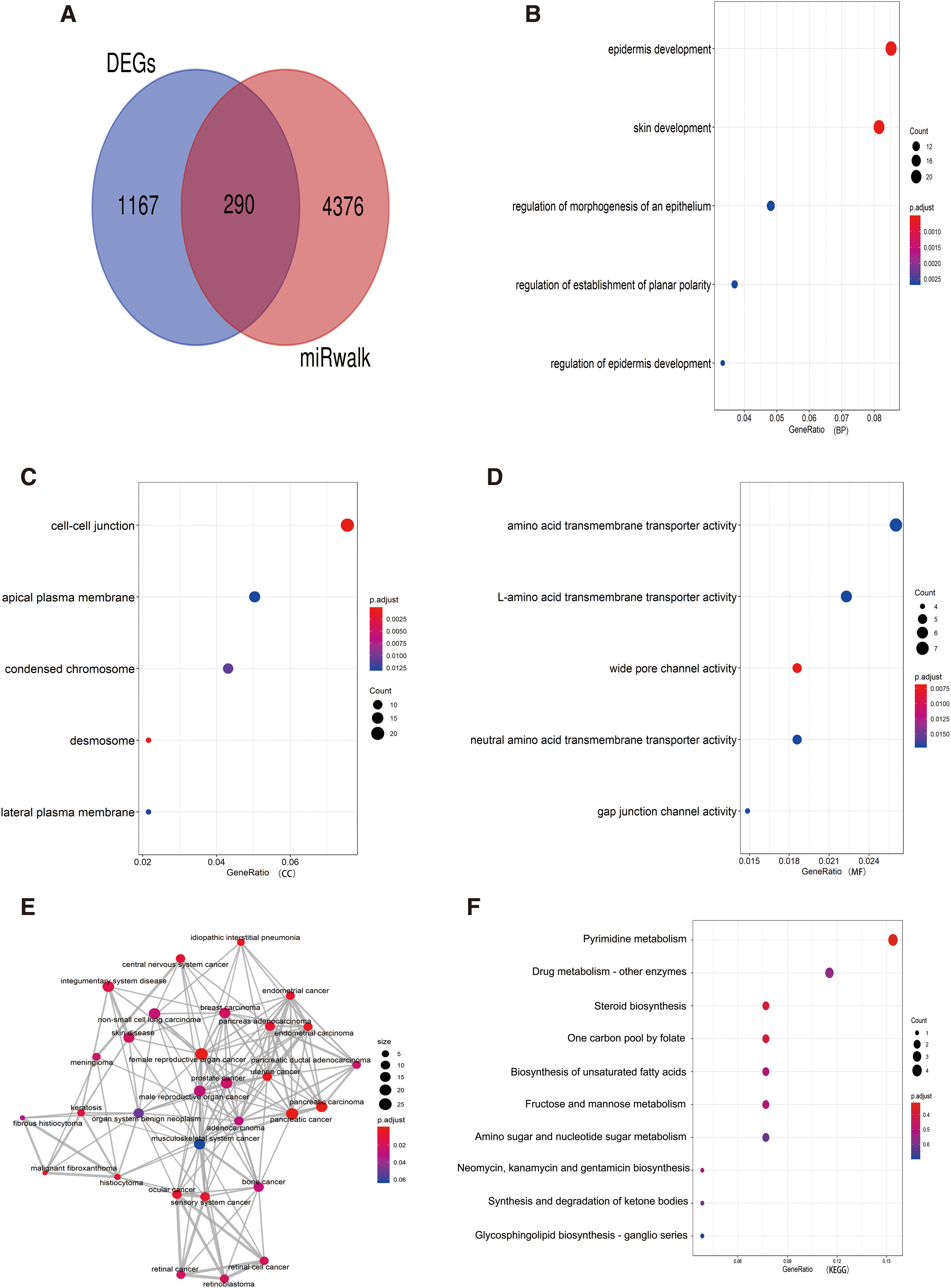
Twelve online prediction algorithms were utilized to identify promising targets for miR-125b-5p. Only the 4666 potential target genes that appeared no fewer than four times among all 12 algorithms were analyzed further. The use of R software to analyze the DEGs of LUSC from microarray datasets and RNA sequencing revealed 1457 upregulated genes among the DEGs from 1 RNA sequencing and 20 microarray datasets. In total, 290 genes were eventually deemed candidate targets of miR-125b-5p. These appeared to be retro-regulated and are worth further discussion (Fig. 6A).
Functional analysis of target genes.
Functional analysis of promising target genes
The 290 potential target genes were used to implement GO, DO, and KEGG pathway analysis with the R package. For the GO analysis, the authors selected the terms of most functional enrichments (FDR <0.05), and the potential target genes were mainly assembled using the following biological process (BP) terms: skin development (GO:0043588) and epidermis development (GO:0008544; Fig. 6B). Enriched terms for the cellular component (CC) were desmosome (GO:0030057) and cell–cell junction (GO:0005911; Fig. 6C). The target genes were also clustered with wide pore channel activity (GO:0005911) for molecular function (MF) terms (Fig. 6D).
The authors also conducted DO annotation of the potential target genes and drew the network shown in Figure 6E. The potential target genes were mainly assembled at pancreatic carcinoma, female reproductive organ cancer, and uterine cancer (all p < 0.05).
The most significant pathway in the KEGG analysis was pyrimidine metabolism (Fig. 6F). Figure 7A shows the results of the PPI network of the top assembled KEGG pathway obtained using the online STRING database. RRM2, UMPS, UCK2, and CTPS1 were involved in the pyrimidine metabolic pathway and were regarded as crucial target genes. MiR-125b-5p binds to the 3′UTR of RRM2, UMPS, UCK2, and CTPS1, and their complementary sequences are shown in Figure 7B–E. The authors also calculated the edges of the four genes that interacted with all 290 latent target genes and found RRM2 to have the most edges, with 29. Consequently, RRM2 was eventually deemed a key target of miR-125b-5p.

Potential target genes of miR-125b-5p.
The calculation results for SMD to elucidate the mRNA levels of RRM2 in LUSC are shown in Figure 8A. RRM2 had a higher mRNA expression in LUSC than in noncancerous control tissues (SMD = 2.62; 95% CI: 2.03–3.20; p < 0.001). The authors also utilized the online HPA to validate the protein level of RRM2, UMPS, UCK2, and CTPS1 in LUSC. Simultaneously, marked increases of the expression levels of UMPS, UCK2, and CTPS1 in LUSC tissues were shown compared with adjacent lung samples (Fig. 8B–D). The immunostaining results of RRM2, UMPS, and CTPS1 in normal lung and LUSC tissues are displayed in Figure 9. The staining intensities of RRM2, UMPS, and CTPS1 were medium in LUSC tissues, while low or not detected in noncancerous lung tissues. However, due to the limited sample size from HPA, statistics analysis was not possible. Immunohistochemistry of UCK2 was not provided by HPA.
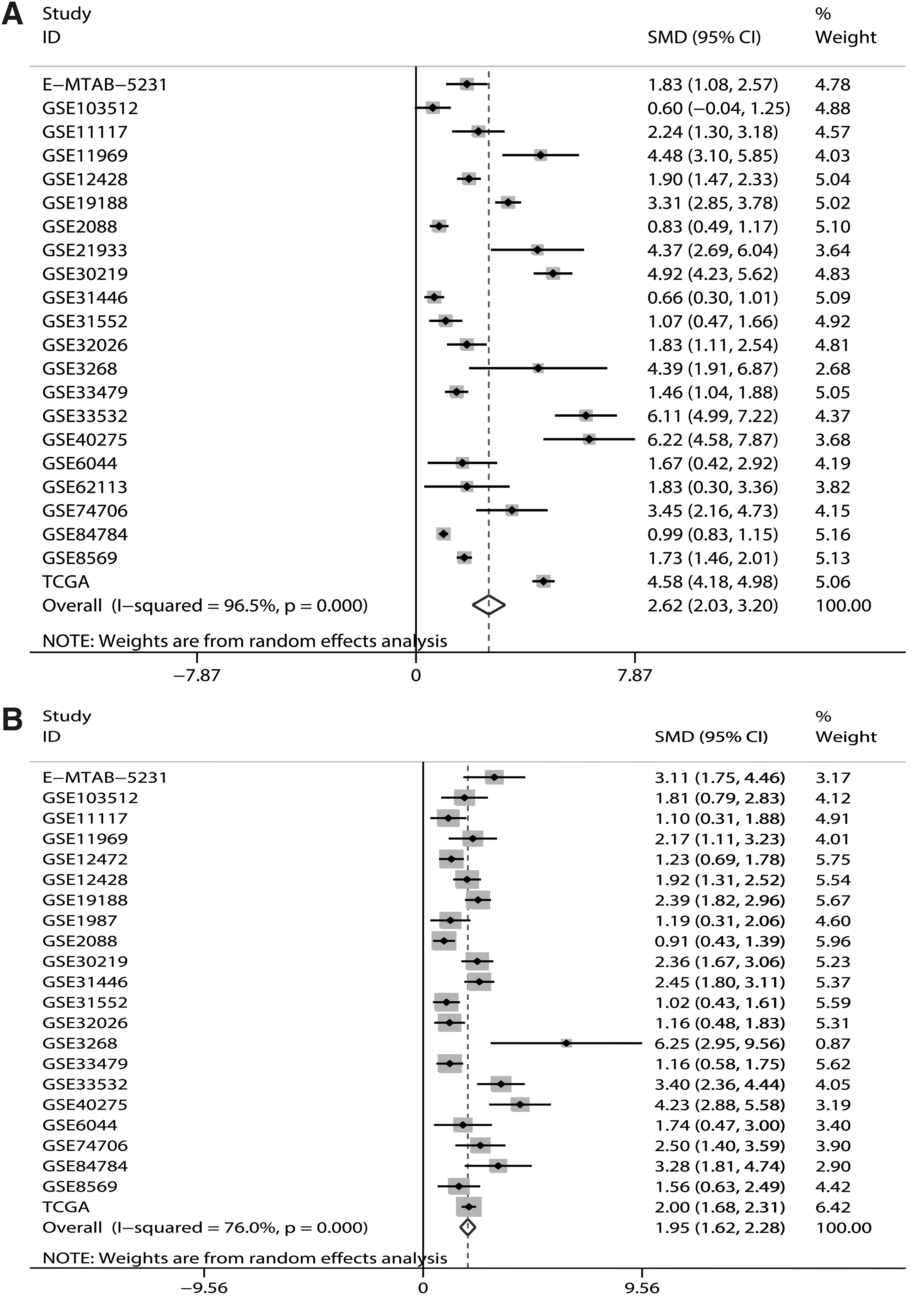
Verification of RRM2, UMPS, UCK2, and CTPS1 level in LUSC. The mRNA expression of RRM2

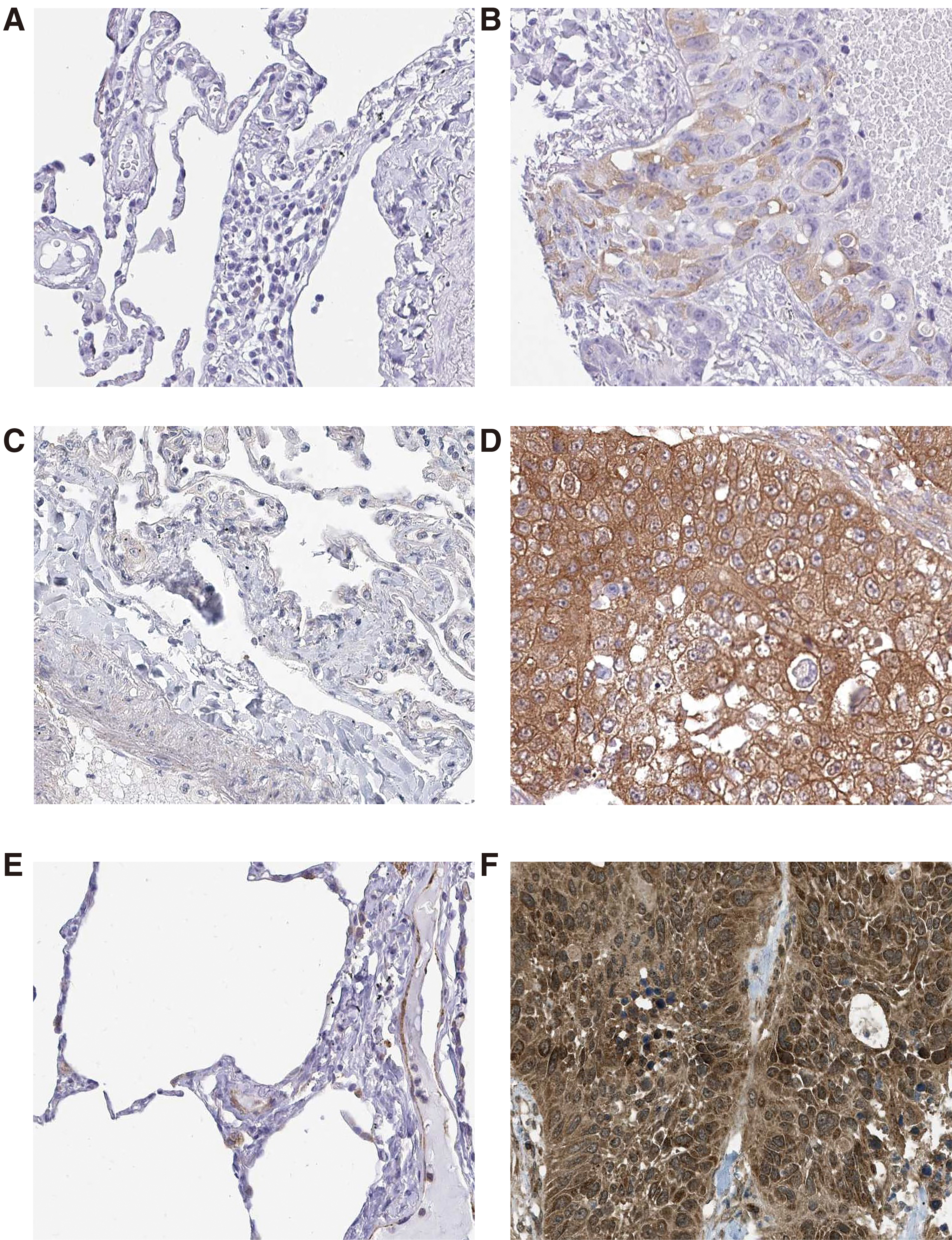
Immunohistochemistry of RRM2, UMPS, and CTPS1 in normal lung and LUSC tissues. RRM2 expression in normal
Discussion
Previous studies 28 have discussed miR-125b expression in NSCLC quite extensively, but miR-125b-5p expression in LUSC has never been reported. The authors found expression levels for miR-125b-5p in LUSC tissues and adjacent noncancerous tissues of 2.177 ± 1.147 vs. 6.039 ± 2.231 (p < 0.001; in-house qRT-PCR) and 583.767 ± 360.765 vs. 835.672 ± 255.248 (p < 0.001; miRNA-seq), indicating a distinct decline of miR-125b-5p in LUSC. The large number of samples collected from in-house qRT-PCR, literature searches, high-throughput datasets, and microarrays in this systematic investigation gave an SMD value of −0.97 (95% CI: −1.36 to −0.58, p < 0.001), indicating that the lower miR-125b-5p level in LUSC was consistent across these cases. Notably, the sROC curve demonstrated that miR-125b-5p might have a moderate ability to discriminate between LUSC patients and noncancerous individuals (AUC = 0.88). A lower expression level of miR-125b-5p also appeared to have a close relationship with larger tumor size, increased clinical stage, and easier LNM, suggesting that miR-125b-5p may play a cancer-suppressive role in LUSC.
LUSC is a highly aggressive and prevalent subtype of NSCLC 38 and lacks effective therapeutic target therapy. LUSC is also strongly resistant to traditional chemotherapy, 39 resulting in poor outcomes for patients with this form of NSCLC. Therefore, finding new therapeutic targets is an urgent challenge to improve the prognosis for patients with LUSC. Overall, the data presented in this study indicate that miRNA studies may open up new avenues for identifying these targets. The miRNAs, as novel noncoding RNA molecules that modify gene expression at the post-transcriptional level, 40 have been extensively acknowledged to play crucial roles in various cancers. Therefore, in the future, the miR-125 family is expected to deliver promising biomarkers and therapeutic targets for LUSC treatment.
Several previous publications have confirmed the vital role of miR-125b in NSCLC. For example, Yuxia et al. 41 reported a notable increase in the circulating miR-125b level in patients with NSCLC when compared to healthy controls. Similarly, Cui et al. 42 confirmed that circulating levels of miR-125b negatively affected the chemotherapy response and prognosis in NSCLC patients, indicating that circulating miR-125b may be a prognostic and therapeutic biomarker for NSCLC. Yu et al. 43 also found a reduction in miR-125b levels in NSCLC tissue, and found that miR-125b expression could decrease LNM and pathological stage, while increasing early progression-free survival, which is in agreement with the authors' findings. In contrast, Li et al. 44 reported that transfection with miR-125b inhibitors suppressed the in vitro adhesion and invasiveness of NSCLC cells. These contradictory results could reflect differences in sample size (Li et al. only collected 20 paired clinical samples to evaluate the expression level of miR-125b in NSCLC, whereas this study evaluated 686 LUSC and 298 noncancerous tissue samples), possible technical errors or reagent problems associated with in vitro and in vivo experiments, or possible analytical bias (Li et al. enrolled 31 patients with NSCLC, 29 of whom were diagnosed with LUAD). In contrast, the authors mainly focused on LUSC patients and carried out comprehensive research to avoid deviations.
Numerous studies have illustrated a relationship between expression levels of miR-125b-5p and its mechanisms in other tumors. For example, Li et al. 18 reported that, by targeting KIAA1522, miR-125b-5p suppressed cell growth, the capacity of cell motility, and invasiveness of breast cancer cells. Similarly, overexpression of miR-125b-5p suppressed the multiplication and apoptosis of LSCC cells through the miR-125b-5p-HK2 axis. 17 Mei et al. 14 found that upregulation of miR-125b-5p profoundly prevented the motility and infiltration of esophageal squamous cells by repressing the expression of HMGA2. The observation that miR-125b-5p may bring about the initiation and progression of some cancers 45,46 supports its action as a tumor suppressor, which is in agreement with the results of this study.
One strength of this study was the use of computational biology to explore the underlying mechanism of miR-125b-5p effects on LUSC. The GO terms of skin development (BP), desmosome (CC), and wide pore channel activity (MF) were highly enriched, while the KEGG pathways indicated an involvement with pyrimidine metabolism. So far, a study has described the role of pyrimidine metabolism pathway in LUSC. 47 This study showed that LKB1 loss (KL) could increase the vulnerability of pyrimidine metabolism, induce the depletion of pyrimidine, and cause the death of lung cancer cells.
According to the authors' KEGG pathway analysis, the candidate genes included a combination of RRM2, UMPS, UCK2, and CTPS1, which appear to be regulated by miR-125b-5p. It has been reported that the higher expression of RRM2 in LUSC could lead to worse prognosis. 48,49 In the study of NSCLC, LINC00667/miR-143-3p was found to promote the growth of NSCLC cells by targeting RRM2, 50 and the prognosis of patients with higher expression of RRM2 was poorer. 48 As for UMPS, overexpression of UMPS increased the sensitivity of NSCLC cells to fluorouracil. 51 Although the above researches about NSCLC were entirely based on LUAD cell lines, the findings could provide a certain reference to consider the effect of RRM2 and UMPS in LUSC, as each subtype of NSCLC might share certain biological commonness. The authors also found that RRM2 and UMPS were enriched in pyrimidine metabolism pathway, and at the same time, these two genes have complementary sequences to combine with the 3′-UTR of miR-125b-5p. Taking all the evidence together, they speculate that RRM2 and UMPS in the pathway of pyrimidine metabolism may serve as potential targets of miR-125b-5p and play specific roles in the progression and prognosis of LUSC. Due to the lack of research on biological function of these genes in LUSC, the detailed mechanisms of RRM2 and UMPS in LUSC are worth further study. Unfortunately, there has been currently no research on UCK2 or CTPS1 in LUSC, or even in NSCLC. Whether miR-125b-5p can target UCK2 and CTPS1 needs further confirmation. In general, these four genes might therefore represent promising therapeutic targets and prognostic biomarkers that could facilitate accurate and individualized therapy.
The authors' DO analysis also suggested that these target genes were assembled in pancreatic carcinoma, female reproductive organ cancer, and uterine cancer. Achievements made in the understanding of these diseases could therefore provide guidance for further research in LUSC.
The PPI network analysis was used to further investigate the interactions of the RRM2, UMPS, UCK2, and CTPS1 genes. The authors concluded that miR-125b-5p action in lung squamous cell carcinogenesis might involve downregulation of these overexpressed genes. Of these four genes, RRM2, with 29 edges, most frequently interacted with other candidate genes, and was regarded as a key target gene. This was confirmed by data mining of the current microarray information, which verified a role for RRM2 in LUSC.
The RRM2 (ribonucleotide reductase regulatory subunit M2) gene plays a significant part in cell growth, apoptosis, and cancer progression, 52,53 and is upregulated in several carcinomas, including bladder, 54 cervical, 55 and pancreatic 56 cancer. RRM2 has also been recognized as an essential target of miR-99a-3p in renal cell carcinoma cells and it promotes resistance to sunitinib. 57 Suppression of RRM2 in human neuroblastomas repressed cell growth and enhanced the arrest of the cell cycle. 58 One study on NSCLC indicated that RRM2 expression increased cellular chemotherapy resistance by activation of the RRM2/EGFR/AKT signaling pathway, 59 but whether this same BP is affected by RRM2 in LUSC requires further exploration and verification. These data, based on the SMD of mRNA-seq and immunohistochemical staining for RRM2, confirmed that RRM2 was upregulated in LUSC tissues. However, the findings of this study are still preliminary, as the authors have not yet conducted experiments to detect changes in pyrimidine metabolism or to evaluate the biological role of RRM2 in LUSC. The specific molecular regulation mechanism therefore needs further research.
Conclusion
The authors' integrated analysis of in-house qRT-PCR and microarray data implicated a low expression of miR-125b-5p in LUSC. A tumor-suppressive role of miR-125b-5p is proposed, based on its effects on LUSC tumor growth, clinical stage progression, and LNM. Further functional analysis of miR-125b-5p might therefore provide new perspectives for LUSC research.
Footnotes
Acknowledgments
The authors would like to thank the Guangxi Degree and Postgraduate Education Reform and Development Research Projects, China (JGY2019050), 2019 Guangxi Medical University Education and Teaching Reform Project (2019XJGZ04), Guigang Scientific Research and Technological Development Plan (No: Guikegong1701008), and Plan of Cultivating Excellent Reserve Talents of the Second Affiliated Hospital of Guangxi Medical University (HBRC201804).
Authors' Contributions
S.-P.H., Y.-F.J., and M.-T.L.: performing the experiments, analyzing all data, and drafting the manuscript; L.-J.Y., J.Y., H.-F.Z., J.L., D.-P.Y., and W.-J.M.: supervising the data analysis and interpretation, and correcting the manuscript; G.C., L.S., and T.-Q.G.: designing and revising the paper; all authors: approval of the version to be published.
Disclosure Statement
There are no existing financial conflicts.
Funding Information
This work was supported by the Guangxi Degree and Postgraduate Education Reform and Development Research Projects, China (JGY2019050), 2019 Guangxi Medical University Education and Teaching Reform Project (2019XJGZ04), Guigang Scientific Research and Technological Development Plan (No: Guikegong1701008), and Plan of Cultivating Excellent Reserve Talents of the Second Affiliated Hospital of Guangxi Medical University (HBRC201804).