
Abstract
Background:
Cervical cancer is identified as the fourth most common female malignancy worldwide. Recently, Linc00319 was reported to play an important role in the development and progression of cervical cancer. However, little is known about the molecular mechanism and clinical significance of Linc00319 in the carcinogenesis of cervical cancer. This study aims to reveal the biological function and molecular mechanisms of Linc00319 in cell proliferation, invasion, and migration of cervical cancer.
Materials and Methods:
In the current study, gene expression levels of Linc00319, miR-147a, and IFG1R were detected by quantitative real-time PCR in clinical tissue samples and cervical cancer cell lines. Protein levels were also determined by western blot assay in cervical cancer cells. CCK-8, transwell, and wound healing assays were used to test the proliferation, invasion, and migration of cervical cancer cell lines in vitro. Target genes were predicted through bioinformatics methods and then verified by gene engineering technology.
Results:
The authors' results showed that Linc00319 was upregulated in cervical cancer tissues and cell lines, while Linc00319silencing could inhibit cervical cancer cell proliferation, invasion, and migration. Further investigations showed that Linc00319 interacted with miR-147a and inhibited its expression, unregulated IGF1R to induce progression of cervical cancer.
Conclusions:
Their research indicated that Linc00319 might play an oncogenic role in cervical cancer and regulate the progression of cervical tumor growth by inhibiting the expression of miR-147a and activating IGF1R-related pathway. The findings suggest a novel molecular biomarker and therapeutic target for cervical tumor and may provide a novel therapeutic strategy for preventing the metastasis of cervical cancer.
Introduction
Cervical cancer ranks the fourth most common female malignancy in both incidence and mortality, accounting for 14% of all female malignancies worldwide,1–3 with an estimated 570,000 cases and 311,000 deaths in 2018. 4 Cervical cancer is a serious public health problem, especially in developing countries. However, the etiology and pathogenesis of cervical cancer have not been fully elaborated for a long time. Epidemiology studies indicated that the risk factors for the development of cervical cancer include low economic status, poor personal sexual hygiene, smoking, early age of sexual activity, multiple sexual partners, and human papilloma virus (HPV).5,6 Moreover, it was reported that genetic and epigenetic alterations are implicated in the development and progression of cervical cancer, especially long noncoding RNAs (lncRNAs) and microRNAs (miRNAs). 7
LncRNAs are defined as a group of noncoding RNAs transcribed from the genome, with more than 200 nucleotides in length and lack protein-coding potential. 8 However, LncRNAs were involved in regulating various biological processes such as gene expression and cellular proliferation through epigenetic silencing, mRNA splicing, lncRNA–miRNA interaction, and so on.9,10 It has been demonstrated that lncRNAs11,12 play significant roles in cervical cancer.13,14 For example, Linc00319 located at chromosome 21q22.3, was reported to play the oncogenic role in various tumors such as ovarian cancer, 15 lung cancer, 16 nasopharyngeal carcinoma, 17 cutaneous squamous cell carcinoma, 18 and glioma. 19 Linc00319 could act as the sponge for miRNAs or interact with miRNAs/protein directly to regulate tumorigenesis by influencing cancer cell proliferation, migration, and invasion. Although the tumorigenesis promoting effects of Linc00319 were verified in various human carcinomas, the detailed mechanisms of Linc00319 in cervical cancer remain to be investigated. Activation of insulin-like growth factors 1 receptors (IGF-1R) can further activate multiple downstream signaling cascades, such asPI3K/Akt and RAS/mitogen-activated protein kinase (MEK)/extracellular regulated kinase (ERK) signaling pathways.20–22 IGF1R also plays critical roles in mediating cell proliferation and survival. 23 Thus, IGF1R axis has been shown to promote cancer development, including ovarian cancer, breast cancer, and cervical cancer. 24 In addition, IFG1R can be regulated by various miRNA at the level of translation directly or indirectly.
To investigate the effects of Linc00319/IGF1R axis in the development of cervical cancer, the gene expression levels of Linc00319 in cervical cancer patients and its function on tumor prognosis and development were analyzed in this study. Further, the downstream pathway was explored by the prediction of its miRNA target by using miRDB database and their connection was confirmed in vitro. Moreover, the molecular signal IGF1R was identified. In conclusion, the research illuminated the function of Linc00319-related lncRNA/miRNA/IGF1R axis in cervical cancer development, which may provide a novel molecular biomarker and therapeutic target for cervical tumor.
Materials and Methods
Cell lines
Normal ectocervical epithelial cell line Ect1/E6E7 and human cervical cancer cell lines HeLa, SiHa, Caski, C33A, and Me180 were obtained from Type Culture Collection of the Chinese Academy of Sciences, Shanghai, China. Cells were cultured with Dulbecco's Modified Eagle's Medium (DMEM) containing 10% fetal bovine serum and double antibiotics penicillin plus streptomycin at 37°C in 5% CO2.
Patients and tissue samples
Sixty cervical cancer samples and adjacent control tissues were obtained from Affiliated Hospital of Beihua University according to World Medical Association and approved by the Ethics Committee of Affiliated Hospital of Beihua University. Clinical samples were taken out and frozen in liquid nitrogen immediately and then transferred into −80°C for long-term storage or analysis. All the obtained clinical materials were complied with the informed written consents from patients.
RNA extraction and real-time polymerase chain reaction
Total RNAs were extracted from clinical tissues or cells using TRIzol reagent (cat.R701, Vazyme, China) according to the protocol. First-strand cDNA was reverse- transcribed from RNAs with reverse transcriptase kit (Vazyme, R101, China). qRT-PCR was performed using SYBR Green PCR Master Mix (Takara; RR047A). The relative expression of target genes was calculated with the 2−ΔΔCt method. The PCR primer sequences of r Linc00319, miR-147a, and IGF1R are listed in Table 1.
Primers for Quantitative Reverse Transcription-Quantitative Polymerase Chain Reaction

qRT-PCR, quntitative reverse transcription-quantitative polymerase chain reaction.
Cell proliferation assay
Cervical cancer cells were seeded onto 96-well plate. Cell proliferation was measured by Cell Counting Kit-8 (CCK-8, Dojindo, HY-K0301) according to the kit's instruction. The optical density (OD) value was detected by microplate reader at 450 nm.
Wound healing assay
Transverse line was drawn on the reverse side of the six-well plates by a marker pen, and 5 × 10⁵ cells were seeded into each well. After 24 h, a streak was made on the bottom of the plates according to the line drawn before. The cells were washed three times with 1 × PBS and cultured at 37°C 5% CO2 for another 24 h. Then the invaded cells were observed under a microscope and analyzed by ImageJ software.
Transwell migration
The Matrigel was diluted with serum-free culture medium and coated onto each well of the transwell plates. SiHa and Caski cells were seeded onto the upper chamber with serum-free culture medium and the conditioned medium was placed in the lower chamber. After incubated for 24 h, the plates were washed for three times with calcium-free PBS cells, fixed with methanol for 30 min and stained with crystal violet for 1 h. Then, the migrated cells were photographed under a light microscope, and the number of cells was recorded for analysis.
Dual-luciferase reporter assay
The dual luciferase reporter gene system was applied to explore the direct connection of Linc00319 and miR-147a as well as miR 147a and IGF1R. In brief, the fragments of Linc00319 and 3′UTR of IGF1R containing the predicted wild-type or mutated binding sites of miR-147a were amplified, purified, and inserted into the dual-luciferase vector pGL3-basic (Promega). Subsequently, the authors cotransfected pGL3-basic wild-type Linc00319 or pGL3-basic mutant Linc00319 with miR-147a mimics or mimic NC into HEK293 cells using lipofectamine 3000. The pGL3-basic wild-type IGF1R or pGL3-basic mutant IGF1R was transfected in a similar way. According to experimental scheme, the pRL-TK plasmid, which carries the Renilla luciferase gene, was cotransfected into HEK293 cells and incubated for 48 h. Cells were harvested and lysed by PLB lysis buffer. The Renilla luciferase activity was detected by dual-luciferase reporter assay system (Promega; E1910).
Cell transfection
1 × 10⁵ cells were added into each well of six-well plates and cultured at 37°C 5% CO2. After 24 h, recombined vectors with Linc00319, miR-147a mimic, or miR-147a inhibitor were transfected into cells using LP 2000 Transfection Reagent (Thermo Fisher Scientific; 11668019) according to the protocol.
RNA immunoprecipitation
RNA immunoprecipitation (RIP) experiment was conducted in consistent with the related studies before. 25 In brief, SiHa and Caski cells were transfected with miR-147a or control miRNAs and cultured for 48 h, RIP assay was conducted according to the instruction of Magna RIP™ RNA-Binding Protein Immunoprecipitation Kit (Millipore; 17-700) and AGO2 antibody (Proteintech; 10686-1-AP) according to the manufacturer's instruction. The expression level of Linc00319 was analyzed by quantitative PCR (qPCR). Murine IgG and anti-snRNP70 were used as negative control and positive control, respectively.
Western blotting
Western blotting was performed according to a conventional protocol, which is well known. 26 Primary antibodies against the following target protein were used: IGF1R (CST, 30275), PCNA (CST, 2586S), P21 (CST, 2947S), E-cadherin (CST, 14472), vimentin (CST, 3932), MMP2 (CST, 4022), and GAPDH (CST, 2118). All the antibodies were purchased from Cell Signaling Technology and diluted according to the instructions. The protein bands were visualized using an electrochemiluminescence system (Amersham Pharmacia Biotech, United Kingdom).
Statistical analysis
Results are presented as mean ± SEM. Two-tailed unpaired Student's t-test was performed to analyze the statistical difference between two groups, p-value less than 0.05 was considered as statistically significant.
Results
Linc00319 was significantly upregulated in cervical cancer tissues and cell lines
To explore the function of Linc00319 in the progression of cervical cancer, the authors collected 60 patients' tumor tissue and adjacent tissues from Affiliated Hospital of Beihua University. The relative expression level of Linc00319 in clinical tumor tissues and adjacent tissues was determined by qRT-PCR. They found a significant increase of Linc00319 expression level in cervical cancer tissues (Fig. 1A, p < 0.0001). Furthermore, clinic pathological analysis of cervical cancer patients' prognosis indicated that higher Linc00319 expression level was related to the low survival rate (Fig. 1B), tumor size, clinical stage, and metastasis distant (Table 2). Similar results were verified in human cervical cancer cell lines (including HeLa, SiHa, Caski, C33A, and Me180) compared with normal ectocervical epithelial cell lines Ect1/E6E7, respectively (Fig. 1C).
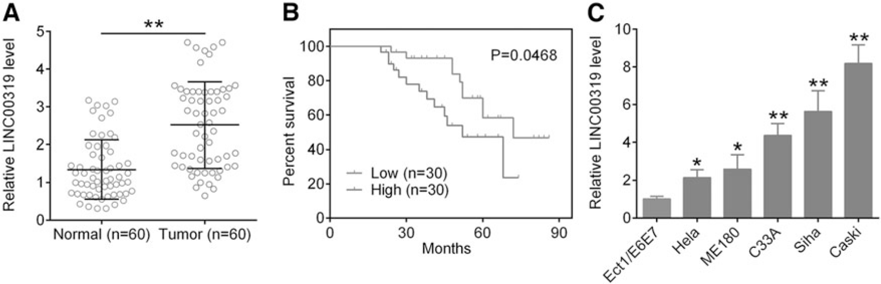
Enhanced expression of Linc00319 in cervical cancer cells.
Associations Between Linc00319 Expression and Clinicopathological Characteristics in Cervical Cancer Patients

The median expression level of Linc00319 was used as the cutoff. The p value was used to analyze the correlation between the expression levels. *p < 0.05.
The above data demonstrated that the expression level of Linc00319 is abnormally elevated in cervical cancer tissues and cervical cancer cell lines.
Linc00319 silencing inhibited the proliferation of cervical cancer cells
To explore the function of Linc00319, two different Linc00319 small hairpin RNA (shRNA, shRNA-1, shRNA-2) were transfected into SiHa and Caski cells and the silencing efficiency was verified by real-time PCR technology. As expected, Linc00319 was remarkably downexpressed in Linc00319 shRNA (shRNA-1, shRNA-2) groups, especially in the Linc00319 shRNA-2 group, which would be used in the following analysis (Fig. 2A). As shown in Figure 2B, Linc00319 silencing inhibited the proliferation of SiHa and Caski cell lines. The Brdu-positive cells were also decreased significantly in Linc00319 shRNA-2 cervical cancer cells compared with Sh-NC group (Fig. 2C). Therefore, Linc00319 may promote the proliferation of cervical cancer cells.
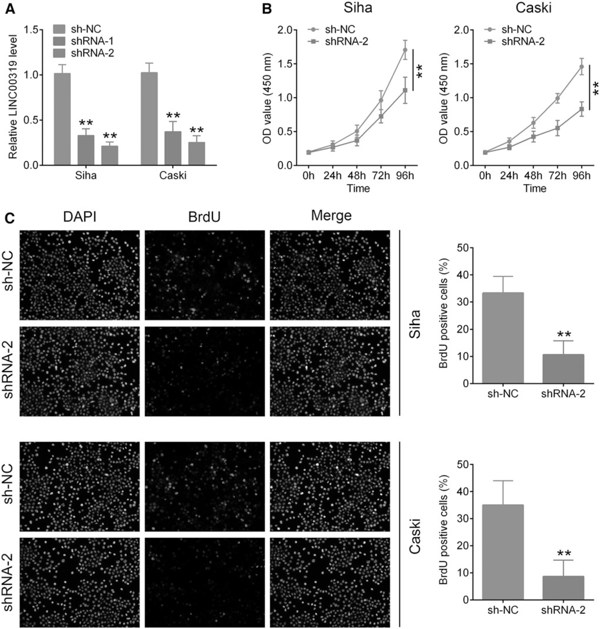
The effect of Linc00319 on the proliferation of cervical cancer cells.
Linc00319 silencing inhibited the migration and invasion of cervical cancer cells
To identify the detailed function of Linc00319 in the migration and invasion of cervical cancer cells, the authors evaluated the migration ability of Linc00319 silencing cells by the wound healing assay. A significant inhibited wound closure was observed compared to control group (Fig. 3A). The invasive capacity of SiHa and Caski cells were also impaired by Linc00319 silencing in the transwell assay (Fig. 3B) compared with control groups. All the results suggest that Linc00319 silencing inhibited the migration and invasion ability of cervical cancer cells.
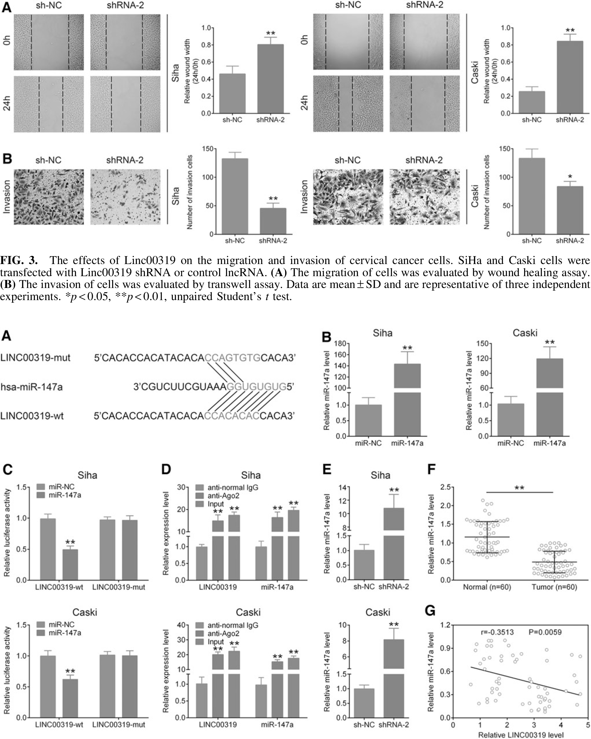
The effects of Linc00319 on the migration and invasion of cervical cancer cells. SiHa and Caski cells were transfected with Linc00319 shRNA or control lncRNA.
Linc00319 could target and downregulate miR-147a expression
As it has been known that lncRNA could regulate the expression level of miRNA via binding it directly, the candidate target miRNAs were predicted by miRDB database (

MiR-147a is targeted by Linc00319.
These above results indicated that Linc00319 could directly bind to miR-147a and negatively regulate its expression in cervical cancer progression.
Linc00319 promotes IGF1R expression via downregulating miR-147a
To investigate the target protein of miR-147a, the authors consulted the TargetScan database (

MiR-147a targets to IGF1R for the downregulation.
Linc00319 activates IGF1R pathway and enhances the development of cervical cancer
For further validating the mechanism of Linc00319/miR-147a/IGF1R axis in cervical cancer, an increased mRNA level of IGF1R was observed in clinical cervical tumor tissues compared to corresponded adjacent tissues (Fig. 6A), which was also positively correlated with the expression level of Linc00319, while negatively correlated with miR147a particularly (Fig. 6B, C). Recent studies indicated that the proliferating cell nuclear antigen (PCNA), which acts as an IGF1R binding partner may play a critical role in DNA replication and replication-associated processes in cancer cell. 28 In addition, IGF1R could activate protein vimentin29,30 and matrix metalloproteinase 2 (MMP2) pathways thus accelerate the process of tumor growth, invasion, migration, and poor prognosis severely. 31 On the contrary, E-cadherin 32 and cyclin-dependent kinase inhibitor p21 have been proved to be sensors and effectors of multiple antiproliferative signals in cancers. 33 As Figure 6D demonstrated, the protein expression levels of PCNA, Vimentin, and MMP2 were remarkably upregulated in IGF1R overexpressing cells compared with Linc00319 silencing cells, indicating a higher proportion of proliferation, invasion, and migration of cancer cells. However, P21 and E-cadherin protein levels were markedly downregulated (Fig. 6D), reflecting a confined tumor inhibition. Taken together, the data suggested that Linc00319 promotes the progression of cervical cancer cells by modulating miR-147a/IGF1R cascade.
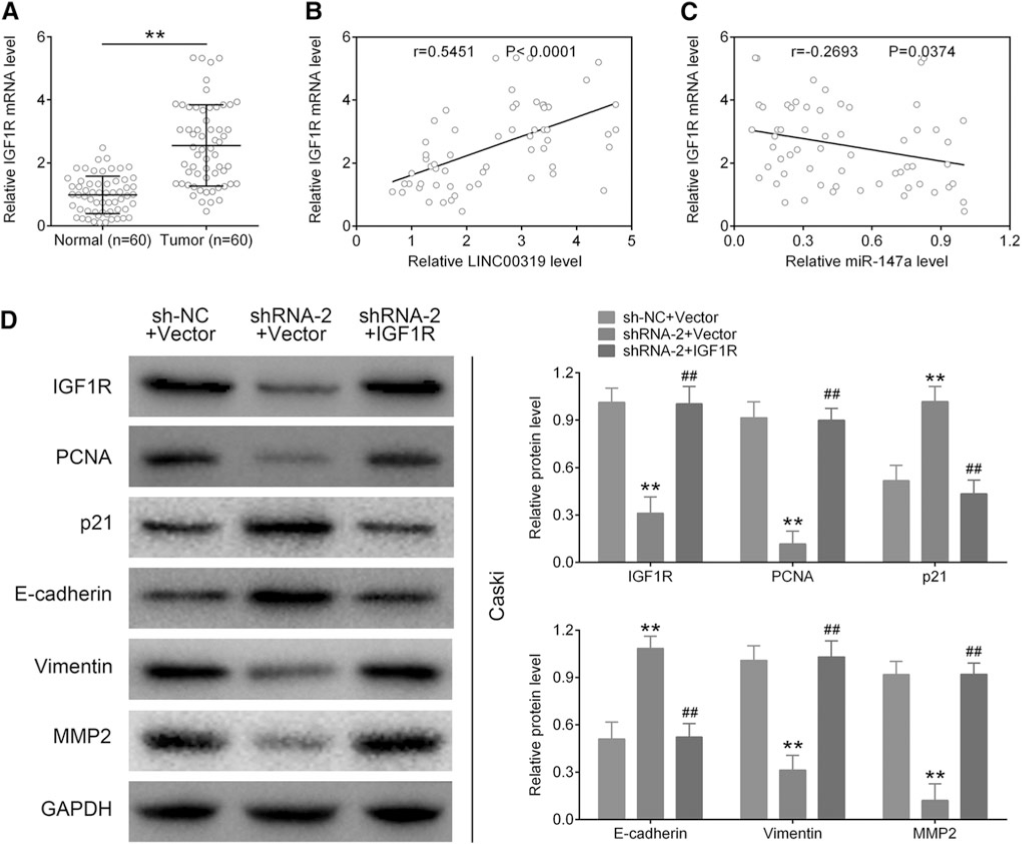
Linc00319 positively regulates the IGF1R signaling.
Discussion
Cervical cancer is a major health problem of women worldwide, but its nosogenesis has not yet been fully elucidated. Several previous studies about cervical cancer suggest that genetic mutation plays an important role in cervical cancer development. Recent clinical and epidemiologic studies discovered a strong association between epigenetic alterations and cervical carcinogenesis. Recently, lncRNA has received more and more attention. Many reports have pointed out that lncRNAs are important in the generation and progression of tumor via regulating cell proliferation, migration, invasion, and biological functions directly or indirectly. 34 LINC00319 was reported to promote the carcinogenesis of several cancer types through targeting special miRNAs and related signaling pathway. 18
In this study, the authors identified a significantly upregulation of Linc00319 in cervical cancer tissue, which implied an remarkable role of Linc00319 in cervical cancer patients. In addition, they found that Linc00319 silencing restrained the proliferation, migration, and invasion ability of cervical cancer cells, which suggested that Linc00319 may promote the deterioration of cervical cancer. Furthermore, their studies confirmed that Linc00319 could bind to miR-147a directly and inhibit the expression of miR-147a. These results are consistent with the findings from previous studies. To explore the related molecular signaling, IGF1R was predicted to be the targeted molecular and miR-147a could bind to the mRNA 3′ UTR of IGF1R. The inhibited expression of miR-147a could increase the expression of IGF1R strongly.
Previous studies indicated that IGF1R could bind to IRS1 and activate the PI3K-AKt-mTOR pathway through PCNA, which was in consistent with their study demonstrated in Figure 6D. This pathway was related to the function of protein synthesis, antiapoptosis, and cell survival 35 in the early stage of tumorigenesis. Therefore, these results implied that IGF1R may activate the DNA replication or related cell growth of cervical cancer through PCNA/PI3K/AKt signaling, 36 but still need more evidence. In addition, IGF1R could stimulate the MEK/ERK pathway 37 or c-Jun N-terminal kinase (JNK)/Mitogen-activated protein kinase (MAPK) pathway 38 through adaptor protein Shc severally and modulate transcription factors such as c-Jun, c-Fos, c-Myc, and ELK and finally resulted in cell proliferation, cell survival, and epithelial–mesenchymal transition (EMT), which was represented by the downregulation of E-cadherin and upregulation of vimentin and MMP2 (Fig. 6D). Therefore, the authors' results might uncover the molecular mechanism of Linc00319/miR-147a/IGF1R that activated EMT progression. However, the mechanism of how IGF1R signaling affects cervical cancer in vivo or in vitro was not explained in their study and that will be carried out in their following work.
Above all, the authors found that Linc00319 was significantly upregulated in cervical cancer tissues and cell lines. Linc00319 could inhibit the expression of miR-147a and activate IGF1R-related pathway to regulate the proliferation, migration, and invasion of cervical cancer cells. Their study identified that Linc00319 as an important oncogene and may provide a novel molecular biomarker and therapeutic target for cervical tumor.
Ethics Approval and Consent to Participate
All procedures performed in studies involving human participants were approved by the Ethics Committee of Affiliated Hospital of Beihua University.
All the clinical materials matched informed written consents from patients before studies, respectively.
Availability of Data and Materials
All data generated or analyzed during this study are included in this published article.
Footnotes
Author Disclosure Statement
No competing financial interests exist.
Funding Information
No funding was received for this article.
Authorship Confirmation Statement
L.L. conceived and designed the experiments, Z.M. and Y.F.C. analyzed and interpreted the results of the experiments, and L.M.Z. and C.C.T. performed the experiments.