
Abstract
Background:
Evidence from previous investigations points to a rising trend in the incidence of colorectal cancer (CRC) worldwide. The mortality resulting from this cancer is high. Unlike nonsmall cell lung cancer for which LINC01123 has been investigated, there are few reports on how this long noncoding RNA (lncRNA) regulates CRC.
Materials and Methods:
The authors evaluated the expression of LINC01123 in CRC tissues by quantitative real-time polymerase chain reaction. Its impact on cancer cell behavior was analyzed with cell counting kit-8 (CCK-8), colony formation, and Transwell invasion assays. To establish the mechanisms of LINC01123 in CRC they carried out RIP and luciferase reporter assays.
Results:
The results show that LINC01123 expression is abnormally elevated in CRC tissues and cell lines. High LINC01123 expression closely correlates with poor prognosis, advanced TNM stage, and lymph-node metastasis. The authors also show that knockdown of LINC01123 inhibits proliferation and invasion in CRC cells. In mechanism, it is revealed that LINC01123 may function as competitive endogenous RNA (ceRNA) against miR-625-5p to promote LIM and SH3 protein 1 (LASP1) expression.
Conclusions:
The data indicate that high LINC01123 exerts its oncogenic roles by regulating the miR-625-5p/LASP1 axis in CRC progression.
Introduction
Evidence from previous investigations points to a rising trend in the incidence of colorectal cancer (CRC) worldwide. The mortality resulting from this cancer is high. 1,2 This cancer is mainly managed surgically, using radiotherapy, chemotherapy, and adjuvant therapy. 3 Although several management strategies have put in place to reduce the disease burden of CRC, prognosis of patients with an advanced CRC remains poor. 4,5 Novel diagnostic biomarkers and therapeutic targets are, therefore, needed for improved patient outcomes.
Long noncoding RNA (lncRNA) has length exceeding 200 nucleotides. 6 Dysregulation of lncRNAs accelerates the development cancers. 7,8 It has been reported that MALAT1 upregulation correlates with poor prognosis and advanced clinical features in renal cell carcinoma. 9 The lncRNA, HOTTIP, has been reported to promote chemoresistance in osteosarcoma cells by activating the Wnt signaling. 10 HOXA11-AS is thought to promote invasiveness and expansion of renal tumor cells through the miR-146b-5p/MMP16 pathway. 11 However, the roles of lncRNAs in tumor is largely elusive.
Recently, an oncogenic role was reported for LINC01123 in lung cancer where it mediates proliferation and glycolysis. 12 However, its function in CRC has not been investigated. In this study, the authors focused on investigating the pivotal roles of LINC01123 in CRC. Results suggest that it facilitates CRC progression by sponging miR-625-5p and thus upregulating LIM and SH3 protein 1 (LASP1) expression. This study reveals that targeting this lncRNA may offer a new treatment strategy against CRC.
Materials and Methods
Clinical samples
Thirty-nine pairs of CRC tissues with corresponding adjacent nontumor tissues were obtained at The First Affiliated Hospital, Zhejiang University School of Medicine. Written informed consent was acquired from all patients. This study obtained the approval of the Ethics Committee of the First Affiliated Hospital, Zhejiang University School of Medicine.
Cell culture and transfection
The following human CRC cell lines were bought from the Chinese academy of science cell bank: HCT116, SW480, HT29, LOVO, and SW620, normal human colon epithelial cell line NCM460. Cell culture was carried out in a humidified incubator using Dulbecco's modified Eagle's medium (DMEM; Gibco) supplemented with 10% fetal bovine serum at 37°C, 5% CO2.
SiRNA targeting LINC01123 (si-LINC01123#1/2/3) and scrambled siRNA (si-NC) were bought from Shenggong Bioengineering Co. (Shanghai, China). MiR-625-5p mimics, miR-NC, si-LASP1, and si-NC were acquired from Genechem (Shanghai, China). Lipofectamine 2000 (Invitrogen, Carlsbad) was applied to facilitate plasmid transfection. Sequences for the oligos used are as follows: si-LINC01123#1: 5′-CUGAACGUCUUGCAACAGUTT-3′; si-LINC01123#2: 5′-GCCCUAGGAAAUCCGUAAUTT-3′. si-LINC01123#3: 5′-UGAGACAGAUCAGCAACAATT-3′; si-LASP1: GAACTACAAGGGCTACGAGAA.
Quantitative real-time polymerase chain reaction analysis
Cells were treated with Trizol reagent (Invitrogen, CA) to extract total RNA. This RNA sample was used to synthesize cDNA with Reverse Transcription kit (TaKaRa, Japan). Quantitative real-time polymerase chain reaction (RT-qPCR) was done using SYBR Premix EX Taq (TaKaRa) using the following cycling program: predenaturation at 95°C for 10 min, 40 cycles of denaturation at 95°C for 15 s, annealing at 60°C for 1 min, and final extension at 72°C for 30 s. Fold change expression was estimated using the 2−ΔΔCt formula using GAPDH or U6 was used as reference genes. The following primers were used LINC01123: F 5′-ACAGTGGCCGCACGCATAGCTG-3′, R 5′-CTGACGACCGAGGTGACAACGATGA-3′; miR-625-5p: F 5′-GGGGAGGGGGAAAGTTCTA-3′, R 5′-GTGCGTGTCGTGGAGTCG-3′; miR-625-5p: F 5′-CTGTCTCTGCCTTATAGCAACAC-3′, R 5′-CATCTCGAACCTGGCTGTTTG-3′; GAPDH F 5′-GCACCGTCAAGGCTGAGAAC-3′, R 5′-TGGTGAAGACGCCAGTGGA-3′. U6 F 5′-CTCGCTTCGGCAGCACA-3′ and R 5′-CTCAACTGGTGTCGTGGA-3′.
Assessment of cell proliferation
Cells were prepared for Cell Counting Kit-8 (CCK-8; Dojindo, Japan) assay as follows: In brief, pretransfected cells were cultured in 96-well plates (1 × 10⁴ cells/well) for 24, 48, and 72 h, and subsequently 10 μL of CCK-8 reagent was added per well and incubated for 4 h. Absorbance was then read at 450 nm using a microplate reader (Bio-Tek, MA).
Assessment of colony formation ability of cells
5 × 10³ pretransfected cells were seeded on six-well plates cultured for 14 days with media change every 3 days. After washing with PBS, colonies fixed with methanol and stained with crystal violet. Colonies were counted manually.
Transwell invasion assay
The invasive capacity of cells was assessed in 8 μm, 24-well Transwell chambers (BD Biosciences, CA). A total of 1 × 10⁵ pretransfected cells in 100 μL DMEM were incubated in upper compartment of precoated with Matrigel. Twenty-four hours later, nonmigratory cells were gently discarded using a cotton-tipped swab. Cells that passed through the membrane were fixed in methanol and stained with 0.1% crystal violet and imaged under an inverted optical microscope (Olympus, Tokyo, Japan).
Cytoplasmic and nuclear fractionation
Cytoplasmic and nuclear RNA kit (Norgen, Thorold, ON, Canada) was used to isolate the cytoplasmic and nuclear fractions from CRC cells. Expression of LINC01123, GAPDH (cytoplasmic control), and U6 (nuclear control) were examined by RT-qPCR assay.
Luciferase reporter assay
The recombination luciferase plasmids, LINC01123-WT, LINC01123-Mut, LASP1-WT, and LASP1-Mut, containing LINC01123 or LASP1 3′-untranslated region (UTR) full-length sequences with wild type (WT) or mutant (Mut) miR-625-5p binding sites were constructed. The luciferase constructs were then cotransfected with miR-625-5p mimics or miR-NC into cells using Lipofectamine 3000. After 24 h, luciferase activity was analyzed using a dual-luciferase reporter assay kit (Promega, Madison, WI).
RNA immunoprecipitation
Binding between LINC01123 and miR-625-5p in CRC cells was subjected to RNA immunoprecipitation (RIP) with Magna RIP RNA-binding protein immunoprecipitation kit (Millipore, Bedford, MA) as previously reported. 13
Statistical analysis
Statistical analyses were done on SPSS statistical suite version 20.0 (IBM). Data are presented as mean ± SD. Student's t-test or one-way ANOVA was used to test significance of differences between two or multiple groups. p-Value <0.05 was considered statistically significant.
Results
LINC01123 is highly expressed in CRC
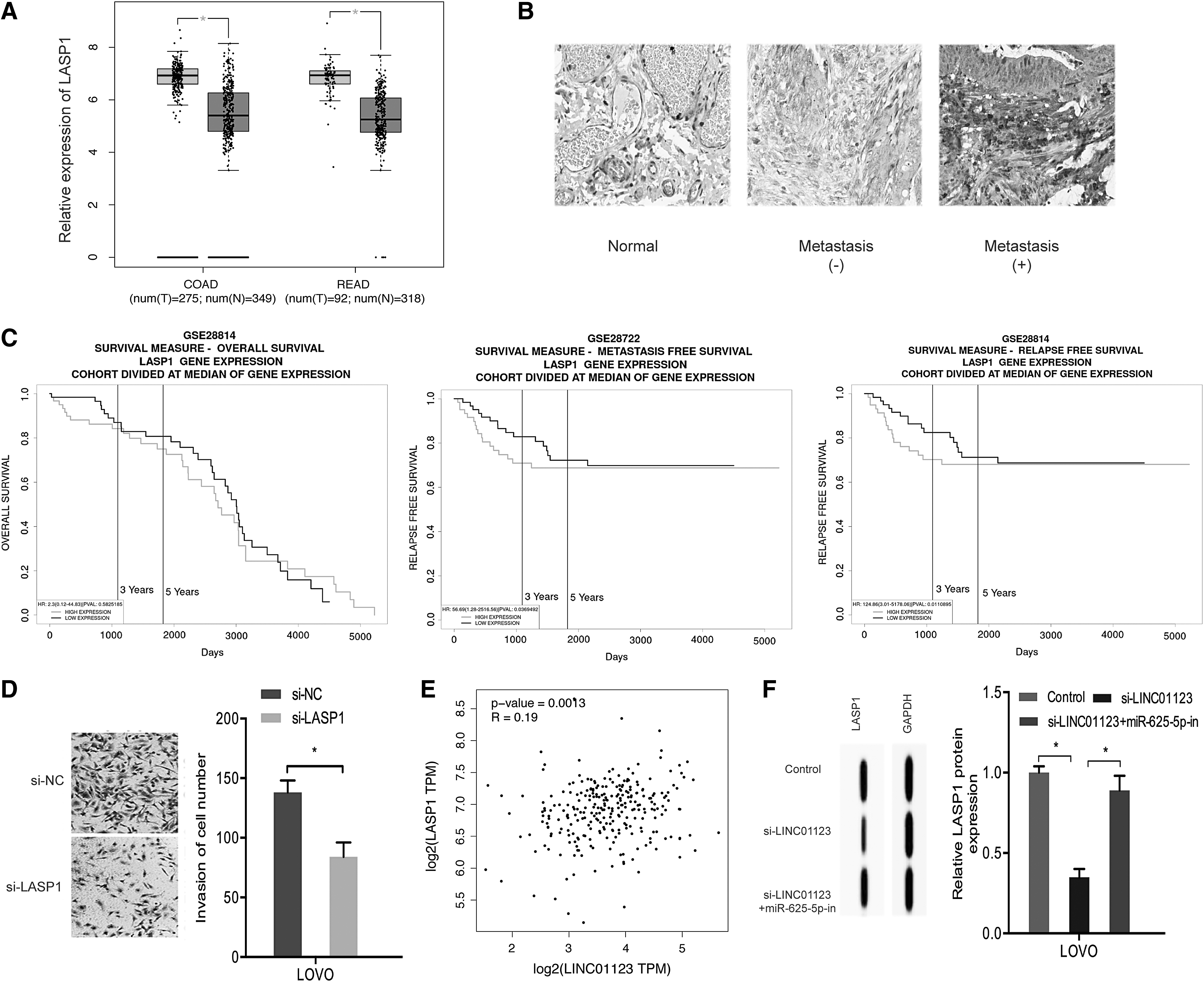
GEPIA analysis revealed that LINC01123 expression is significantly elevated in CRC tissues (Fig. 1A). Kaplan–Meier analysis indicated that high LINC01123 expression highly correlates with overall survival and disease-free survival in CRC patients (Fig. 1B, C). RT-qPCR uncovered that LINC01123 expression is markedly elevated in 39 paired CRC tissues relative to the control tissue (Fig. 1D). High LINC01123 levels were linked with advanced TNM stage and lymph-node metastasis in CRC (Fig. 1E, F). Together, these data suggested that LINC01123 might be involved in CRC tumorigenesis.
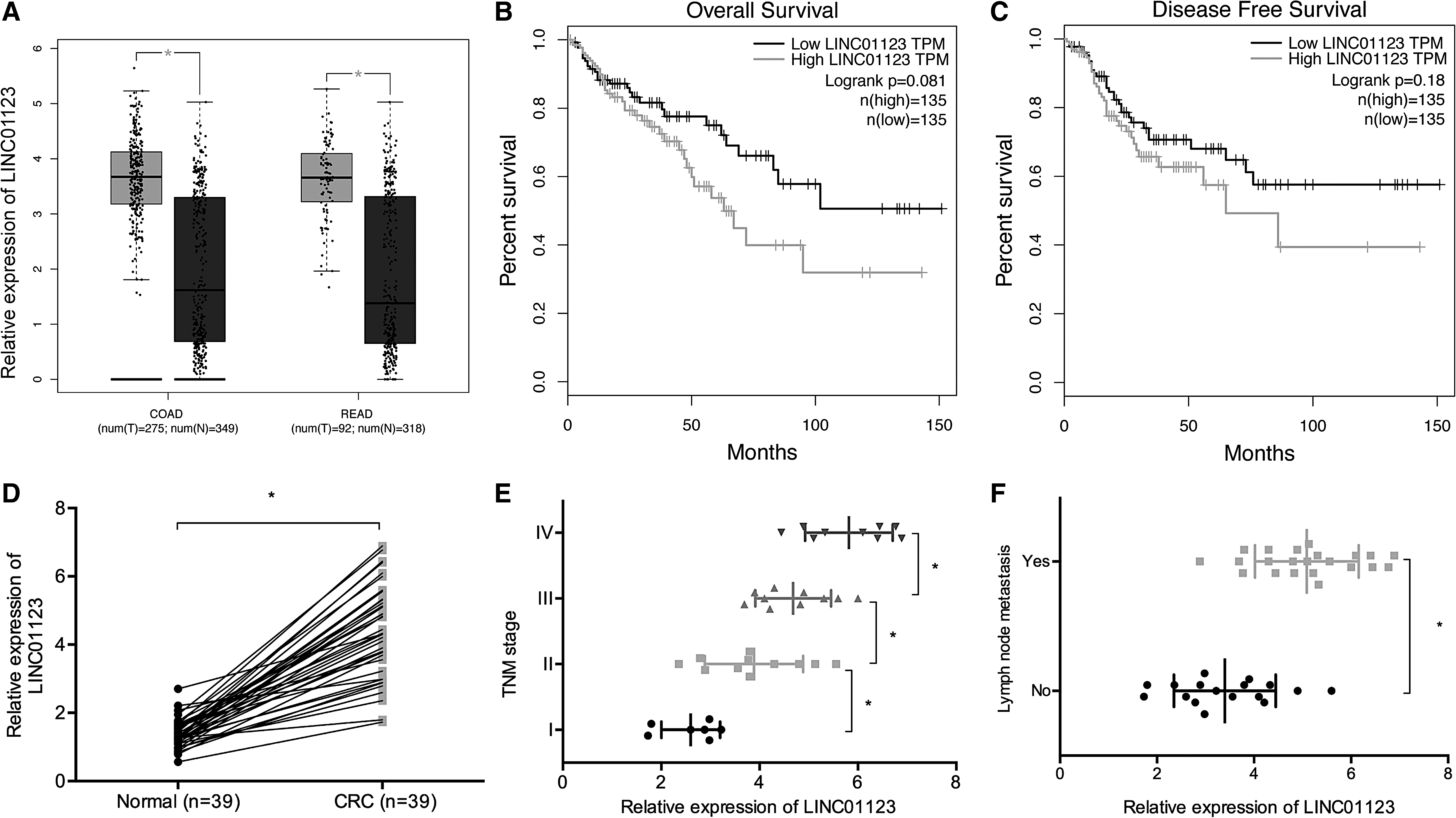
LINC01123 is upregulated in CRC.
LINC01123 promoted CRC cells proliferation and invasion
To elucidate how LINC01123 regulates CRC, the authors analyzed LINC01123 levels in CRC cell lines with RT-qPCR and found it to be upregulated in CRC cell lines (HCT116, SW480, HT29, LOVO, and SW620) relative to the normal human colon epithelial cells NCM460 (Fig. 2A). Next, they transfected si-LINC01123 into SW480 and LOVO cells (Fig. 2B). CCK-8 and colony formation assays demonstrated that LINC01123 inhibition suppresses SW480 and LOVO cells proliferation (Fig. 2C–F). Transwell invasion assay showed that knockdown LINC01123 expression reduces SW480 and LOVO cells invasion (Fig. 2G, H). Together, these results indicated that LINC01123 may be an oncogene in CRC.
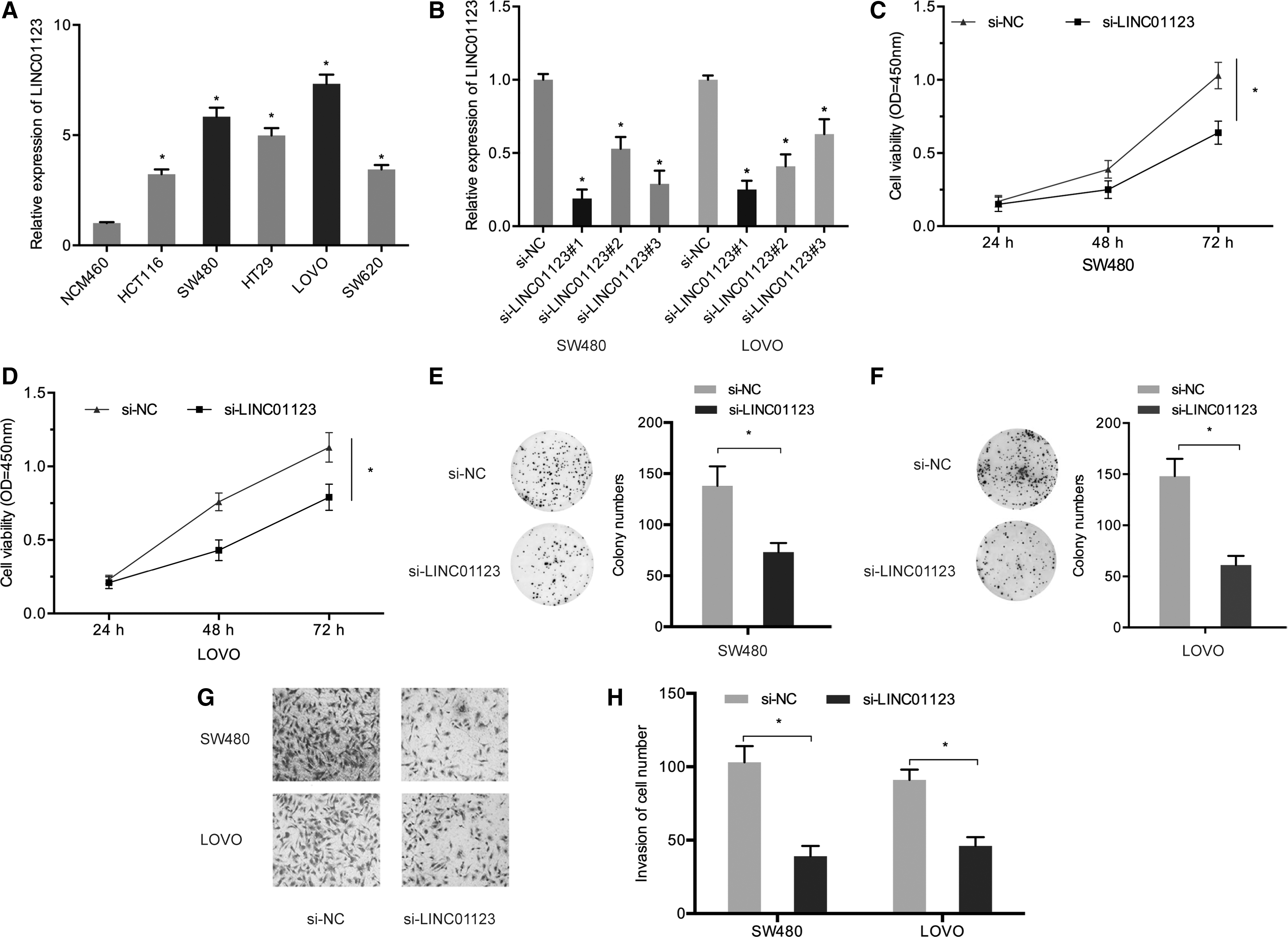
LINC01123 inhibition suppresses CRC cells proliferation and invasion.
LINC01123 bound to miR-625-5p
Next, the authors analyzed the molecular mechanism by which LINC01123 might contribute to CRC. To this end, they performed a nuclear-cytoplasmic fractionation assay and observed that in CRC cells, LINC01123 was enriched in cytoplasm (Fig. 3A). The authors then used bioinformatics to predict potential target microRNAs (miRNAs) of LINC01123. Out of seven candidate miRNAs, miR-625-5p attained the highest score and therefore was, studied further (Fig. 3B–D). Luciferase reporter analysis revealed that miR-625-5p mimics reduce luciferase activity in LINC01123-WT-transfected cells. No reduction in luciferase activity was observed in cell transfected with LINC01123-MUT (Fig. 3E). RIP assay showed that both LINC01123 and miR-625-5p were enriched in Ago2 (Fig. 3F).
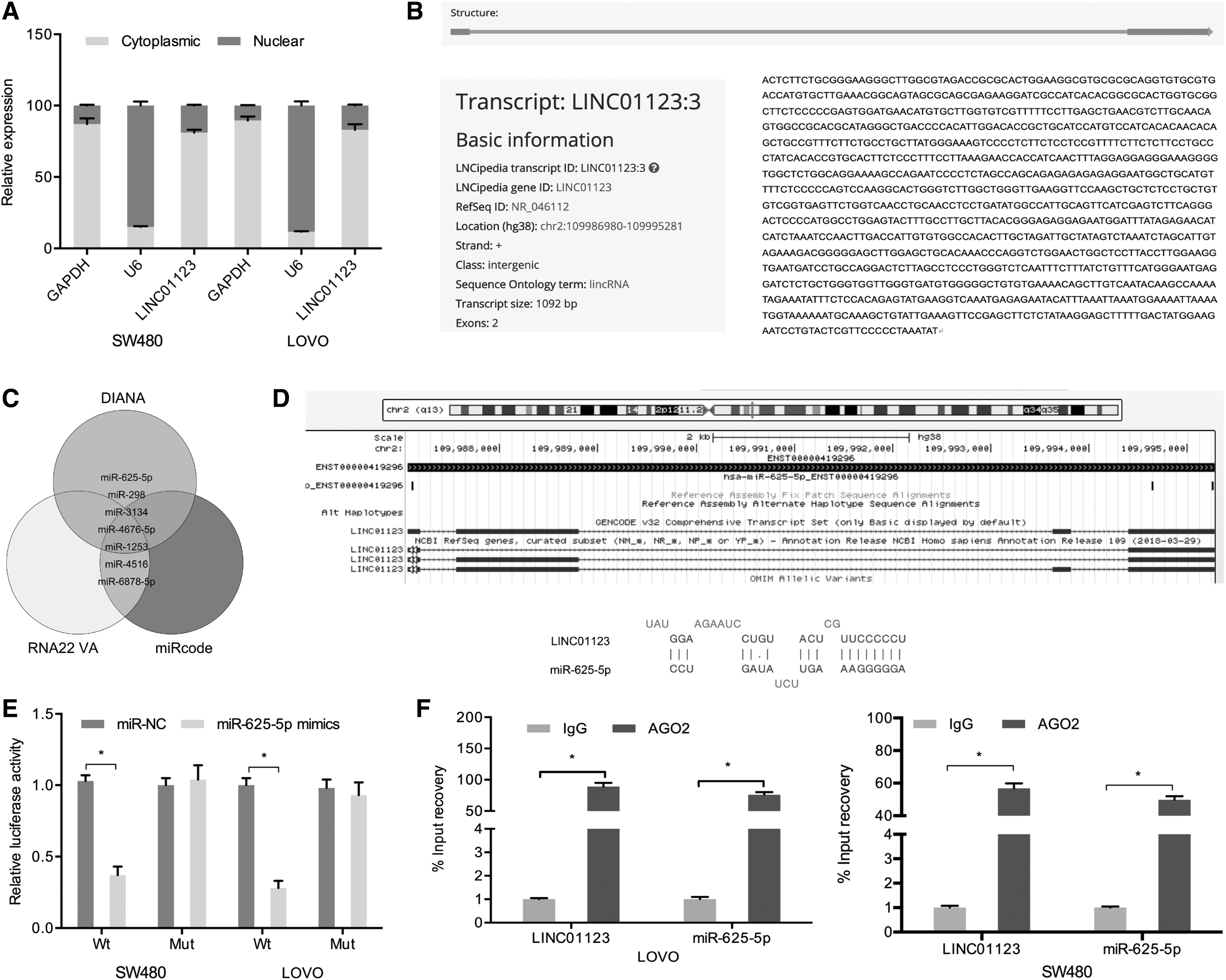
LINC01123 acts as a molecular sponge for miR-625-5p in CRC.
LASP1 acted as a target gene of miR-625-5p
Next, the authors explored the underlying mechanism of miR-625-5p in CRC. Bioinformatics analysis suggested the 3′-UTR of LASP1 mRNA bears sequences complementary to the seed region of miR-625-5p (Fig. 4A, B). RT-qPCR and Western blot assays showed that LASP1 expression was significantly lower in CRC cells transfected with miR-625-5p mimics (Fig. 4C, D). Luciferase reporter assays showed that miR-625-5p mimics reduce luciferase activity of the LASP1-WT group (Fig. 4E).
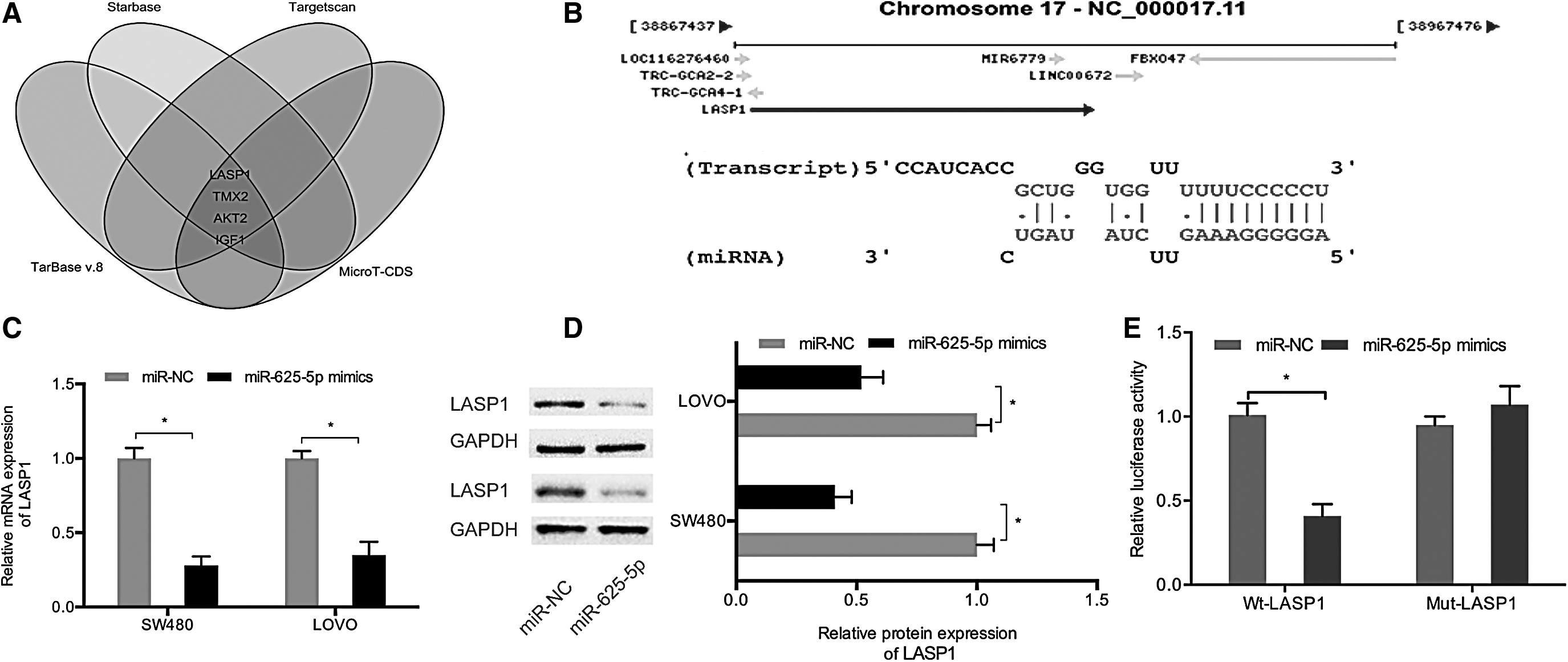
LASP1 acts the target of miR-625-5p in CRC.
The authors then explored the role of LASP1 in CRC. Data mining on TCGA indicated that LASP1 expression is significantly elevated in CRC tissues (Fig. 5A), an observation that was confirmed by immunohistochemistry (Fig. 5B). Kaplan–Meier analysis showed that high LASP1 level markedly correlates with poor CRC prognosis (Fig. 5C). LASP1 inhibition significantly suppressed invasion by CRC cells (Fig. 5D). Subsequent correlation analysis indicated that LINC01123 expression positively correlates with LASP1 expression in CRC tissues (Fig. 5E). Moreover, miR-625-5p inhibitors rescued the effects of si-LINC01123 on LASP1 protein expression in LOVO cells (Fig. 5F). Taken together, these data suggest that LINC01123 may be a competing endogenous RNA (ceRNA) for miR-625-5p to facilitate LASP1 level in CRC.
Roles of LASP1 in CRC.
Discussion
Evidence from previous investigations points to a rising trend in the incidence of CRC worldwide. The mortality resulting from this cancer is high. 1,3 Mounting evidence shows that lncRNAs play important roles in CRC tumorigenesis. For example, high lncRNA Sox2ot expression has been associated with poor CRC prognosis and has been shown to promote cells proliferation and motility. 14 The lncRNA PlncRNA-1 has been suggested to promote CRC progression through PI3K/Akt signaling. 15 ABHD11-AS1 promotes CRC invasiveness and proliferation through miR-1254/WNT11 axis. 16 In this study, the authors found LINC01123 is upregulated in CRC tumors and cell lines. The data point to an association between high LINC01123 expression and poor prognosis, lymph-node metastasis and advanced TNM stage. In addition, LINC01123 suppression suppressed CRC cells proliferation and invasion in vitro.
ceRNAs are indispensable nucleic acids that regulate gene expression. 17 Multiple studies indicate that lncRNAs exert their effects by “sponging” miRNA through competitive binding to miRNA, thereby preventing them from repressing their target mRNA. 18,19 Herein, the authors identified miR-625-5p as a target of LINC01123. miR-625-5p suppresses tumor growth in many types of cancer. For example, the miR-625-5p/PKM2 axis has been shown to negatively modulate glycolysis in melanoma. 20 LINC00511 has been shown to promote gastric cancer progression through miR-625-5p/NFIX pathway. 21 LINC00958 has been demonstrated to enhance the proliferative ability of cervical cancer cells as well as its metastatic potential by sponging miR-625-5p, thereby upregulating LRRC8E. 22 In this study, the authors find that LINC01123 may “sponge” miR-625-5p in CRC.
LASP1 acts an adhesion adaptor and scaffold protein. 23,24 LASP1 oncogenic function has been observed in multiple cancers. For example, it has been reported that LASP1 promotes nasopharyngeal carcinoma progression by negatively regulating the tumor suppressor, phosphatase and tensin homolog. 25 LASP1 has is thought to promote nonsmall-cell lung cancer progression through FAK-AKT signaling. 26 Moreover, miR-133a has been shown to repress CRC progression by targeting LASP1 and inhibiting MAPK signaling. 27 In this study, the authors find that LASP1 was markedly elevated in CRC that predicted unfavorable prognosis. LASP1 inhibition significantly suppresses invasion by CRC cells in vitro. Furthermore, they found that LASP1 to be an effector of miR-625-5p in CRC, whereas miR-625-5p inhibitors rescue the impact of LINC01123 silencing on LASP1 expression in CRC.
Therefore, this study has uncovered that LINC01123 is overexpressed in CRC and drives CRC proliferation and invasiveness by sponging miR-625-5p, thereby upregulating LASP1 level. These findings highlight the potential of LINC01123 as a potential therapeutic avenue against CRC.
Footnotes
Author Contributions
W.B.C. conceived and designed the experiments. T.S. and X.K.Z. analyzed and interpreted the results of the experiments. T.S., X.K.Z., and W.B.C. performed the experiments.
Declarations
Disclosure Statement
This article has not been published elsewhere in whole or in part. All authors have read and approved the content and agree to submit for consideration for publication in the journal. The authors declared no conflict of interest.
Funding Information
No funding was received for this article.