
Abstract
Interleukin-22 (IL-22), secreted by tumor infiltrated lymphocytes, is identified as a tumor-promoting factor in certain cancers, which was secreted by tumor infiltrated lymphocytes. However, the role of IL-22 in breast cancer remains conflicting. In this study, we assessed the expression of IL-22, IL-22 receptor 1 (IL-22R1), CD4, CD8, FOXP3, and CD68 in breast cancer by immunohistochemistry. IL-22 expression was exhibited in 105 (69.1%) cases in tumor cells (tIL-22), whereas only 24 (15.8%) samples displayed IL-22 expression in stromal cells. Multivariate analysis showed that tIL-22 expression was a poor prognostic factor for overall survival (OS) (p = 0.04). Meanwhile, IL-22R1 was predominantly presented in tumor cells (84.9%), which was associated with tIL-22 expression. The CD68-positive tumor-associated macrophages (TAMs) displayed the highest infiltration rate (50.7%) compared with CD4−, CD8−, and FOXP3-positive cells. Kaplan–Meier analysis confirmed patients with high TAM infiltration displayed significantly worse relapse-free survival (RFS) compared with low TAMs group (p = 0.017). TAM infiltration was also positively associated with tIL-22 and IL-22R1 expression. Furthermore, tIL-22 expression together with high TAM infiltration displayed the worst prognosis outcomes both in OS (p = 0.039) and RFS (p = 0.008). Instead of lymphocytes, our data indicated that tumor cells express IL-22 in breast cancer that is associated with IL-22R1, high TAM infiltrating, and poor prognosis.
Introduction
Interleukin-22 (IL-22) which belonged to the IL-10 cytokine superfamily was first found in IL-9 activated murine T cells. 1 In human homeostasis, immune cells is the main cellular source of IL-22 including CD4+ T cells, CD8+ T cells, γδ T cells, and innate lymphoid cells. 2,3 The functional performance of IL-22 depends on the combination with its receptor complex composed of IL-22 receptor α chain 1 (IL-22R1) and IL-10 receptor β chain 2 (IL-10R2). Of interest, although immune cells are the main source of IL-22 production, they would not effected by IL-22 owing to the nonexpression of its specific receptor IL-22R1. 4 This distinct characteristic of IL-22 makes it an attractive therapy target with less side-effect on immune system.
Besides autoimmune and inflammatory diseases, IL-22 was found to play an important role in tumor progression and cancer metastasis. 5 Compared with nontumor tissue and healthy control, gastric cancer patients displayed increased intratumoral IL-22-producing CD4(+) T cells and Th22 cells that correlated with cancer progression and poor patient survival. 6 In serum, elevated IL-22 positively correlated with colon cancer progression was well explored in rodent models and patients. 7,8 In addition to lymphotic lineage-derived IL-22, high expression of IL-22 and IL-22R1 by cancer cells were demonstrated in patients with pancreatic ductal adenocarcinoma, which also predicted poor prognosis and a suitable independent prognostic marker. 9
Breast cancer is the most common malignant tumor and the second leading cause of cancer death in women worldwide. 10 In this study we analyzed IL-22, IL-22R1, CD4, CD8, FOXP3, and CD68 expression in human breast cancer tissues, aiming to further understand the role of IL-22 in pathogenesis and provide possible therapy methods for these patients.
Materials and Methods
Patients and patient-derived samples
A total of 152 cases of patients diagnosed with primary breast invasive ductal carcinoma treated at the Fourth Hospital of Hebei Medical University (152 cases) (collected from January 2012 to December 2014) were eligible to be included in this study. All were adult female patients 20 years old or older with stage I–III breast carcinoma. Analyses for ER, PR, HER2 expression, and molecular subtypes were conducted. All patients were followed up after diagnosis until May 2019. This study was approved by the institutional ethnics committee of the Fourth Hospital of Hebei Medical University and Tumor Hospital of Hebei Province, China. The authors had access to information that could identify individual participants during or after data collection. Approval number: 201107209.
Immunohistochemistry
Paraffin-embedded specimens were sectioned (4 μm) and studied by immunohistochemistry. IL-22 (clone ab18499, 1:100 dilution, Abcam, Cambridge, United Kingdom) and IL-22R1 (clone ab5984, 1:100 dilution; Abcam) expression was quantified by using the Histo-score (H-score) system. The intensity (0 = no staining, 1+ = weak, 2+ = moderate, 3+ = strong) was scored and the percentage of staining tumor cells was evaluated in the whole section. The H-score was calculated using the following formula: (3 × percentage of cells with strong staining) + (2 × percentage of cell with moderate staining) + (1 × percentage of cells with weak staining). IL-22 expression was classified into two groups according to a cutoff H-score of 100. 11 For CD4 (clone BM4263, 1:50 dilution; Boster, Wuhan, China), CD8 (clone BM4379, 1:30 dilution; Boster), FOXP3 (clone ab20034, 1:50 dilution; Abcam), and CD68 (clone BA3639, 1:100 dilution; Boster) immunohistochemistry staining, expression in nuclei of lymphocytes was defined positive. The density of these infiltrating immune cells was categorized as high or low relative to the median of 11 cells per 0.0625 mm2 as the cutoff value within the slide at high power ( × 40 objective lens).
To perform the immunohistochemistry staining, paraffin-embedded sections were treated in xylene and rehydrated by gradient of ethanol. Antigen retrieval was carried out by heating slides for 15 min at 95°C in citrate buffer (pH 9.0). Endogenous peroxidase activity was blocked with 3% H2O2 for 30 min. Blocking with 5% goat serum (except goat antihuman primary antibodies) was performed. Specimens were incubated with primary antibody above at 4°C overnight. Nuclei were counterstained with hematoxylin (Dako). Rabbit immunoglobulin G (IgG) and goat IgG were used as isotype controls.
For evaluation of CD68 immunohistochemistry staining, expression in nuclei of peri-tumor immune cells infiltrated in stromal tissues was defined positive. The density of these infiltrating immune cells was categorized as high or low relative to the median of 11 cells per 0.0625 mm2 as the cutoff value within the slide at high power ( × 40 objective lens).
Statistical analysis
Statistical analysis was performed using the SPSS 21.0 and GraphPad Prism 5 software. Data are given as mean ± SD. The associations between IL-22 expression and immune cell infiltration with pathologic characteristics was examined using chi-square statistical tests. Overall survival (OS) and relapse-free survival (RFS) were estimated by Kaplan–Meier method and compared with log-rank test. The correlations between IL-22 expression or CD68 infiltration with clinical survival was evaluated by COX regression in both univariate and multivariate analyses. All tests were two-sided; values of p < 0.05 were considered significant.
Results
Expression of IL-22 by tumor cells in breast cancer
We first detected IL-22 expression in breast cancer tissues by immunohistochemistry staining. A total of 152 cases of patients diagnosed with primary breast invasive ductal carcinoma were eligible to be included in this study. tIL-22 expression was exhibited in 105 (69.1%) cases (Fig. 1A–D), whereas only 24 (15.8%) samples displayed IL-22 expression in stromal cells (Supplementary Table S1). The baseline clinical and pathological characteristics are given in Table 1. Meanwhile, tIL-22 expression was associated with poor prognosis indicators including increased tumor size (p = 0.021), negative hormone receptor status (p < 0.001), positive HER-2 status (p = 0.008), and invasive molecular subtypes (p < 0.001) (Table 1), but not lymph nodes status, histological grade, diseases stage, and vessel tumor embolus.
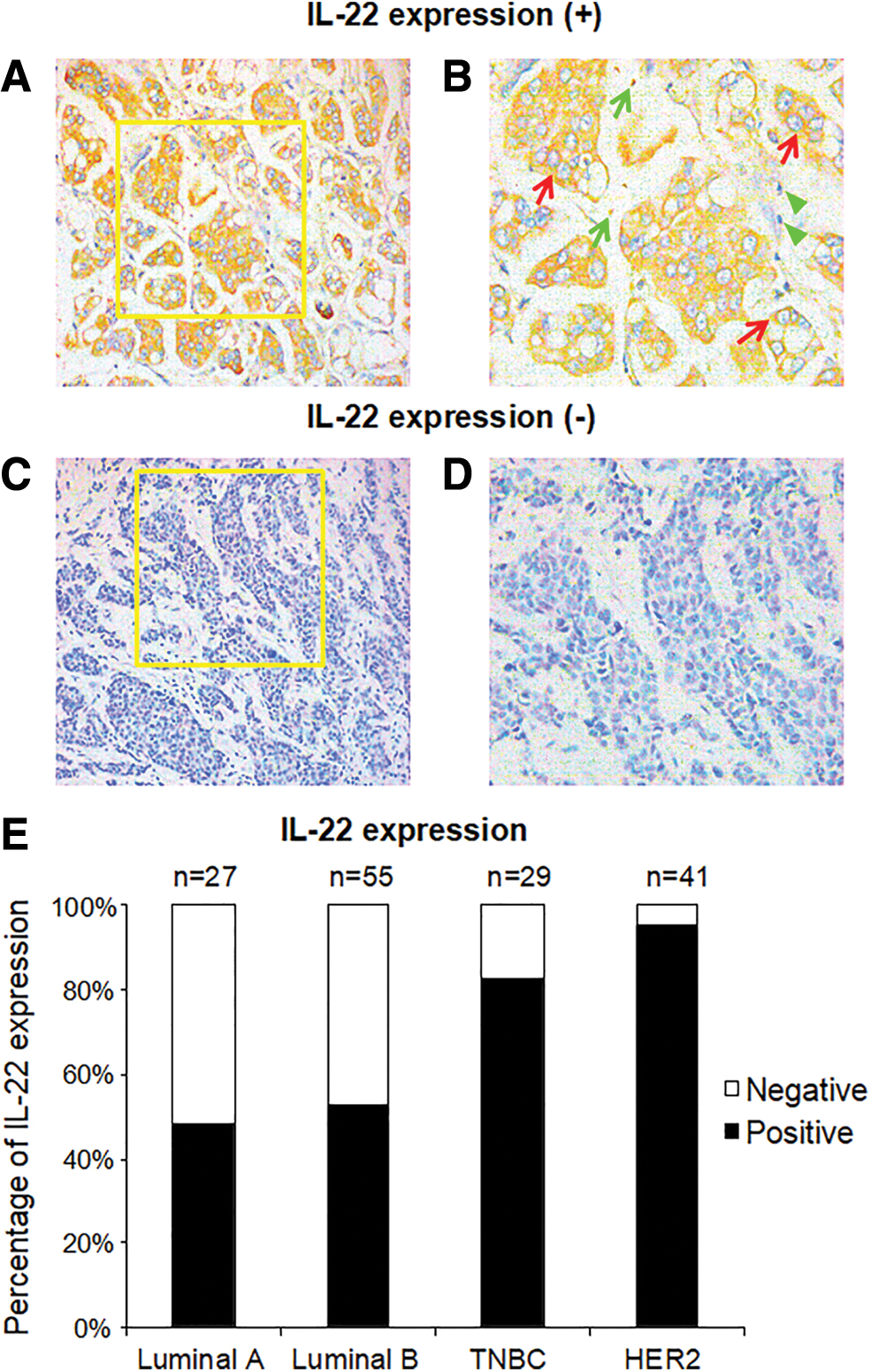
Tumor cells IL-22 expression in breast carcinoma tissues. Immunohistochemistry staining of representative tumor cells IL-22 expression within cytoplasmic are given in
Association of Tumor Cell IL-22 Expression with Clinicopathological Characteristics in Breast Cancer
p values are in italic, and values with statistic significance are indicated in bold.
Within four molecular subtypes of breast cancer, HER2-enriched subtype displayed the highest tIL-22 expression rate (39/41, 95.1%), which was significantly higher than in other subtypes (p < 0.001 vs. Luminal A; p < 0.001 vs. Luminal B; p = 0.089 vs. triple-negative breast cancer [TNBC]). Meanwhile, TNBC also displayed high tIL-22-positive percentage (24/29, 82.8%), whereas the proportions of tIL-22 expression were 52.7% (29/55) in Luminal B and 48.1% (13/27) in Luminal A (Table 1) (Fig. 1E).
tIL-22 expression associated with poor prognosis
A total of 152 patients were followed up for 9–77 months and the median follow-up time was 66 months. During this period, the follow-up rate was 97.4% with 23 cases being dead, 7 cases had recurrence or metastatic diseases, and 4 cases were lost. RFS and OS were estimated by Kaplan–Meier analysis. tIL-22-positive group (65.49 months, 95% confidence interval [CI] = 61.95–69.02) showed a worse RFS compared with tIL-22 negative cases (72.89 months, 95% CI = 69.30–76.48), although no significant difference was found (p = 0.051) (Fig. 2A). Log-rank analysis showed that patients with tIL-22 expression (67.38 months, 95% CI = 64.02–70.73) displayed a significantly worse OS compared with tIL-22 negative ones (76.66 months, 95% CI = 76.00–77.32), p = 0.003 (Fig. 2B). In the entire cohort, univariate analysis confirmed tIL-22 expression, tumor size, ER, PR status, and molecular subtypes as significant prognostic factors (Table 2). In the case of multivariate analysis, only tIL-22 expression was found to be the independent prognostic factors related to OS, p = 0.04 (95% CI = 0.014–0.907).
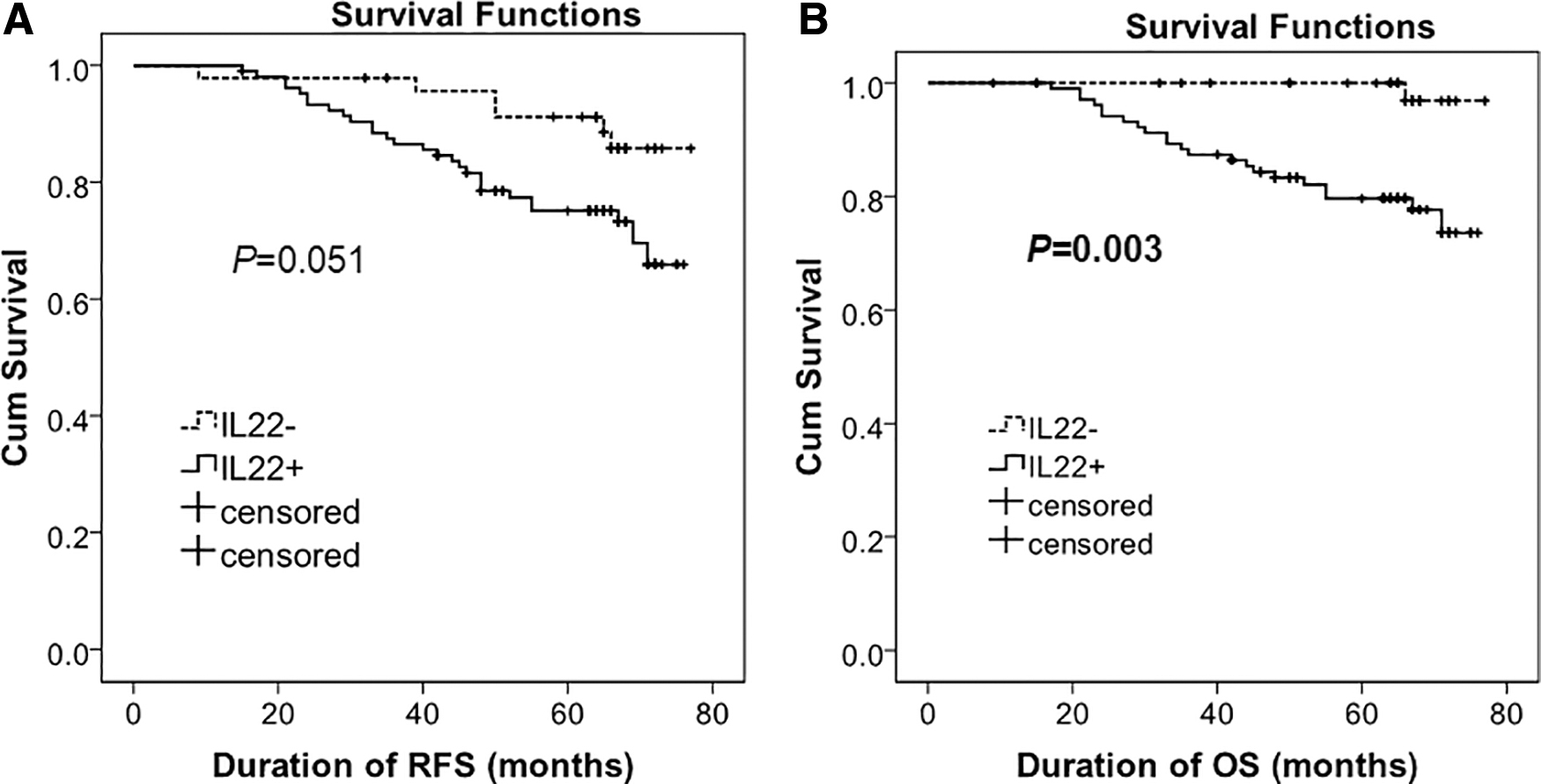
Prognostic impact of tumor cell IL-22 expression in breast cancer.
Univariate and Multivariate Analyses of Associations Between Clinical Parameters and Tumor Cells IL-22 Expression and Tumor-Associated Macrophage Infiltration with Overall Survival in Patients with Breast Carcinoma
p values are in italic, and values with statistic significance are indicated in bold.
tIL-22 expression associated with high TAM infiltration
We analyzed the expression of IL-22-specific membrane receptor IL-22R1 in breast cancer tissues. IL-22R1 staining was present in the cytoplasm and cellular membrane of tumor cells (Fig. 3A–3D). As given in Supplementary Table S1, IL-22R1 was predominantly expressed by tumor cells in 84.9% cases (129/152). Meanwhile, tIL-22R1 presentation was associated with tIL-22 expression (p = 0.007) (Table 3). Since our data displayed tIL-22 associated with poor clinical outcomes, tumor cells IL-22-IL-22R1 axis expression predicted not only an autocrine effect, but also a paracrine role in breast cancer progression.

The presence of IL-22 receptor 1 (IL-22R1) and CD68 expression in breast cancer tissues. Representative immunohistochemistry staining images are given in
Association of Tumor Cell IL-22 Expression with Stromal IL-22, IL-22R1 Expression and Immune Cells Infiltration in Breast Cancer Tissues
p values are in italic, and values with statistic significance are indicated in bold.
Tumor infiltration immune cells including CD4, CD8, FOXP3, and CD68-positive cells were assessed by immunohistochemistry. We analyzed the associations between peritumor immune cell infiltration with tIL-22 expression. Among all these immune cell markers, pan-macrophage marker CD68 displayed the highest infiltration rate (44.1%) compared with CD4+ cells (35.5%), CD8+ (40.1%), and FOXP3+ cells (28.3%) (Supplementary Table S1). As given in Table 3, high associated macrophages (TAMs) infiltration (CD68) was associated with tIL-22 expression (p = 0.002), whereas no association was found with other T cell populations (Fig. 3E–H).
Of interest, the tendency of TAM infiltration among different molecular subtypes was in accordance with the tIL-22 expression (Fig. 3I). HER2-enriched subtype also displayed the highest percentage of CD68+ infiltration rate (65.9%) within four molecular subtypes, whereas the Luminal A subtype still showed the lowest one (22.2%) (Supplementary Table S2).
Furthermore, high infiltration of TAMs (CD68) was also associated with negative hormone receptor status (p < 0.001), positive HER-2 status (p < 0.001) and molecular subtypes (p < 0.001), and lymph nodes involvement (p = 0.024) (Supplementary Table S2). Kaplan–Meier analysis confirmed patients with high TAM infiltration (64.42 months, 95% CI = 60.03–68.81) showed a worse RFS (70.69 months, 95% CI = 60.03–68.81), p = 0.017 (Fig. 4A). High TAM infiltration group (68.65 months, 95% CI = 64.76–72.54) also displayed a worse OS (71.72 months, 95% CI = 68.62–74.81), although there was no significant difference, p = 0.302 (Fig. 4B). Univariate analysis showed that high CD68 infiltration was a clinical characteristic affecting OS (Table 2).
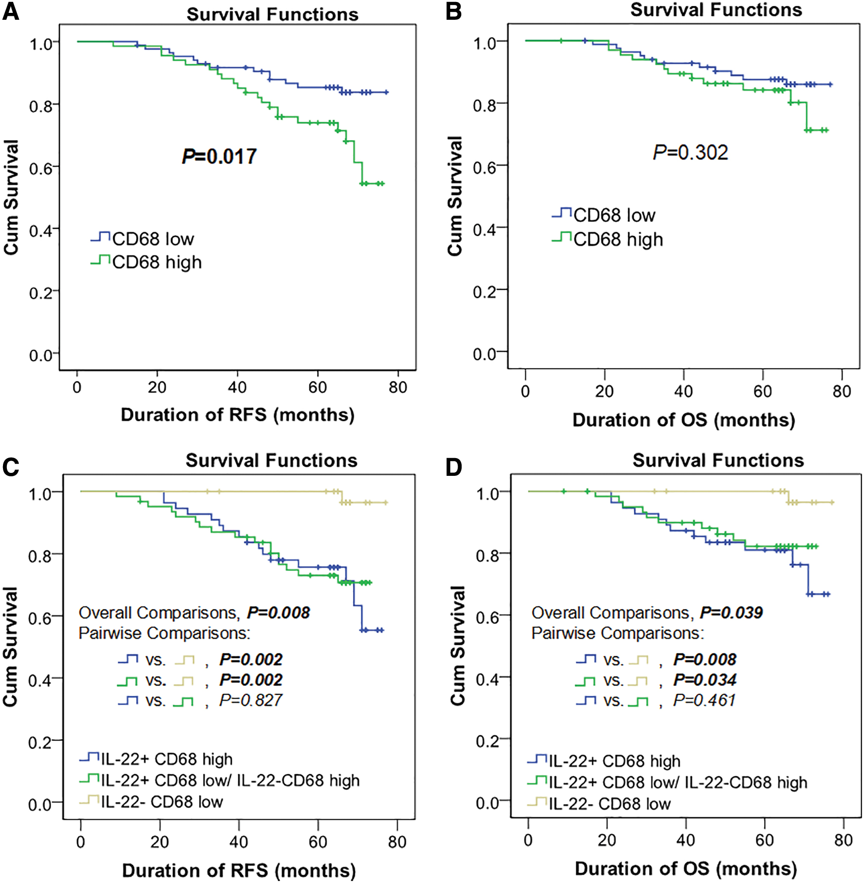
Survival analysis of CD68 and its synergistic expression with IL-22 in breast cancer. Kaplan–Meier estimates of RFS and OS based on CD68 expression are given in
We divided the patients into three groups: tIL-22+ CD68 high, tIL-22− CD68 low, and tIL-22+ CD68 low/tIL-22− CD68 high. Patients in tIL-22+ TAMs high group displayed the worst prognostic outcomes compared with the other two, both in RFS (65.11 months, 95% CI = 60.42–69.80) (p = 0.008) and OS (67.26 months, 95% CI = 62.71–71.80) (p = 0.039) (Fig. 4C, D). In Figure 4C and D, p-values of overall comparisons among these three groups by log-rank test were 0.008 and 0.039, separately. The pairwise comparisons between IL-22-CD68 low and the other two groups were also significantly different, as indicated in Figure 4.
Discussion
Tissue IL-22 level, presumably derived from lymphocytes, also positively correlated with MAP3K8 expression in breast cancer. Meanwhile, breast cancer could induce IL-22 production from memory CD4+ T cells and release IL-1 to promote tumor growth. 12 In contrast, IL-22-induced cell cycle arrest in the G2-M phase of a murine breast cancer cell line EMT6 cells was reported. Up to now, no report addressed the prognostic value of IL-22 expression by primary breast cancer tissues. In this study we confirmed that instead of lymphocytes, tumor cells commonly express IL-22 (105/152, 69.1%), whereas only 24 (15.8%) samples displayed stromal IL-22 expression. Meanwhile, tIL-22 expression is a poor prognosis factor for patients with invasive breast carcinoma. Positive tIL-22 expression is associated with tumor cells IL-22R1 presence and TAM infiltration.
In addition to its role in autoimmune and inflammatory diseases, IL-22 have also been proved to be important in cancer development. 13 In this study, multivariate analysis proved that tIL-22 expression was a poor prognostic factor for OS. The tIL-22 expression was also significantly associated with poor prognosis indicators including increased tumor size and negative hormone receptor status. Furthermore, tIL-22 presence was associated with HER-2 status (p = 0.008), which plays a crucial role in molecular subtype classification and breast cancer targeting therapy. As expected, tIL-22 expression was also associated with invasive molecular subtypes of breast cancer (p < 0.001). HER2-enriched subtype displayed the highest percentage of tIL-22 expression, whereas Luminal A and Luminal B showed the lower percentage.
Previous reports found that IL-22 transgenic and overexpression mice did not develop spontaneous tumors. 14 It demonstrates the less effect on tumor cell proliferation by IL-22-IL-22R1 system. We assessed the CD4, CD8, FOXP3, and CD68 infiltration in breast tumor microenvironment, aiming to further understand the role of IL-22. Among these infiltrating immune cells, expression of CD68, the marker for TAMs, displayed the highest presenting rate (44.1%) and was associated with RFS. Increased tumor TAM infiltration induced immune tolerance and attenuated antitumor therapy has been reported in some tumors. 15 In our data, high CD68 infiltration was only associated with worse RFS. No significant association with OS was found, indicating the insufficiency of using TAM infiltration alone as a prognostic factor for breast cancer. When combined IL-22 expression with TAM infiltration, we found the synergistic prognostic significance of tIL-22 expression and high TAM infiltration revealed the significantly poor prognosis outcomes both in RFS (p = 0.008) and OS (p = 0.039) compared with other groups.
One of the limitations in our data is the link between IL-22-IL-22R1 system and TAM infiltration. Previous studies confirmed that attracting macrophage infiltrate into tumor microenvironment is the critical step for TAM-induced tumor progression. 16 However, more studies are required to confirm the effect of TAM infiltration in human breast cancer cells.
In summary, our data demonstrated that IL-22 expressed by breast cancer cells was an independent prognostic factor that is related to poor clinical outcomes. Positive tIL-22 expression was associated with tumor cell IL-22R1 presence and TAM infiltration in the entire cohort. IL-22-IL-22R1 system may play an important role in TAM infiltration and tumor metastasis. Targeting tumor cell IL-22-IL-22R1 system may provide new therapeutic method for breast cancer treatment.
Footnotes
Acknowledgment
The authors thank Huichai Yan and Qiushuang Ding for excellent technical assistance, and all the investigators for participating in this study.
Disclosure Statement
No competing financial interests exist.
Funding Information
This work was supported by the Hebei Province Department of Science and Technology Research Grants: No. 162777249 and No. 17277747D.
Supplementary Material
Supplementary Table S1
Supplementary Table S2
References
Supplementary Material
Please find the following supplemental material available below.
For Open Access articles published under a Creative Commons License, all supplemental material carries the same license as the article it is associated with.
For non-Open Access articles published, all supplemental material carries a non-exclusive license, and permission requests for re-use of supplemental material or any part of supplemental material shall be sent directly to the copyright owner as specified in the copyright notice associated with the article.