
Abstract
Background:
Glioblastoma is a malignant and very aggressive brain tumor with a poor prognosis. Despite having chemotherapy concomitant with surgery and/or radiation therapy, the median survival of glioblastoma-affected people is less than 1 year. Temozolomide (TMZ) is a chemotherapeutic used as a first line treatment of glioblastoma. Several studies have reported that resistance to TMZ due to overexpression of O6-methylguanine-DNA methyltransferase (MGMT) is the main reason for treatment failure. Several studies described that pulsed-electromagnetic field (EMF) exposure could induce cell death and influence gene expression.
Materials and Methods:
In this study the authors assessed the effects of EMF (50 Hz, 70 G) on cytotoxicity, cell migration, gene expression, and protein levels in TMZ-treated T98 and A172 cell lines.
Results:
In this study, the authors show that treatment with a combination of TMZ and EMF enhanced cell death and decreased the migration potential of T98 and A172 cells. The authors also observed overexpression of the p53 gene and downregulation of cyclin-D1 protein in comparison to controls. In addition, T98 cells expressed the MGMT protein following treatment, while the A172 cells did not express MGMT.
Conclusion:
Their data indicate that EMF exposure improved the cytotoxicity of TMZ on T98 and A172 cells and could partially affect resistance to TMZ in T98 cells.
Introduction
Glioblastoma is a malignant and very aggressive brain tumor with a poor prognosis. Although more effective treatment in clinical application chemotherapy is associated with surgery and/or radiation therapy, the median survival of glioblastoma-affected patients is less than 1 year. 1,2 Temozolomide (TMZ) is an important alkylating chemotherapy drug used today as the first line of glioblastoma treatment. 3 This drug could cross the blood–brain barrier and induce tumor cell death by introducing methyl groups into the O6 position of guanine. 4 O6-methylguanine leads mispairs with thymine, which first causes single- and double-strand DNA breaks and then activates the p53 protein. Eventually, P53 induces senescence mechanisms and apoptosis in glioblastoma cells. 5,6 However, some mechanisms such as the upregulation of DNA repair genes reduce the effect of chemotherapy drugs. 7 Several studies have reported that resistance to TMZ due to overexpression of O6-methylguanine-DNA methyltransferase (MGMT) is the main reason for treatment failure. 8,9 MGMT is known as a DNA repair enzyme that eliminates O6-methylguanine and protects the genome against the mutagenic effects of alkylating drugs. 10 MGMT deficiency sensitizes glioblastoma to alkylating drugs. 11 In the last decade, some researchers studying in the field of cancer therapy have focused on the biophysical effects of electromagnetic field (EMF) exposure. 12,13 Exposure time and physical parameters of the applied EMF, including amplitude, frequency, and waveform, could affect a lot of biological responses in different cells. 14,15 Several studies described that EMF exposure could induce cell death and influence gene expression. 16,17 T98 and A172 cells are glioblastoma cell lines that were used routinely in most of the basic in vitro studies on nervous system tumors. Previous studies have reported that cotreatment with EMF and chemotherapy drugs improves the efficacy of the drug and attenuates tumor cell progression. 18,19 Akbarnejad et al. found that T98 cell viability decreased by about 22% at day 6 (144 h) after EMF exposure alone (100 Hz, 100 G). 16
Achievement of an innovative and novel approach to target metastasis of nervous system tumors is essential. Considering that the effects of EMF on tumor cell migration have been less addressed and importance of EMF exposure on gene expression, in this study the authors have aimed to investigate the effects of EMF (50 Hz, 70 G) on cytotoxicity, cell migration, gene expression, and protein levels in TMZ-treated T98 and A172 cell lines.
Materials and Methods
Cell culture
All materials were provided by Sigma Company (Sigma-Aldrich) unless otherwise indicated. The T98 and A172 (human glioblastoma) cell lines (Pasteur Institute, Iran) were cultured in 75 cm2 flasks in Dulbecco's modified Eagle's medium (DMEM) supplemented with 50 mg/mL streptomycin, 100 IU/mL penicillin, and 10% fetal bovine serum (Gibco). All flasks were incubated (37°C, 5% CO2) and the culture medium refreshed every other day.
TMZ treatment and EMF exposure
The authors prepared a primary TMZ stock solution with a concentration of 1.6 mM, and fresh 0.2, 0.4, and 0.8 mM TMZ solutions were obtained by diluting in DMEM. Based on a pilot test (data are not shown in the present study) IC50 of TMZ was assessed, and eventually, doses of 0.8 and 1.6 mM were selected for the experiments. The EMF exposure device has been described previously. 20 Briefly, this device was calibrated according to a magneto-therapy system (Fisioline Co, Italy) to produce continuous waves (50 Hz, 70 G). Some technical features of this device were as follows: (1) technical classification: medical device class I type BF (93/42/EEC directive); (2) power: single-phase 230 V; (3) size: 156 × 220 × 100 mm; (4) line frequency: 50–60 Hz; (5) magnetic field's peak intensity: 100 G for each applicator; (6) MF's mean intensity: 50 G for each applicator; (7) output number: 2; (8) applicators number: 2; (9) weight: 2 kg; (10) applicable regulations: EN60601-1 (IEC-60601-1); (11) emission frequency: 1–100 Hz; (12) waveform: square wave; and (13) CE certified: this instrument fully complies with specifications dictated by the European Community under 93/42/EEC and 89/336/EEC directives and further modifications. T98 and A172 cells were randomly divided into four groups: (1) Control, (2) TMZ treatment (TMZ), (3) EMF exposure (EMF), and (4) coexposure to TMZ and EMF (TMZ+EMF). The second and fourth groups consisted of two subsets at doses of 0.8 and 1.6 mM. The EMF exposure was done 6 h/d. The experiments were replicated at least thrice in the same conditions.
Cell viability
MTT assay
T98 and A172 cells were seeded into 96-well plates (5000 cells/well) in 100 μL culture medium in triplicate. After 24 h incubation, the medium was refreshed, and the cells were treated with TMZ and EMF alone and as combination of both (for 24, 48, 72, 96, and 120 h). The T98 and A172 cells of different groups were incubated with MTT (1 mg/mL, 2 h) and then washed twice with phosphate-buffered saline (PBS). After solubilizing MTT formazan crystals in dimethyl sulfoxide, 20 the optical density of wells was read at 570 nm (ELISA Reader; Pharmacia Biotech, Sweden).
Trypan blue staining
T98 and A172 cells were seeded onto 35 mm culture plates (1 × 10⁵ cells/dish). Forty-eight and 96 h after treatment, the cells were collected in sterile tubes and centrifuged at 1200 rpm for 5 min. Then, 10 μL of the sample was mixed with 10 μL of trypan blue stain (0.5%). Finally, the viable and dead cells were counted using a Neubauer slide and optical microscope (Nikon, Japan). 21,22 The experiments were repeated three to five times.
In vitro scratch assay
To assess the effects of TMZ and EMF on cell migration, the human glioblastoma cells were plated onto 35 mm culture plates (4 × 10⁵ cells/dish). After 24 h incubation, the cells of each group were scratched in a straight line using a 200 μL pipette tip. Then, the plates were washed by PBS, and scratch borders were marked. 22,23 The treatment for different groups was performed, and all plates were kept in an incubator. Scratch healing was evaluated every 24 h.
RNA isolation and quantitative real-time-polymerase chain reaction
Total RNA was extracted from T98 cells at 48 and 96 h (RNeasy Mini Kit; Qiagen, United Kingdom) and then, based on primary RNA integrity, the cDNA was synthesized using Script RT Kit (Qiagen). 24 quantitative real-time-polymerase chain reaction (qRT-PCR) for p53 was performed in triplicate on each sample of cDNA. The setup of the PCR system was 35 cycles of 95°C for 40 s followed by 65°C for 1 min. The primers used in this study are presented in Table 1. Based on the 2−ΔΔCT method, the relative gene expression was calculated and GAPDH was used as the housekeeping gene. 25,26
The Primer Sequences Used for quantitative Real-Time-Polymerase Chain Reaction
MGMT and cyclin-D1 expression analysis by Western blots
Thirty micrograms of total protein from cell lysate was separated by sodium dodecyl sulfate–polyacrylamide gel electrophoresis and then electrotransferred onto polyvinylidene fluoride (PVDF) membranes (Merck Millipore). 16,20 The authors analyzed protein levels using the following antibodies MGMT and cyclin-D1 (sc-56432, sc-70899; Santa Cruz). Specific antibody binding was detected by 2 h incubating the membranes with horseradish peroxidase-conjugated secondary antibody (sc-2005; Santa Cruz). Then PVDF membranes were washed twice with tris-buffered saline and scanned by the ChemiDoc™ imaging system (Bio-Rad). Finally, optical density-based quantification was carried out by ImageJ Software (version 1.51j8, MD). Western blots of Beta-actin (sc-81178; Santa Cruz) were used as controls.
Light microscopy
The cells of different groups were observed after each treatment using an inverted microscope (Nikon, Japan) to examine the morphological changes.
Statistical analysis
In the present study all data were expressed as the mean ± SD. In addition, one-way analysis of variance followed by Tukey post hoc was used for data analysis. p < 0.05 was considered statistically significant.
Results
Cell viability
In the present study the authors evaluated the Time course (24, 48, 72, 96, and 120 h) of cytotoxicity of two different concentrations of TMZ (0.8 and 1.6 mM), EMF alone, and TMZ+EMF on T98 and A172 cells with the MTT assay (Fig. 1). Their findings indicate that the T98 cell viability at all time points in TMZ and TMZ+EMF groups was significantly reduced in comparison to the control group. T98 cell viability in EMF, TMZ (0.8 mM), and TMZ (1.6 mM) groups was reduced by about 13%, 22%, and 39% after 72 h of treatment, respectively. In addition, when the cells were exposed to the TMZ and EMF simultaneously, the T98 cell viability in the TMZ (0.8 mM)+EMF and TMZ (1.6 mM)+EMF groups reduced by 39% and 41%, respectively. A notable finding of T98 cells was that the TMZ (0.8 mM)+EMF group had the same effect as the TMZ (1.6 mM) group. Furthermore, the rate of T98 cell death at 48 and 72 h after treatment was almost the same. However, at 96 h the cells became more TMZ resistant, and overall, cell death decreased by about 16% compared to 48 and 72 h (Fig. 1A). Concerning the A172 cell line, the results show that the cell death rate at 72, 96, and 120 h in the TMZ and TMZ+EMF groups was significantly higher than that in the control group. In addition, cell viability in TMZ (1.6 mM)+EMF group reduced by 52% after 96 h of treatment (Fig. 1B).
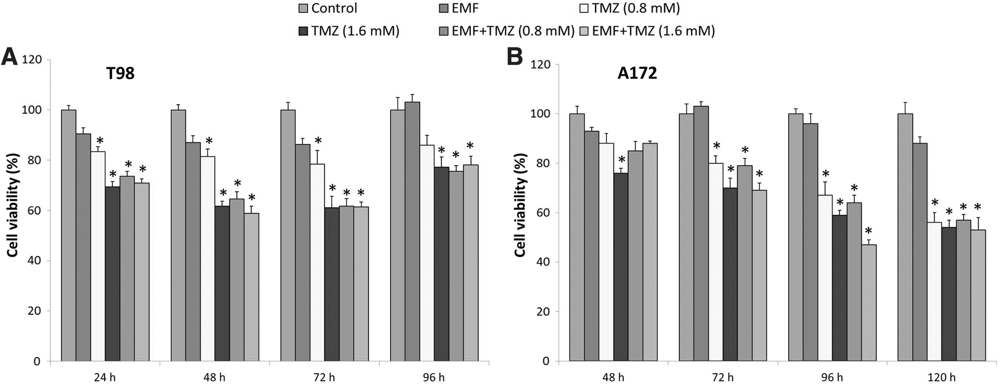
MTT assay of T98
The results of trypan blue staining show that coexposure to TMZ and EMF significantly reduced cell viability and increased the number of dead cells for both cell lines (Fig. 2A, B). Furthermore, the morphological changes, including apoptotic cells (bleb formation) (Fig. 5B) and rounded soma (Fig. 2C), were observed following TMZ and TMZ+EMF treatment. It seems that the T98 cells were slightly more sensitive to TMZ and coexposure to TMZ+EMF in comparison to A172 cells.
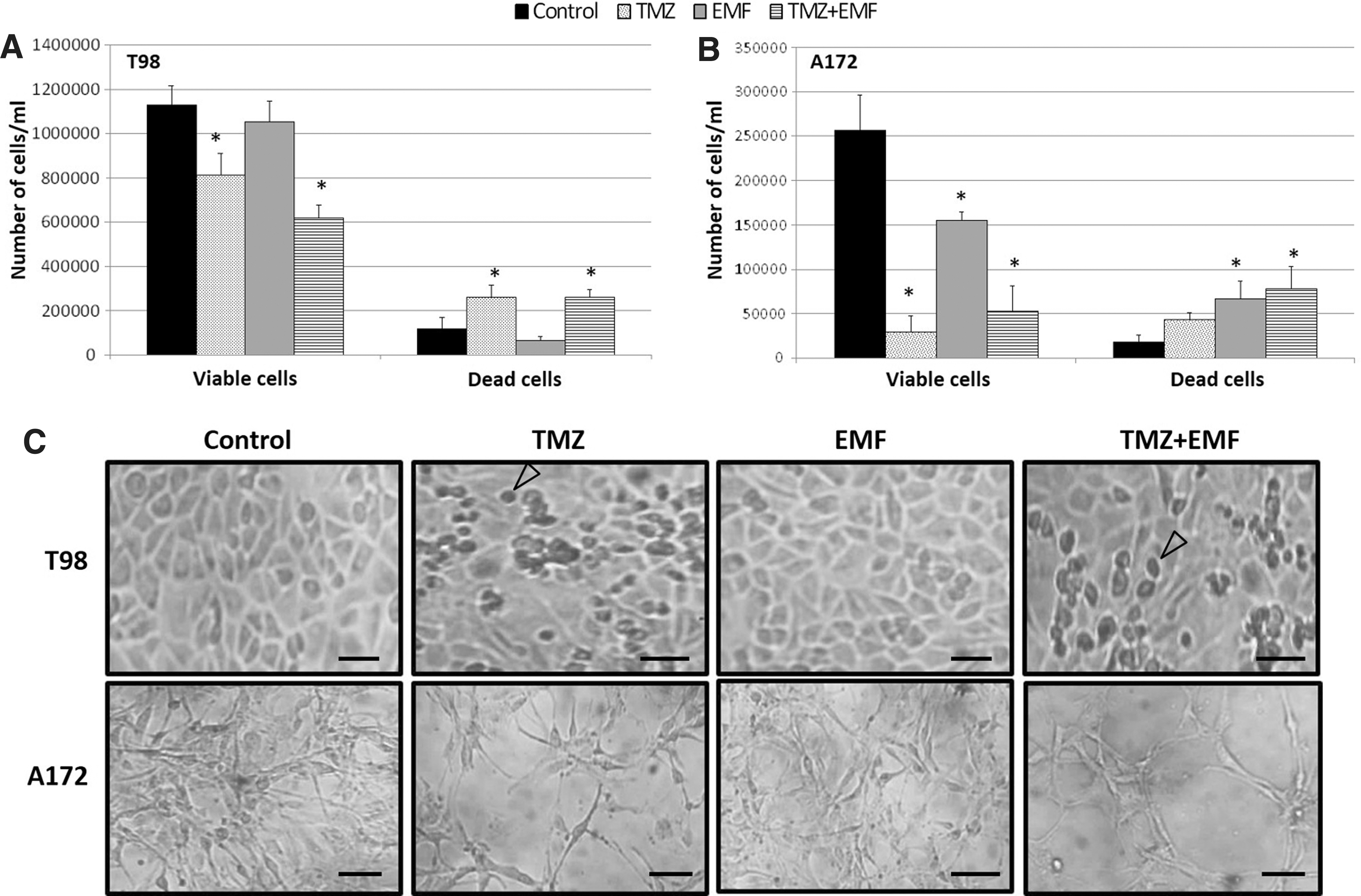
Trypan blue assay. Number of viable and dead cells/mL for T98
Scratch assay and cell migration
Their findings revealed that more than 90% of the distance between the scratch borders was repaired at 48 and 72 h in control groups of T98 and A172 cells, respectively (Fig. 3). Scratch renovation in TMZ and TMZ+EMF groups was 15%–20% less than the control group for both cell lines at 24 h after the treatment. Moreover, the scratch margins in all groups of T98 cells were joined at 96 h. However, scratch renovation in TMZ and TMZ+EMF groups was considerably less than the control group at 96 h for A172 cells.
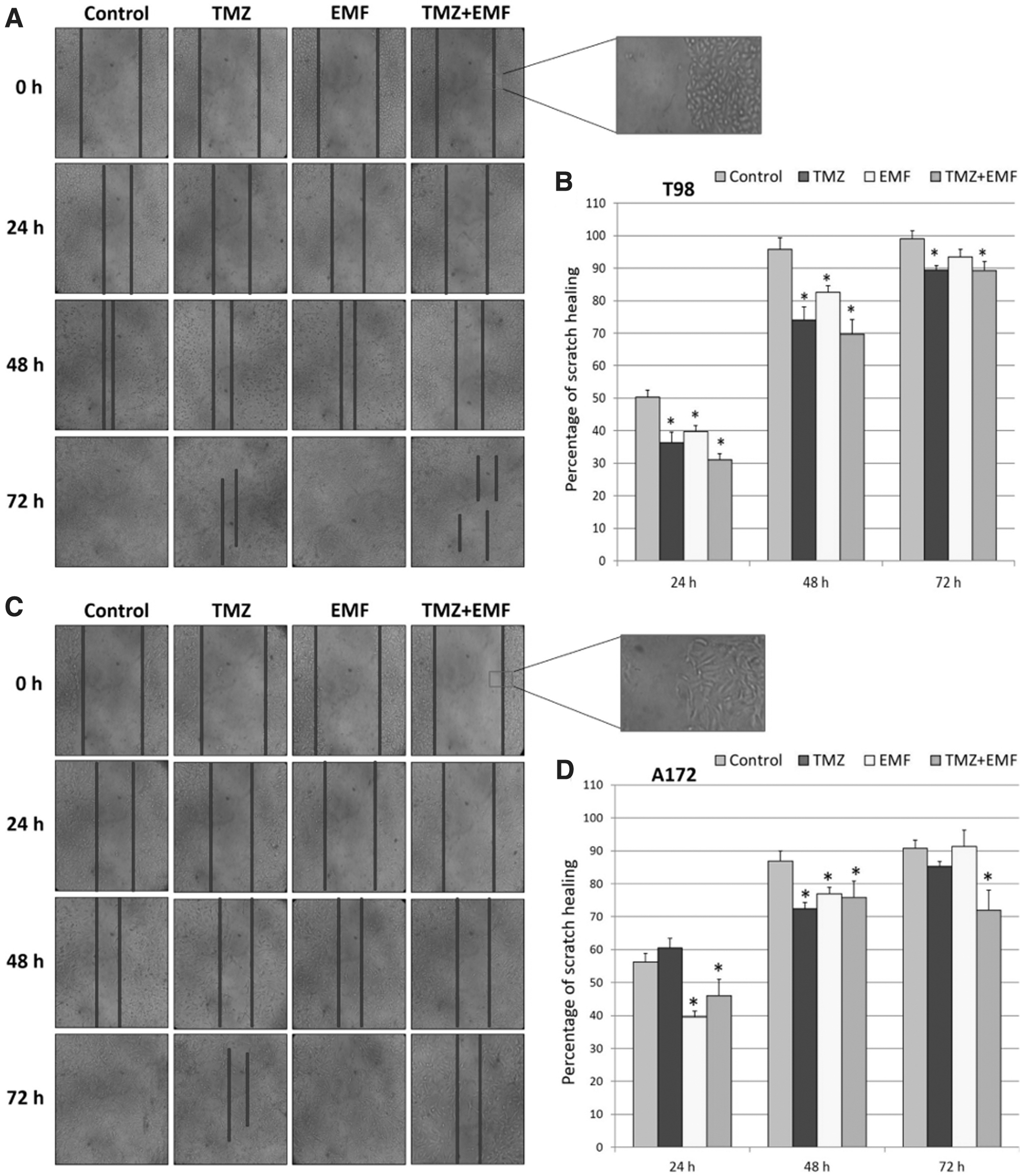
Evaluation of scratch healing in different time points. The percentage of scratch renovation was significantly decreased at different time points in comparison to control group for T98
MGMT and cyclin-D1 protein expression
Their present data indicated that the expression of the cyclin-D1 protein in TMZ and TMZ+EMF groups at 48 h for T98 cells was decreased by 20% and 31%, respectively, compared with the control group. However, there was no significant difference in the expression of this protein between groups at 96 h (Fig. 4A, D). About the A172 cells, the cyclin-D1 was significantly downregulated in all treated groups compared to the control group. Furthermore, the highest decrease of cyclin-D1 expression was observed in TMZ+EMF group by about 69% (Fig. 4B, D). In addition, T98 cells express higher MGMT protein following TMZ and TMZ+EMF treatment at 96 h, whereas A172 cells did not express MGMT.
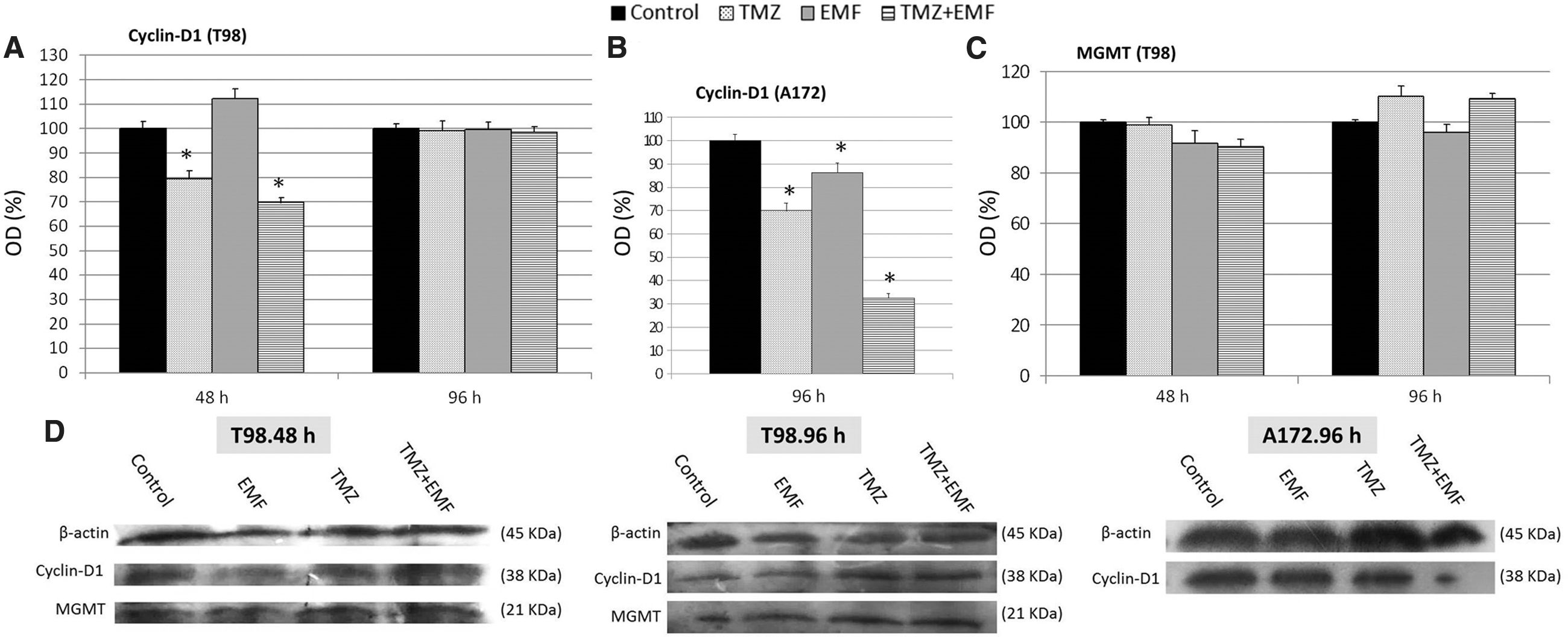
The expression of cyclin-D1 and MGMT proteins (Western blot) in T98 and A172 cells
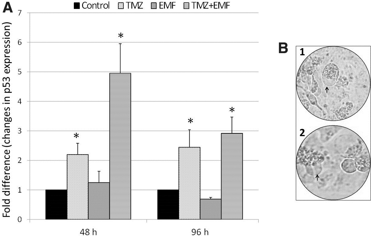
Evaluation of p53 expression by qRT-PCR
Their present data demonstrated that the expression of p53 in the TMZ group at 48 and 96 h was significantly increased compared with the control group (Fig. 5A). In addition, cotreatment of TMZ and EMF resulted in overexpression of the p53 gene at 48 and 96 h after treatment.
Analysis of qRT-PCR.
Discussion
In vitro evaluation of different factors that can affect the expression of DNA repair genes provides useful findings for understanding molecular mechanisms involved in chemotherapy drug resistance. In this study the authors attempted to investigate the effects of EMF (50 Hz, 70 G) on cytotoxicity, cell migration, gene expression, and protein levels in TMZ-treated T98 and A172 cell lines. Their results represent 24 h after initiation of treatment; significant cell death was observed in T98 cells for both concentrations of TMZ. Furthermore, the rate of T98 cell death at 48 and 72 h after treatment was almost the same. However, at 96 h the cells became more TMZ resistant, and overall, cell death decreased compared to 48 and 72 h. Concerning the A172 cell line, initiation of significant cell death was observed at 48 h only in a concentration of 1.6 mM. Overexpression of HO-1 in T98 cells following TMZ treatment has been reported, 16 which is associated with drug resistance in some cancer cells. 27 HO-1 expression may lead to resistance mechanisms for T98 cells after 72 h.
Their data also demonstrate that cotreatment with TMZ and EMF can considerably enhance the cytotoxic effect of TMZ. In addition, about the A172 cells, TMZ decreased cell viability in the dose and time-dependent flow. Increased A172 cell death after TMZ treatment in an exposure time-dependent way has been reported previously by Kanzawa et al. 28 They also found that T98 cells presented TMZ resistance with increasing time, which agrees with their data. The data here reported clarify the influence of TMZ and EMF coexposure on the mRNA level of p53. It has been suggested that p53 has multifunctional roles. Based on the condition of its activation, it could result in differentiation, cellular senescence, inhibition of cell cycle progression, accelerated DNA repair, and apoptosis by upregulating Bax and downregulating Bcl-2 expression. 29,30 Jiang et al. showed that TMZ treatment for 72 h could increase reactive oxygen species (ROS) production and apoptosis in glioblastoma cells. 31 Glioblastoma cells have characteristics such as self-renewal and multilineage differentiation ability and express CD133 (cancer stem-like cell marker) and Nestin (neural stem/progenitor marker) at a high level that makes them a malignant and very aggressive brain tumor. 32 It has been found that cotreatment with TMZ and EMF resulted in a reduction of CD133 and Nestin expression, which shows depletion of stem-like cell pool. In addition, a combination of EMF and TMZ could improve the overexpression of glial fibrillary acidic protein (a marker of differentiated astrocytes). In other words, cotreatment prevents tumor progression not only through the reduction of cell proliferation but also by enhancing glioma cell differentiation. 33,34 In another study, Akbarnejad et al. investigated the effect of TMZ and EMF on glioma cells and described that cotreatment synergistically improved TMZ cytotoxicity by decreasing the expression of Bcl-2 and increasing that of Caspase-3 and Bax. 16
Moreover, their study elucidated the influence of EMF on the migration potential of glioblastoma cells for the first time. The wound healing studies using EMF have emphasized the enhanced cellular activities, including cytokine secretion and cell proliferation, following exposure of fibroblast into the different frequency and intensity. 35,36 Their findings show that EMF exposure alone, TMZ, and TMZ/EMF exposure significantly decreased scratch renovation. These data indicated that depending on the basal metabolic rate, the type of cell that is exposed to EMF is very important in the obtained result. Moreover, different parameters of the EMF, including frequency, amplitude, and exposure time, can trigger different biological responses. Filipovic et al. 37 reported that functional electrical stimulation disrupts microtubule polymerization during cell migration and decreased cell motility. Considering the electric and magnetic components of EMF, this phenomenon could describe the decreased scratch renovation observed under EMF exposure in their study. Gliomas are tumors with high aggressiveness. 38 Concerning glioma migration and invasion, TMZ and TMZ+EMF displayed the effect of reducing cell viability and preventing migration. It has been shown that treatment with a combination of TMZ and EMF enhanced ROS production and apoptosis in T98 and U87 cells. 16 This issue might partly describe the greater inhibition of cellular migration in TMZ+EMF group compared with other groups.
Their data also elucidated that cyclin-D1 protein expression significantly decreased following coexposure to TMZ and EMF. The expression of this protein is essential for the progression of cell cycle G1/S transition. 39 In addition, Neumeister et al. reported that cyclin-D1 could induce cell migration. 40 Therefore, the reduction of cyclin-D1 expression leads to a decrease in the migration potential of glioma cells.
Recently, the effects of EMF exposure on different cells have attracted the attention of cancer therapy field researchers, due to its nonionizing, noninvasive, and nonthermal properties and its remarkable potential to influence cell activity and especially gene expression. 41
Concerning the biostimulatory mechanism of EMF exposure, Park et al. described that EMF exposure (50 Hz) could increase ROS production and enhance neural differentiation of stem cells through phosphorylation of epidermal growth factor receptor. 42 Moreover, it has been reported that Ca2+ ion flux, superoxide dismutase, and Notch signaling were dramatically affected following exposure to EMF. 43 Previous studies have shown that EMF exposure with different frequency and intensity may induce different cellular responses, including changes in cellular morphology through cytoskeletal rearrangements, 44 charge transfer along DNA strands, and separation of base pair. 45 As most of the activity of the neural cells are mediated by charged ions and electric currents and considering the electric and magnetic components of EMF, 15,46 the biomodulation and biostimulation effects of EMF exposure on cells mentioned in literature are presumable. In addition, based on Western blot data, T98 cells express higher MGMT protein following TMZ and TMZ+EMF treatment at 96 h, whereas A172 cells did not express MGMT. It has been shown that over 70% of gliomas express MGMT. 47 Natsume et al. suggested that coexposure to interferon-β and TMZ could downregulate the MGMT expression and sensitize glioblastoma cells to TMZ. 8 Moreover, it demonstrated that levetiracetam (an antiepileptic drug) improves glioblastoma sensitivity to TMZ by p53-mediated MGMT inhibition. 48 Furthermore, Kohsaka et al. reported that STAT3 inactivation resulted in the downregulation of MGMT and could enhance overcoming to glioblastoma TMZ resistance. 9 Considering that inhibition or reduction of MGMT expression is vital for overcoming drug resistance, further studies are needed to clarify the cellular and molecular mechanisms associated with this process.
Conclusion
In summary, for the first time, the effects of EMF and EMF+TMZ of decreasing T98 and A172 glioma cell migration were described. In addition, their data indicate that EMF exposure enhanced the cytotoxicity of TMZ on T98 and A172 cells and could partially affect resistance to TMZ in T98 cells through modulating the expression of MGMT, Cyclin-D1, and p53. However, to achieve a novel clinical approach for treating glioblastoma cells, further investigation is needed.
Authors' Contributions
S.H.E.-V. and M.A.-Z. conceived and designed the experiments, A.B. and L.M.-g. analyzed and interpreted the results of the experiments, and S.D.-S. and P.V. performed the experiments.
Footnotes
Disclosure Statement
No competing financial interests exist.
Funding Information
This study was financially supported by Vice-chancellor for research of Kerman University of Medical Sciences, Kerman, Iran, Grant no. [97000958]. This work was part of the PhD thesis of Samereh Dehghani-Soltani at the anatomical sciences department.