
Abstract
Background:
Colorectal cancer (CRC) has seriously endangered human health. Despite significant advances in clinical treatment of CRC in recent years, clinically effective treatment options for CRC patients remain rare. Therefore, reducing the incidence and mortality of CRC is still a worldwide concern. This study aims to explore the clinical significance of lactamase beta (LACTB)-like expression in CRC tissues.
Materials and Methods:
The expression of LACTB in CRC tissues and adjacent tissues in The Cancer Genome Atlas database was analyzed and the analysis results were verified by immunohistochemistry. The correlation between the expression level of LACTB and pathological factors and prognosis was analyzed.
Results:
There was statistical difference in the expression of LACTB in CRC tissues and adjacent tissues (p < 0.01). The expression of LACTB in CRC tissues was correlated with clinical stage (p < 0.01). The expression of LACTB in CRC patients with lymph node metastasis was significantly lower than that in CRC patients without lymph node metastasis (p < 0.01). Low expression of LACTB contributed to the poor prognosis of CRC patients. The 5-year survival rate of CRC patients with low LACTB expression was significantly lower than that of CRC patients with high LACTB expression (p = 0.010, p = 0.047).
Conclusions:
The expression of LACTB in CRC tissues was significantly lower than that in normal tissues, and it was significantly correlated with clinical prognosis, suggesting that LACTB could inhibit the CRC invasion and metastasis. This indicated to some extent that LACTB could be used as a prognostic marker and a new therapeutic target for CRC.
Introduction
Colorectal cancer (CRC) is one of the most common digestive tract tumors that threatens human health. According to GLOBOCAN 2018 data, there were ∼690,000 CRC-related deaths and ∼2 million new cases in 2018, ranking CRC the third most deadly and fourth most commonly diagnosed cancer in the world. 1 The proportion of morbidity and mortality of CRC in China's malignant tumors continues to rise with the development of China's economy and changes in living habits. 2 Despite significant advances in clinical treatment of CRC in recent years, it remains one of the leading causes of cancer-related death worldwide. Clinically, CRC patients have limited options for effective treatment. At present, the most effective treatment for CRC is surgery, supplemented by radiotherapy, chemotherapy, and targeted therapy. A meta-analysis made by Delbeke et al. 3 found that 70%–80% of patients with CRC could undergo radical resection, but the recurrence and metastasis rate were as high as 30%–40% within 1 year and 80% within 2 years. Therefore, exploring the pathogenesis and finding new treatment of CRC are worthy of attention.
Lactamase beta (LACTB) is an intermembrane protein of mitochondrial derived from penicillin-binding β-lactamase proteins (PBPs), which is mainly located in mitochondrion and cytoplasm of different mammalian tissues. LACTB was previously detected only in bacteria, while now it is proved to be expressed in all vertebrates. 4 Recently, LACTB was found involved in the synthesis of peptidoglycan and formed fibrous mitochondrial membrane in the intermembrane space. 4 PBP in mitochondria can affect the transformation of phosphatidylserine to phosphatidylethanolamine, which directly or indirectly regulates the metabolism of mitochondrial phospholipids. Mitochondrial phospholipid metabolism is closely related to cell proliferation and tumorigenesis, indicating that LACTB expression is closely associated with tumorigenesis. 5,6
Recently, there were increasing evidence that proved that LACTB inhibited tumor proliferation and invasion. 7 –11 Keckesova et al. showed that LACTB could change mitochondrial lipid metabolism and reorganization, and regulated the differentiation of cancer cells. 12 However, there are very limited studies on the clinical significance of LACTB expression in CRC tissues. Therefore, in this study, it was aimed to explore the relationship between the expression level of LACTB and CRC prognosis.
Materials and Methods
Tissue samples
A total of 94 primary CRC and corresponding normal colorectal tissue paraffin-embedded specimens were obtained from the pathology department of East Hospital of Qingdao Municipal Hospital between January 2010 and September 2010. All cases were diagnosed as CRC by 2 pathologists. Histological grading and tumor-node-metastasis (TNM) staging were carried out according to TNM8 (from 2017) and WHO 5th edition (from 2019). General data including name, gender, and age were collected. The patients were followed up by telephone and outpatient service, and their survival time was recorded. Data were censored at last known date of death, and patients lost to follow-up were excluded. The overall survival (OS) was calculated from the operation date to death or the end of follow-up. This study has been approved by the ethics committee of East Hospital of Qingdao Municipal Hospital (Ethics No.: XGLY2020-03-28) and conformed to the declaration of Helsinki.
Immunohistochemical staining
Paraffin tissues were sliced cut into 4-μm sections, dewaxed, and hydrated. Triton X-100 was used to permeate after phosphate-buffered saline (PBS) wash, followed by boiling in citrate buffer (pH 6.0) for antigen retrieval. After inhibition of endogenous peroxidase activities for 30 min with 0.3% H2O2, the sections were blocked with 5% goat serum albumin (PBS dilution) for 10 min at room temperature and incubated overnight at 4°C with rabbit anti-LACTB antibody (1:250, Cat. No. #18195-1-AP; Proteintech Group, Wuhan, China). After rewarming at 37°C for 45 min, and washing five times with PBS, the slides were incubated with secondary antibody at 37°C for 30 min, followed by reaction with DAB Developer (DAB kit; MXB Biotechnologies, Fuzhou, China) and dyeing with peroxidase labeling. Then the slides were rinsed by tap water thoroughly to stop dyeing, restained, dehydrated, transparency, and sealing. The negative control consisted of PBS rather than the primary antibody.
Each slide was evaluated by two pathologists without prior knowledge of the clinical information of the patients. Degree of staining intensity and positive tumor cells were analyzed under 10 high-power fields. The proportion of tumor cells was scored as follows: 0 (<10% positive tumor cells), 1 (10%–25% positive tumor cells), 2 (26%-50% positive tumor cells), and 3 (>50% positive tumor cells). The intensity of staining was graded according to the following criteria: 0 (no staining), 1 (light yellow), 2 (yellow), and 3 (yellow brown). The staining index was calculated as the product of the proportion of positive cells and the staining intensity score, 0–3 (low LACTB expression) and 4–9 (high LACTB expression).
Statistical analysis
All statistical analyses were performed using the SPSS 20.0 statistical software package (SPSS, Inc., Chicago, USA). Measurement data conforming to normal distribution and homogeneous variance are expressed as mean ± standard deviation and analyzed using t-test. Counting data are expressed as constituent ratio (%) or rate (%) and analyzed using χ 2 test or Fisher definite probability method. The log-rank test was used for univariate survival analyses. The Cox proportional hazards regression model was used to determine the hazard ratio and identify factors that independently predict survival. The overall survival rate was calculated using Kaplan-Mier analysis. p < 0.05 was considered as a statistically significant difference.
Results
General data of CRC patients
A total of 94 CRC patients' specimens were enrolled in this study, which were obtained from the pathology department of East Hospital of Qingdao Municipal Hospital on January 2010, solstice on September 2010. Demographics and clinical characteristics of these 94 CRC patients are summarized in Tables 1 and 2. In particular, among the 94 CRC patients, the median age was 64 years old, 57 males (60.6%) and 37 females (39.4%). And 72 tissue samples (74.2%) located in rectum and sigmoid, 11 tissue samples (11.3%) located in left colon, 7 tissue samples (7.2%) located in right colon, and 4 tissue samples (4.1%) located in transverse colon. Postoperative pathology showed that there were 80 cases (82.5%) of tubular adenocarcinoma, 13 cases (13.4%) of mucinous adenocarcinoma, and 1 case (1.0%) of signet ring cell carcinoma. All 94 CRC patients were staged according to TNM8, specifically, there were 11 stage I patients (11.3%), 39 stage II patients (40.2%), 34 stage III patients (35.1%), and 10 stage IV patients (10.3%). In terms of the degree of tumor differentiation, 14 cases with low differentiation accounted for 14.4%, 69 cases with moderate differentiation accounted for 71.1%, and 11 cases with high differentiation accounted for 11.3%.
Association Between β-Lactamase Expression and Other Clinicopathological Features in Colorectal Cancer
Independent sample t-test.
One-way ANOVA.
LACTB, lactamase beta; TNM, tumor-node-metastasis.
Univariate Analysis of Prognostic Factors for Overall Survival of Colorectal Cancer Patients
Correlation between LACTB expression and clinicopathological features of CRC patients
Analysis result of The Cancer Genome Atlas (TCGA) data showed significantly lower mRNA expression of LACTB in CRC tumor nodules than in para-tumoral tissues (Fig. 1), and the analysis result was examined by immunohistochemistry (IHC) (Fig. 2). The staining index analysis result revealed that LACTB expression in cancer tissues was significantly lower than that in normal tissues (2.88 ± 1.47 vs. 6.20 ± 1.82, p < 0.0001). The expression of LACTB was significantly decreased in patients with higher stage and lymphatic metastasis (Fig. 3 and Table 1). No significant correlation was observed among LACTB expression, tumor location, tumor histological types, and tumor differentiation (Table 1).

Analysis result of CRC TCGA data of LACTB mRNA expression LACTB mRNA expression in CRC tissue and paratumoral tissue among the CRC patients from TCGA, p = 0.00016. Data are representative of three independent experiments, and were analyzed by unpaired t-test. Error bars denote SD. ***p < 0.001. CRC, colorectal cancer; LACTB, lactamase beta; SD, standard deviation; TCGA. The Cancer Genome Atlas.
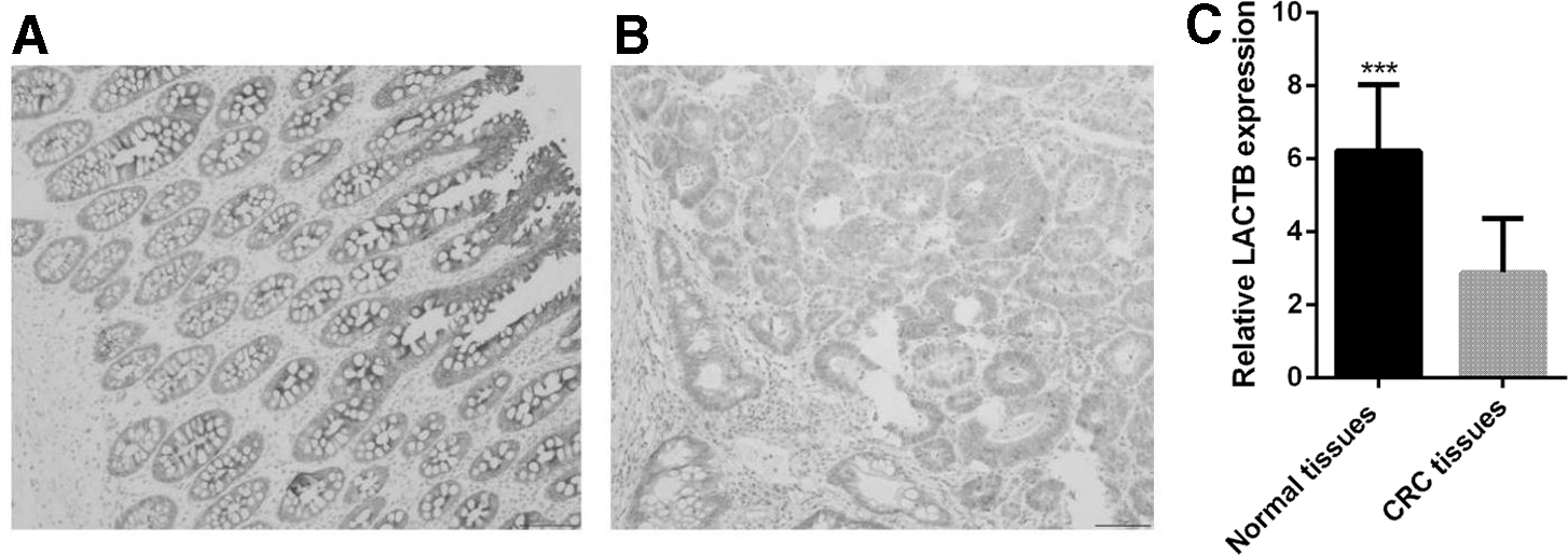
IHC of LACTB expression in CRC tissue.
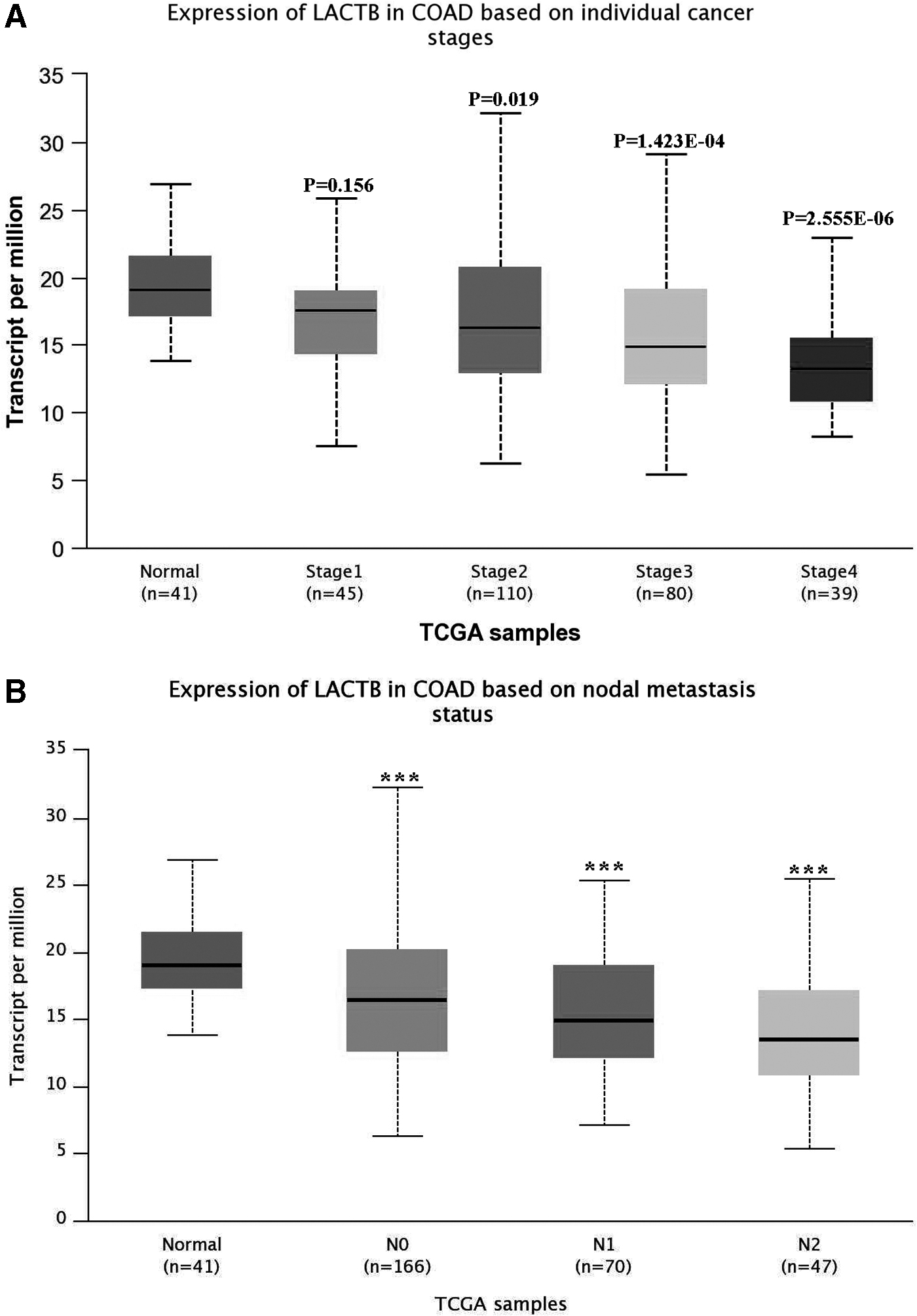
Expression of LACTB in CRC based on metastasis status and nodal metastasis status.
Correlation between LACTB expression and prognosis in CRC patients
Univariate analyses of prognostic factors for OS of these 94 CRC patients are summarized in Table 2. Specifically, univariate analysis showed that the clinicopathological features associated with prognosis of CRC patients were tumor stage, lymphatic metastasis, degree of differentiation, and LACTB expression. There was no significant correlation with tumor histological type and location of tumors (Table 2). Statistically significant factors (tumor stage, lymphatic metastasis, tumor differentiation, and LACTB expression) in univariate analysis were included in Cox proportional hazards regression model for multivariate analysis. The results showed that higher tumor stage (p = 0.005) and lower LACTB expression (p = 0.012) were significantly associated with poor prognosis in CRC patients. Tumor histological type and location of tumors could not predict prognosis of CRC patients (Table 3). In addition, the follow-up durations ranged between 12 and 60 months, the median follow-up time was 45 months, and the time of end of the follow-up was September, 2015. During the follow-up, 44 cases survived, 20 cases were lost, and 30 cases died.
Cox Regression Analysis Between β-Lactamase Expression and Clinicopathological Features in Colorectal Cancer
According to the staining intensity of LACTB in CRC tumor nodules, patients were divided into two groups: high LACTB expression group and low LACTB expression group. As shown in Figure 4A, the 5-year OS of CRC patients with high LACTB expression was significantly better than that in CRC patients with low LACTB expression (p = 0.010), which was consistent with the result of TCGA database analysis (Fig. 4B).

Overall survival of follow-up patients' data and TCGA CRC data.
Discussion
CRC is a common invasive malignant tumor, and there is an urgent need for new specific diagnostic molecular markers and therapeutic targets. 13 Twenty-seven dysregulated LACTB is often closely related to obesity and atherosclerosis, suggesting that LACTB may be involved in metabolism disorders. 14 Berkers et al. 15 reported that the deletion of p53 gene in tumors could enhance glycolysis and biosynthesis, which was beneficial to tumor growth, suggesting a correlation between tumor growth and energy metabolism. Decreased expression of LACTB can be observed in different tumors such as glioma, 16 breast cancer, 13,17 and hepatocellular carcinoma. 11 These studies suggest that low expression of LACTB can promote cell transformation, and LACTB has the characteristics of inhibiting tumors. Consistent with previous studies, it was found that LACTB has lower expression at both mRNA and protein levels. Through TCGA data set analysis, it was found that compared with nontumor tissues, the expression level of LACTB was reduced in CRC tissues, which was confirmed by IHC assay. These results indicate that the expression of LACTB is often reduced in CRC tissues.
Importantly, univariate analysis showed that LACTB downregulation and late TNM stage were independent factors for poor CRC prognosis, suggesting that low LACTB expression is related to CRC poor prognosis. Li et al. reported that in glioma patients, the expression of LACTB was significantly reduced and this decreased expression of LACTB was associated with poor prognosis. 13 Zhang et al. confirmed that the downregulation of LACTB expression can be used as an independent prognostic factor for predicting poor breast cancer patients survival. 9 These results suggested that the downregulated LACTB could be used as a prognostic marker for CRC.
Moreover, it was found that LACTB expression decreased significantly in CRC patients with lymphatic metastasis. The expression of LACTB was not significantly correlated with histological types of tumor tissue. The prognosis of CRC patients was significantly correlated with lymphatic metastasis and tumor differentiation. In addition, the clinicopathological analysis suggested that the decreased expression of LACTB was significantly associated with the TNM stage, histological grade, and OS of CRC patients. Consistent with the results, in breast cancer 9 and glioma, 13 low expression of LACTB was markedly correlated with poor OS. In summary, these results indicated that downregulation of LACTB is involved in tumor progression, and LACTB can be used as a therapeutic target for CRC patients.
In conclusion, downregulation of LACTB expression is significantly associated with poor CRC patient prognosis, suggesting that LACTB may have potential tumor suppressive feature and can be used as a therapeutic target for the treatment of CRC. There are still some limitations in this study: LACTB as a prognostic marker for CRC has not been verified by prospective experiments, whether LACTB can be used as a screening marker for CRC is not confirmed, and the specific regulation of molecular mechanism of LACTB on cancer cells is still not clear.
Compliance with Ethical Standard
All procedures performed in studies involving human participants were in accordance with the ethical standards of the institutional and/or national research committee and with the 1964 Helsinki declaration and its later amendments or comparable ethical standards.
Footnotes
Disclosure Statement
No competing financial interests exist.
Funding Information
No funding was received for this article.