
Abstract
Background:
The aim of the study was to perform PET imaging and radiotherapy with a novel neurotensin derivative for neurotensin receptor 1 (NTSR-1)-positive tumors in an animal model.
Materials and Methods:
A di-DOTA analog of NT(6–13) with three unnatural amino acids was synthesized and radiolabeled with either 64Cu or 68Ga and tested for serum stability and tumor imaging in mice bearing NTSR-1-positive PC3, and HT29 xenografts. A dose–response therapy study was performed with 18.5, 37, and 74 kBq of 225Ac-di-DOTA-α,ɛ−Lys-NT(6–13).
Results:
68Ga-di-DOTA-α,ɛ−Lys-NT(6–13) was >99% stable in serum for 48 h, had an IC50 of 5 nM using 125I labeled NT(8–13) for binding to HT-29 cells, and high uptake in tumor models expressing NTSR-1. 68Ga-di-DOTA-α,ɛ−Lys-NT(6–13) had an average %ID/g (n = 4) at 2 h of 4.0 for tumor, 0.5 for blood, 12.0 for kidney, and <1 for other tissues, resulting in a favorable T/B of 8. Mean survivals of tumor-bearing mice treated with 18.5 or 37 kBq of 225Ac-di-DOTA-α,ɛ−Lys-NT(6–13) were 81 and 93 d, respectively, versus 53 d for controls. Whole-body toxicity was seen for the 74 kBq dose.
Conclusions:
Based on the results of the animal model, di-DOTA-α,ɛ−Lys-NT(6–13) is a useful imaging agent for NTSR-1-positive tumors when radiolabeled with 68Ga, and when radiolabeled with 225Ac, a potent therapeutic agent.
Introduction
There is currently a strong interest in theranostic agents that can be used for both imaging and therapy. 1,2 As an example, somatostatin analogs that bind to somatostatin receptors (SSTRs) conjugated to promiscuous metal chelators, such as DOTA, can be radiolabeled with positron emitters, such as 68Ga for PET imaging, 3,4 and β−-emitters, such as 177Lu for therapy. 5 Although peptides have relatively short in vivo half-lives due to their rapid degradation by serum proteases and rapid excretion by the kidney, 6,7 their high uptake by tumor-specific peptide receptors has led to impressive results in both imaging and therapy studies. 8 –10 In the case of neuroendocrine tumors, 177Lu-DOTATE, a small-molecule analog for SSTRs radiolabeled with 177Lu, has shown impressive therapeutic responses. 5 This success has led to an interest in bringing other small peptides or their analogs to the clinic as theranostic agents. Among them is neurotensin, a 13-amino acid peptide that binds to neurotensin receptors (NTSRs), including NTSR-1 (high affinity), NTSR-2 (low affinity), and NTSR-3 (also known as Sortilin-1; high affinity). Among these, NTSR-1 is highly expressed in tumors of the prostate, 11 colon, 12 pancreas, 13 breast, 14 lung, 15 and head and neck 16 cancers.
A major challenge in developing NT theranostic agents is that natural NT(1–13) has a serum half-life of only a few minutes, 17 preventing its systemic delivery to tumors. Although the truncated version of NT, NT(8–13), retains full binding to its receptors, it also is rapidly degraded in serum. 18 Two approaches have been taken to improve serum stability: incorporation of unnatural amino acids into the structure, 19 –22 and development of small-molecule NTSR-binding drugs. 23,24 Since NTSR-1 binds NT at its C-terminus, derivatives at the N-terminus have had minor effects on the binding of peptide analogs. 25,26 Thus, N-terminal derivatives incorporating chelates that bind radiometals have led to a variety of analogs studied for their promise as imaging agents. For example, 68Ga-DOTA-NT derivatives were evaluated in animal models of colon 27 and pancreatic 28 cancer and a 64Cu-DOTA-NT derivative in a prostate model using PC3 cells. 29 Recently, they demonstrated that a vinylsulfone analog of Cys-NT(6–13) when labeled with 18 F, gave tumor uptake of 0.8%ID/g and a tumor to blood ratio of 6 at 2 h, with excellent tumor imaging in an HT-29 model of colon cancer. 30
Small-molecule NTSR-binding drugs have also shown promise for both imaging and therapy of NTSR-positive tumors. Schulz et al. have shown that the NTSR binder 3BP-227 conjugated to DOTA-imaged HT-29 tumors when radiolabeled with 111In 23 and gave about 50% tumor reduction when radiolabeled with 177Lu. 31 The β−-emitter radionuclide 177Lu conjugated to 3BP-227 is currently being investigated in an ongoing clinical study with promising results. 27,32 A 177Lu-DOTA NT analog was evaluated by Jia et al., 20 but was not used in a therapeutic study. In this study, we evaluated a 225Ac-DOTA analog for therapy given the higher linear energy transfer of α-compared with β−-emitter radionuclides. In fact, the α-emitter 225Ac has been shown to have higher therapeutic efficacy and lower toxicity even in the treatment of solid tumors compared with β−-emitter radionuclides. 33,34
Materials and Methods
Cells
HT-29 and PC3 cells were obtained from ATCC (Rockville, MD). PC3 cells were transfected with NTSR-1 as previously described. 29
Synthesis of NT analogs
See Supplementary Data.
Radiolabeling
64CuCl2 was obtained from Washington University, St. Louis, Mo. with a specific activity of 6260 MBq/μg. 68GaCl3 was eluted with 5 mL of 0.1 N HCl from a 1860 MBqGalliaPharm 68Ge/68Ga generator (Eckert & Ziegler, Berlin, Germany). 225Ac obtained from Oakridge National Laboratory was at secular equilibrium. The authors performed spectral analysis and counted the gamma at 440 keV (26%) arising from the daughter 213Bi (45 min half-life). Di-DOTA-α,ɛ−Lys-NT(6–13) was radiolabeled with 64CuCl2 (20 μg with 45 MBq in 0.1 mL in 0.25 M ammonium acetate, pH 5.5 at 43°C for 30 min), or with 68GaCl3 (40 μg with 148 MBq in 0.9 mL of 1 M ammonium acetate, pH 5.5 at 95°C for 15 min), or with 225Ac (20 μg with 45 kBq in 0.1 mL of 0.25 M ammonium acetate, pH 5.5 at 43°C for 30 min). Radiolabeled peptides were chased with 10 mM DTPA at RT for 10 min followed by ITLC to determine radiochemical purity. Radiochemical purity for 68Ga and 64Cu radiolabeled peptides was between 95% and 99% and for 225Ac 87%. The 225Ac-labeled samples were not further purified.
Absorbed dose calculations
See Supplementary Data.
Cell-binding studies
Cell-binding studies were performed as previously described. 21,29 Briefly, HT-29 cells were incubated with 125I-NT(8–13) (PerkinElmer, Waltham, MA) in 0.2 mL of binding buffer plus inhibitors [NT(8–13) or di-DOTA-α,ɛ−Lys-NT(6–13) or GaCl3+di-Di-DOTA-α,ɛ−Lys-NT(6–13)] for 1 h at 37°C, washed 3 times with 0.2 mL of ice-cold phosphate-buffered saline (PBS), taken up in 0.2 mL of 1 m NaOH and counted in a Gamma Counter. Results were plotted as percent bound versus concentration of inhibitor.
Stability studies
68Ga-di-DOTA-α,ɛ−Lys-NT(6–13) was diluted into 1 mL of normal mouse serum, incubated at 37°C and 100 μL analyzed by SEC (Superdex 75) over time. The percent counts in the peak at 42 min were recorded versus time.
Animal studies
All animal studies were performed in male NCI athymic NCr-nu/nu mice (nude; Charles River) in accordance with IACUC protocols 91037 approved by the City of Hope Institutional Animal Care and Use Committee, in accordance with the NIH Office of Laboratory Animal Welfare guidelines, in an AALAC-approved facility. HT-29 or PC3.NTSR1 cells were injected s.c. (2 × 10⁶ in 0.2 mL of PBS per mouse) and tumor size followed with caliper measurements [TS = 0.5 (L × W 2 )]. Tumors were allowed to reach 200 mm3 for start of imaging studies and between 50 and 100 mm3 for therapy studies.
PET imaging and biodistribution studies
Mice bearing xenografts were anesthetized with 2%–4% isoflurane in oxygen, tail vein injected with 1.85–3.7 MBq (1–2 μg) of 68Ga- or 64Cu-di-DOTA-α,ɛ−Lys-NT(6–13) or DOTA-ɛ−Lys-Lys-NT(10–13) and imaged on a Inveon microPET/CT scanner (Siemens Medical Solutions, Germany). At the terminal time point of imaging, the mice were euthanized, tissues weighed and counted, and the results expressed as %ID/g. Blocking studies included 10 μg of unlabeled di-DOTA-α,ɛ−Lys-NT(6–13).
Therapy studies
Mice bearing xenografts were treated with 18.5, 37 or 74 kBq of 225Ac-di-DOTA-α,ɛ−Lys-NT(6–13). All therapy doses were made up to 2.0 μg NT with the addition of cold NT. Whole-body toxicity was measured by monitoring weight loss of the mouse. The endpoint of mice was determined as the time the mice lost >20% of their initial body weight or when tumor size reach 1500 mm3.
Statistical analysis
One-way analysis of variance (Tukey's multiple comparison test) was used to analyze tumor growth curves, and log-rank Mantel–Cox test for survival curves.
Results
Synthesis of di-DOTA-α,ɛ−Lys-NT(6–13) and DOTA-ɛ−Lys-Lys-NT(10–13)
Based on an NT(6–13) analog with three unnatural amino acids (pip-Gly, pip-AmGly and tBuGly), we synthesized an N-terminal di-DOTA derivative (Fig. 1). The strategy for DOTA conjugation at both the α-amino and ɛ-amino groups of an N-terminal lysine was to prevent amino-peptidases from digesting the peptide, a major problem with many NT analogs. 35

Structures of di-DOTA-α,ɛLys-NT(6–13) and DOTA-ɛ−Lys-Lys-NT(10–13).
We also prepared another NT analog, DOTA-ɛ-Lys-Lys-NT(10-13) (Fig. 1), reported by Maschauer et al. 27 The Maschauer analog was interesting in that it substituted two N-terminal Lys residues for the usual two N-terminal Arg residues of natural NT(6–13), yet their imaging results were excellent at 45-min, comparable to those reported by Alshoukr et al. 36 who performed 68Ga imaging with a different DOTA-NT(6–13) analog that incorporated two unnatural amino acids in the structure.
Radiolabeling and stability studies
Di-DOTA-α,ɛ−Lys-NT(6–13) was >95% radiolabeled with 68Ga and 64Cu, and 87% with 225Ac at ratios of 37 MBq/20 μg, 74 MBq/20 μg, and 37 kBq/20 μg, respectively (Supplementary Fig. S1). When 68Ga-radiolabeled di-DOTA-α,ɛ−Lys-NT(6–13) was incubated in normal serum at 37°C, it gave a single radiolabeled peak on SEC (Supplementary Fig. S2) with no evidence of degradation over 2 h.
Cell-binding studies
The binding of di-DOTA-α,ɛ−Lys-NT(6–13) to the NTSR-1-positive cell line HT-29 was used to evaluate NT binding. 21 Compared with all natural NT(8–13) that had an IC50 of 0.8 nM, the IC50 for di-DOTA-α,ɛ−Lys-NT(6–13) analog was 5.0 nM (Fig. 2). The addition of a molar amount of cold Ga3+ had no effect on the binding (Fig. 2).
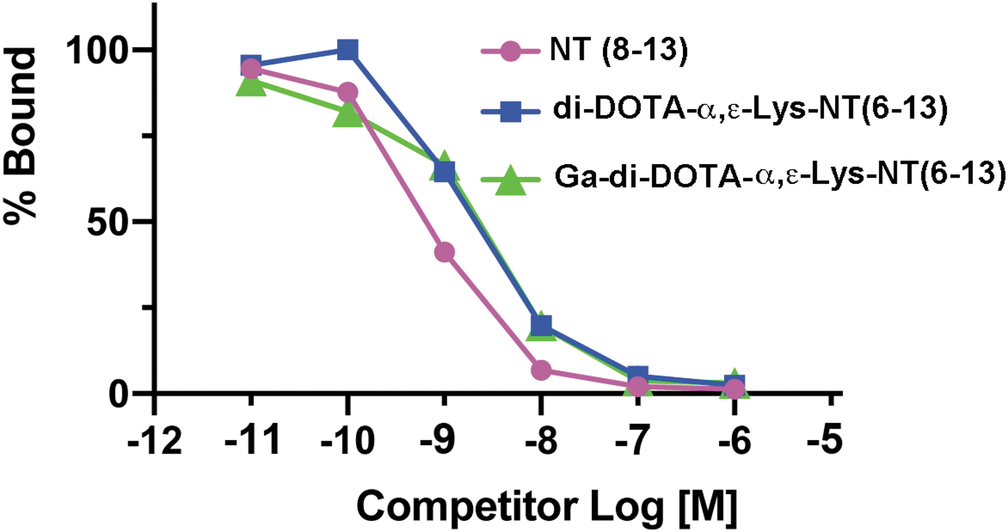
Cell-binding study of NT(8–13), di-DOTA-α,ɛ−Lys-NT(6–13) and Ga-di-DOTA-α,ɛ−Lys-NT(6–13). Competitors were tested against binding of 125I-NT(8–13) to HT-29 cells as described in methods, plotted as percent bound versus molar concentration of competitor.
PET imaging studies with 68Ga-di-DOTA-α,ɛ-Lys-NT(6–13) and 68Ga-DOTA-ɛ-Lys-Lys-NT(10–13)
The NT analogs were radiolabeled at a ratio of 3.7 MBq/μg with >95% radiolabeling (Supplementary Fig. S1). Animals bearing s.c. PC3.NTSR1 xenografts, a prostate cancer model previously described by the authors,
29
gave excellent images of the tumor at both 1 and 2 h postinjection (Fig. 3A

PET images of nude mice bearing PC3.NTSR1 xenografts with 68Ga-di-DOTA-α,ɛ−Lys-NT(6–13).
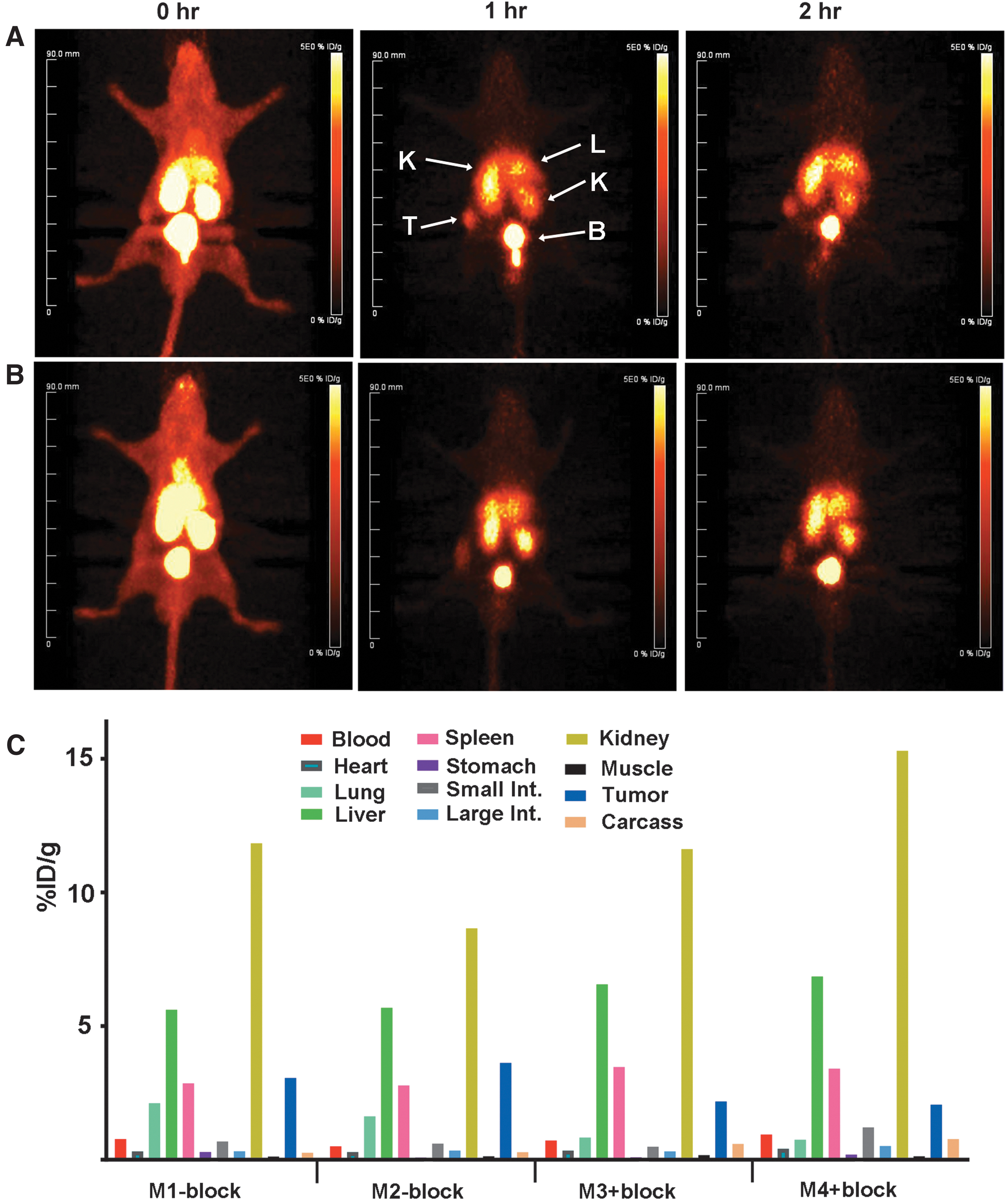
PET images of nude mice bearing PC3.NTSR1 xenografts with 68Ga-di-DOTA-α,ɛ−Lys-NT(6–13) ± blocking peptide.
A follow-up imaging study with 68Ga-di-DOTA-α,ɛ-Lys-NT(6–13) was performed in HT-29 xenografts (Fig. 5A
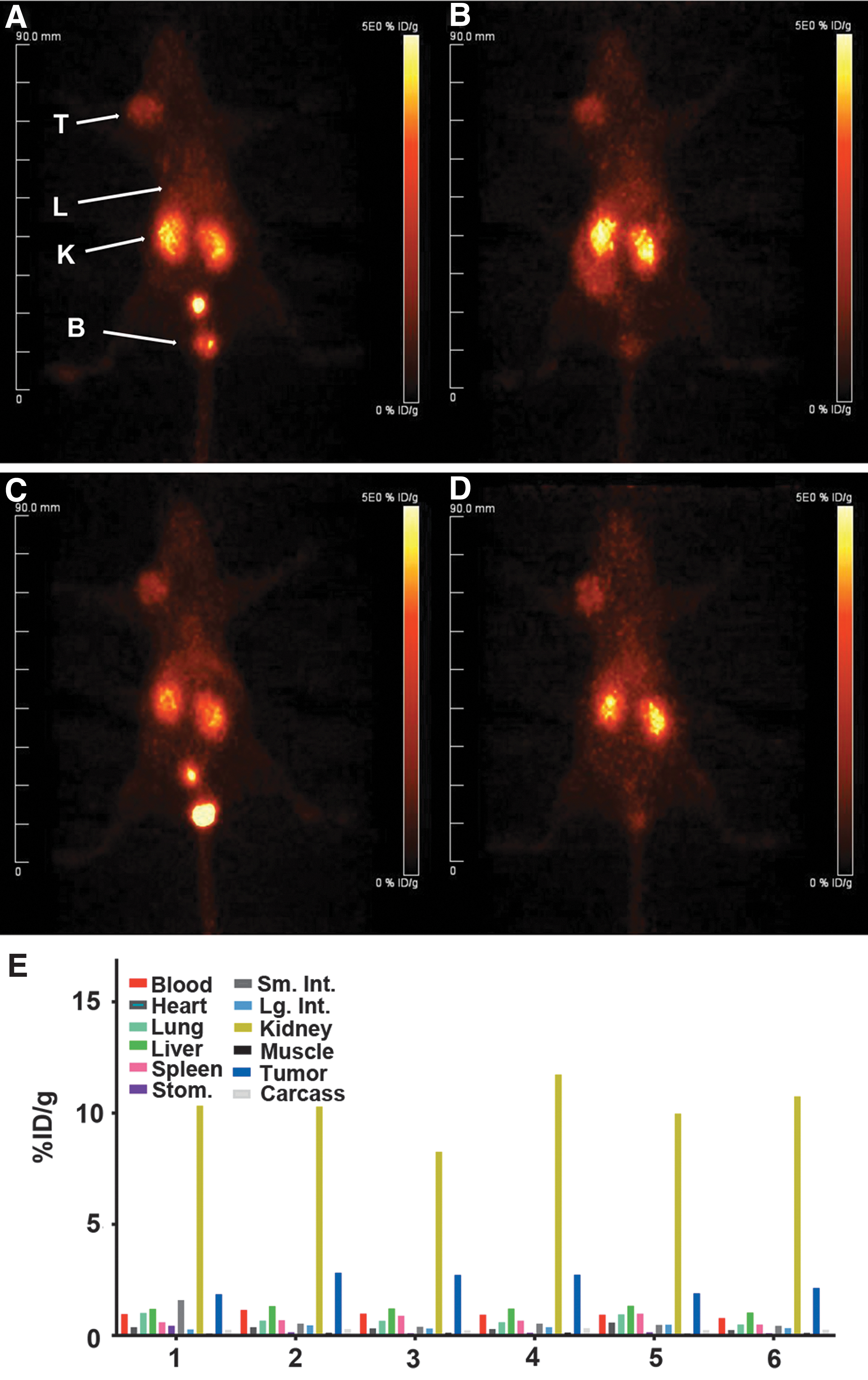
PET images of nude mice bearing HT-29 xenografts with 68Ga-di-DOTA-α,ɛ−Lys-NT(6–13).
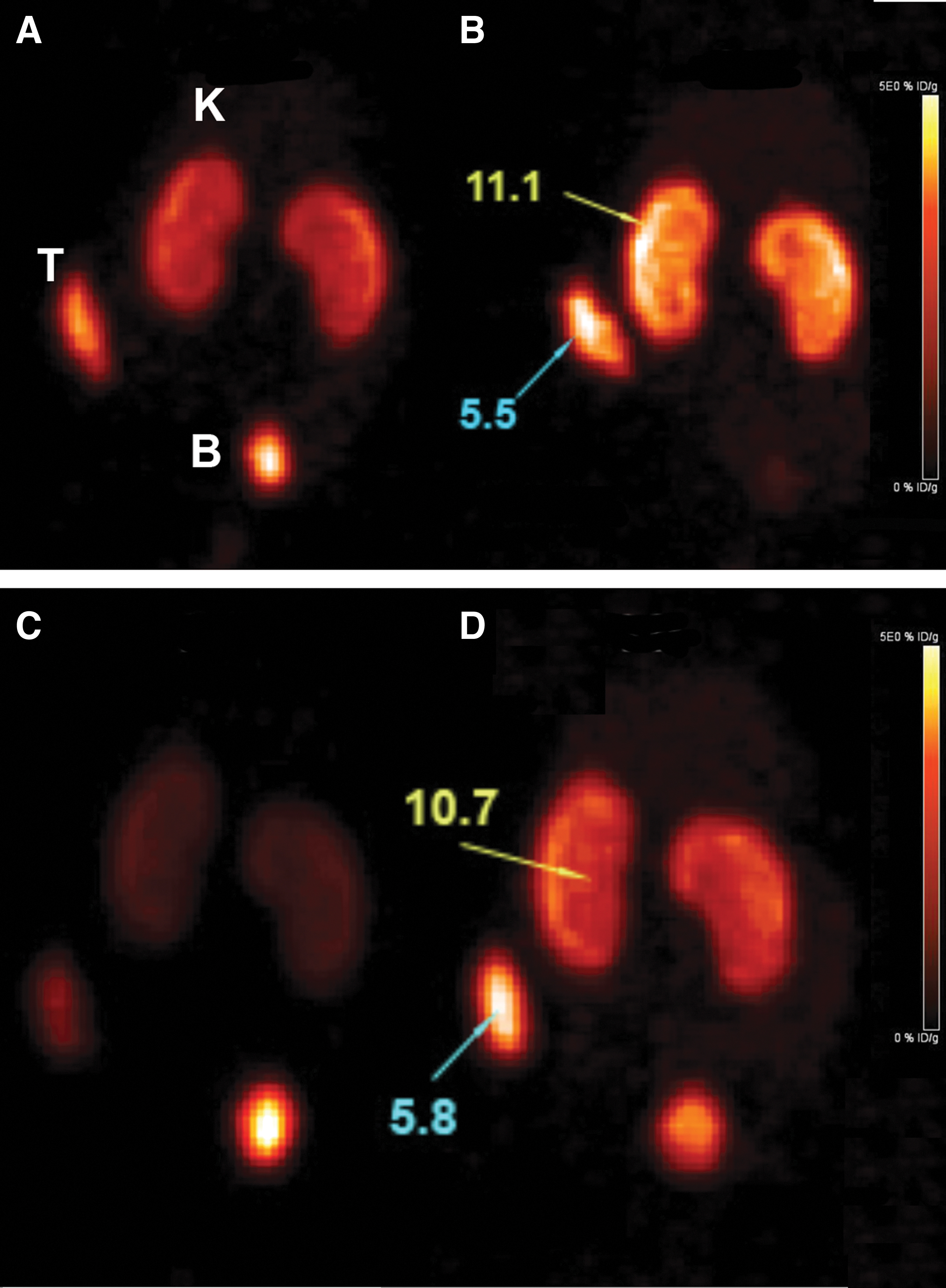
PC3.NTSR1 tumors were also imaged with 64Cu-di-DOTA-α,ɛ-Lys-NT(6–13) at 2 and 4 h (Fig. 6). When using 64Cu instead of 68Ga, the highest tumor uptakes were recorded at 4 h, 5.5–5.8%ID/g. Time activity studies were performed in HT-29 xenografts (Supplementary Fig. S4). A maximum tumor uptake of 3.0%ID/g was observed at 10 min, decreasing to 2.8%ID/g from 15 to 30 min, thereafter exhibiting a linear decrease to 1.2% ID/g at the terminal time point of 140 min. There was a two-phase clearance profile with the t1/2 of the α phase estimated as 5 min and the t1/2 of the β phase as 30 min. Favorable tumor-to-blood ratios were observed from 10 min on, increasing to 4.7 at 30 min and dropping to 4.0 at 60 min. The kidneys had a clearance t1/2 of 10 min. Assuming a similar biodistribution in man, this study suggests that clinical imaging could be performed as early as 30–60 min, preferably with 68Ga rather than 64Cu to reduce radiation exposure to the kidney.
PET images of nude mice bearing PC3.NTSR1 xenografts with 64Cu-di-DOTA-α,ɛ−Lys-NT(6–13).
225Ac-di-DOTA-α,ɛ-Lys-NT(6–13) therapy study
The 225Ac -di-DOTA-α,ɛ-Lys-NT(6–13) antitumor effect was investigated in a PC3.NTSR1 prostate xenograft model with a dose escalation study. There was a significant dose-dependent effect in the tumor growth curves for both the 18.5 and 37 kBq treatment groups, with the 74 kBq treatment group causing complete tumor regression (Fig. 7A). Fifty percent of the mice in the 18.5 kBq treatment group and 100% of the mice in the 37 kBq treatment group maintained reduced tumor growth past day 40 (Supplementary Fig. S5).

Dose escalation of 225Ac-di-DOTA-α,ε−Lys-NT(6–13) therapy in nude mice bearing PC3.NTSR1 xenografts.
Survival among the treatment groups showed a dose-dependent effect for the 18.5 and 37 kBq treatment groups (Fig. 7B). The 18.5 kBq-treated group increased mean survival by 50%, from 52.8 d in the control group to 81.3 d in the treatment group (Table 1). The 37 kBq treatment group almost doubled mean survival to 93.0 d (Table 1). Although the 74 kBq treatment group had a significant antitumor effect, the mean survival was only 27.5 d due to whole-body toxicity, observed as rapid weight loss post-therapy (Table 1 and Fig. 7C). A dose-dependent whole-body toxicity effect was observed, with no significant weight loss seen in the 18.5 kBq-treated group, while the higher doses had increased toxicity (Fig. 7C). Twenty-five percent of mice in the 18.5 kBq-treated group, 67% mice in the 37 kBq-treated group, and 100% mice in the 74 kBq-treated group were euthanized when weight loss reached 20% of their starting weight (Fig. 7C). In addition, mice in the 74 kBq group had a mild-to-severe skin rash (Supplementary Fig. S6). Mice in the 74 kBq treatment group also exhibited a dose-dependent weight loss that did not recover over time. This effect may be due to the effects of peripheral NT on appetite. 37 The absorbed radiation dose in Gray was calculated for the tumor based on the administered dose and organ uptake (Table 1). It can be seen that the highest dose studied, 74 kBq, gave a substantial maximum dose to the tumor (11.4 Gray).
Mean Survival and Total Absorbed Tumor Dose for 225Ac-di-DOTA-α,ɛ-Lys-NT(6–13) Treatment of PC3.NTSR1 Xenografts
Mean survival in days post-therapy.
Doses in Gray. Maximum doses assume 4 α decays at tumor site. Minimum dose assumes 1 α decay at tumor site. Biodistribution data from the 64Cu study was used to estimate the amount of 225Ac delivered to tumor, assuming equivalency between 225Ac- and 64Cu-diDOTA-α,ɛ-Lys-NT(6–13) tumor uptake.
NTSR, neurotensin receptor.
Discussion
Among investigators that have studied 68Ga-labeled NT analogs for PET imaging, Maschauer et al. 27 synthesized a NT(8–13) analog DOTA-ɛ-Lys-Lys-Pro-Tyr-Tle-Leu-OH that had an IC50 of 320 nM without Ga3+ and an IC50 of 19 nM when incubated with cold Ga3+. The 68Ga-labeled analog had a tumor uptake of 1.4%ID/g, blood of 0.15%ID/g and kidney of 35%ID/g at 1 h in nude mice bearing HT-29 xenografts. Using the same analog, they obtained a tumor uptake of 1.2%ID/g at 1 h, but high blood levels prevented good imaging. In contrast to the Maschauer analog, the present di-DOTA-α,ɛ-Lys-NT(6–13) analog gave an IC50 of 5 nM that was unchanged with the addition of Ga3+. When labeled with 68Ga, we obtained a tumor uptake of 2.0%ID/g at 2 h in the HT-29 tumor model, and an even higher tumor uptake at 2 h in the PC3.NTSR1 model (4.0%ID/g).
Alshoukr et al. 36 synthesized several NT(6–13) analogs, one of which Ac-Lys(ɛ-DOTA)-Pro-MeArg-Arg-Pro-Tyr-Tle-Leu-COOH (DOTA-NT20.3) had an IC50 of 14 nM and gave a tumor uptake of 4.7%ID/g, blood 0.4%ID/g, and kidney 7.6%ID/g at 1 h when radiolabeled with 111In in the HT-29 nude mouse model. Although it was stated that the 68Ga biodistributions were similar, those data were not shown. In comparison, di-DOTA-α,ɛ-Lys-NT(6–13) had an IC50 of 5 nM and a calculated tumor uptake of 4.0%ID/g at 1 h in the HT-29 tumor model.
Prignon et al. 28 evaluated the same 68Ga-DOTA-NT20.3 analog in a pancreatic cancer (AsPC-1) model. The maximum tumor uptake of 6%ID/g occurred at 1 h but with blood at 1%ID/g. By 45 min the tumor uptake dropped to 0.2%ID/g, indicating rapid metabolism of the analog by this particular tumor. The other study demonstrates that even when higher tumor uptake can be achieved at earlier time points, the T/B contrast is better at later time points, when blood levels drop well below 1%ID/g, usually in the range of 0.1–0.5%ID/g.
The potential use of 64Cu for PET imaging of DOTA-NT(8–13) analogs were also studied. Deng et al. 29 synthesized DOTA-NT20.3 that gave a tumor uptake of 1.6%ID/g at 1 h and 1.3%ID/g at 4 h that increased slightly (1.8%ID/g) by using the more metabolically stable bridged chelator AmSBaSar in the HT-29 nude mouse model. In comparison with this study, the 64Cu-di-DOTA-α,ɛ-Lys-NT(6–13) analog gave a tumor uptake of about 5.5%ID/g at 4 h in the PC3.NTSR1 model, an improved result that is partially due to the higher expression of NTSR-1 in this tumor model. Nonetheless, it appears that these DOTA analogs perform equally well with 68Ga or 64Cu as the radionuclide. Since maximum tumor uptake occurs in 1–2 h, there is no apparent advantage to the use of 64Cu with its longer half-life, but there is a major disadvantage in terms of the increased dose to the kidney. Although several groups compared DOTA with NOTA and even the macrobicyclic sarcophagine 38 chelates with both radionuclides, there was no statistically clear advantage of one chelate over the other.
In terms of radionuclide-based therapy, most preclinical studies have utilized the β−-emitter 177Lu, in which a beneficial effect was observed with rather high doses, 110–165 MBq per mouse. 31,32 In this study, the authors evaluated the α-emitter 225Ac, which like 177Lu, strongly binds to DOTA-based agents, but unlike 177Lu3+ that decays to stable 177Hf3+, 225Ac will be released from DOTA due to the high-energy recoil associated with its first α-particle decay. 39 The release of subsequent α-particles from its daughters may occur in the vicinity of antibody tumor complex (maximum dose), or may diffuse away (minimum dose) as mentioned in Table 1. In the authors' dose–response study, they found that doses of 18.5 and 37 kBq led to a significant reduction in tumor growth rates and increased survival compared with untreated controls. The 74 kBq dose led to complete tumor regression, but substantial toxicity was observed as measured by weight loss and a skin rash. In the case of the 18.5 kBq treatment group, the maximum tumor dose was 3.57 Gray, whereas for the 37 kBq treatment group, 7.15 Gray. Although the mice received a single dose of 225Ac-labeled NT, the weight loss continued from 80 to 110 d for the 74 and 37 kBq doses, respectively. The failure to regain weight can be explained partially by the 10 d half-life of 225Ac, but also, by the known effects of peripheral NT on appetite. 37 Thus, it is possible that uptake of the α-emitter peptide into NTSR-1-positive neuroendocrine cells of the gut destroyed these cells, abrogating the usual hunger response. Indeed, if this explanation is correct, further studies are required to determine an optimal therapeutic dose that allows recovery of appetite.
Conclusion
The authors have developed a theranostic agent for PET imaging and therapy of NTSR-1-positive tumors in an animal model of prostate cancer using NTSR-1-transfected PC3 cells. The 68Ga-labeled di-DOTA-neurotensin derivative gave excellent PET imaging in animals bearing xenografts of both prostate PC3.NTSR1 and colon HT-29 cells. When radiolabeled with 225Ac, the derivative exhibited tumor reduction at doses of 18.5 and 37 kBq, but showed substantial whole-body toxicity at the highest dose of 74 kBq.
Footnotes
Authors' Contributions
D.L. and J.E.S. supervised the research and drafted the article, M.M. performed the in vivo therapy studies, R.A. performed binding studies, J.B. analyzed the data, J.C. and P.W. performed the imaging studies, N.B. imaging performed the stability studies, and E.P. radiolabeled samples. All authors have read and approved the final version of the article.
Disclosure Statement
No competing financial interests exist.
Funding Information
This research was supported by NIH grant P30 CA033572.
Supplementary Material
Supplementary Data
Supplementary Figure S1
Supplementary Figure S2
Supplementary Figure S3
Supplementary Figure S4
Supplementary Figure S5
Supplementary Figure S6
References
Supplementary Material
Please find the following supplemental material available below.
For Open Access articles published under a Creative Commons License, all supplemental material carries the same license as the article it is associated with.
For non-Open Access articles published, all supplemental material carries a non-exclusive license, and permission requests for re-use of supplemental material or any part of supplemental material shall be sent directly to the copyright owner as specified in the copyright notice associated with the article.