
Abstract
Purpose:
The assessment of HER2 expression has a significant impact on optimizing cancer treatment protocol in patients. The aim of this study was to evaluate the potential usefulness of 99mTc-HYNIC-(Ser)3-LTVPWY peptide for detecting HER2 alteration after paclitaxel therapy of ovarian tumor xenografts in nude mice.
Materials and Methods:
Mice bearing SKOV-3 tumors were treated with paclitaxel and saline. The antitumor efficacy of paclitaxel was compared with the control group in tumor size and histopathological examinations. In biodistribution and imaging studies, the tumor uptakes of radiolabeled peptide were evaluated in mice-bearing ovarian tumors in both groups. The HER2 expressions in transplanted tumors were analyzed by immunohistochemistry (IHC).
Results:
Tumor size gradually increased in all mice during the treatment, whereas tumors had considerably faster growth in the saline group compared to those in the paclitaxel-treated mice. Paclitaxel could suppress ovarian tumor growth and prevent vascular and cell proliferation in the tumoral mass. Biodistribution and imaging results demonstrated nonsignificant radionuclide accumulations in transplanted tumors in the paclitaxel- and saline-treated groups. IHC staining confirmed the HER2 status that was similar in both groups.
Conclusions:
The response of HER2 status to paclitaxel in mice bearing HER2-expression tumors was profitably monitored by HER2 targeted 99mTc-HYNIC-(Ser)3-LTVPWY peptide that was agreement with IHC. The utilization of this radiolabeled peptide may be a valuable probe in evaluating HER2 status after chemotherapy.
Introduction
Ovarian cancer is a common malignancy that causes high death rate in the female reproductive system. 1 The ovaries are located in the pelvic cavity; thus is more difficult to detect and treat at early stage. 2 The human epidermal growth factor receptor 2 (HER2), a member of HER family, plays a crucial role in ovarian cancer cell proliferation. 3 The HER2 oncogene protein expression is involved in regulating cell growth, differentiation, and survival. 4 HER2 status is a predictive biomarker in breast, ovarian, head, and neck cancers, 5 since the HER2 overexpression is evaluated to make appropriate treatment protocol decisions for patients. 6 The rates of HER2 overexpression and/or amplification in ovarian cancer are variable, ranging from 2% to 66%. 7,8 A meta-analysis to systematically review was performed for finding the association between HER2 expression and ovarian cancer prognosis. Thirty-four studies with 5180 ovarian cancer patients were collected for analysis. Authors concluded that HER2 status may be a potential marker to predict poor prognosis ovarian cancer patients, especially for patients with unclassified ovarian cancer. 9
Despite biopsy being an invasive method for HER2 status evaluation, it demonstrates low sensitivity, while radiopharmaceuticals have been used as a noninvasive and effective method. The single-photon emission computed tomography (SPECT) is a sensitive molecular imaging tool for detecting targets for patient stratification. 10 99mTc (γ ray = 142 keV, half-life, 6.02 h) is widely used for labeling of molecules for SPECT imaging. Currently, various radiolabeled probes have been investigated for targeting and imaging of cancers with HER2 overexpression such as 99mTc-HYNIC-trastuzumab Fab, 11 99mTc-Affibody, 12 99mTc-CGGG-LTVSPWY, 99mTc-CSSS-LTVSPWY peptide, 13 99mTc-HYNIC-(Ser)3-LTVPWY peptide, 14 and 99mTc-aptamer. 15,16 Previously, a research group demonstrated that 99mTc-HYNIC-(ethylenediamine diacetate [EDDA]/tricine)-LTVPWY peptide is much better than other reported radiolabeled peptides such as 99mTc-CGGG-LTVSPWY and 99mTc-CSSS-LTVSPWY for targeting and imaging of HER2-overexpression tumor. 13,14
Chemotherapy is one of the most effective protocols for cancer treatment. Paclitaxel, an effective anticancer drug, has been used for various cancer treatment. 17 Mechanistically, paclitaxel stabilizes microtubule, and is a standard treatment protocol for advanced ovarian cancer. 18,19 Paclitaxel has been Food and Drug Administration approved for the therapy of ovarian cancer. 20 HER2 expression assessment in cancer before and after chemotherapy is crucial for choosing the type of adjuvant therapeutic protocol. Evaluation of HER2 expression has become essential in cancer treatment because trastuzumab has been approved for the HER2 overexpression tumor treatment. 10,21 –23 However, the status of HER2 expression before treatment has routinely been evaluated by immunohistochemistry (IHC) and fluorescence in situ hybridization (FISH) in patients. HER2 expression status is not consistently evaluated after chemotherapy treatment, while the change of HER2 expression after therapy is vital for future treatment. 21 –23 IHC and FISH tools have been used for predicting HER2 status, which are time-serving, sample errors, and invasive tools. 24 –27 Furthermore, finding a noninvasive tool for predicting of HER2 status alteration after chemotherapy is the main issue in cancer treatment strategy. In the current study, the potential usefulness of 99mTc-labeled HYNIC-(Ser)3-LTVPWY peptide was investigated for detecting HER2 status after chemotherapy of ovarian tumor xenograft in nude mice. To accomplish this, the study was performed in terms of tumor size, biodistribution, imaging, histopathology, and IHC.
Materials and Methods
Materials and equipment
Paclitaxel was purchased from Sobhan Oncology Pharmaceutical Company (Rasht, Iran). The 99mTcO4Na was eluted from a 99Mo/99mTc radionuclide generator (Parsisotope, Tehran, Iran). HYNIC-(Ser)3-LTVPWY peptide was purchased from ProteoGenix (France). Acetonitrile (high-performance liquid chromatography [HPLC] grade) and sodium acetate were obtained from Merck Company (Darmstadt, Germany); EDDA and N-[tris(hydroxymethyl)methyl]glycine (tricine) and tin (II)-chloride dihydrate were purchased from Sigma Company. The radioactivity in the samples was measured using a NaI (Tl) γ-detector (Delshid, Iran). The distributions of radioactivity on the thin layer chromatography (TLC) strips were quantified and analyzed using a LabLogic Mini-Scan TLC Scanner (Sheffield, United Kingdom). The analytical reverse-phase high-performance liquid chromatography (RP-HPLC) was performed on a Knauer HPLC system (Germany) with a LabLogic γ-detector. HPLC column was Eurospher 100-5 C18, 4.6 × 250 mm with precolumn. The mobile phase consisted of 0.1% TFA in acetonitrile (A) and 0.1% TFA in H2O (B). RP-HPLC elution was performed with a gradient solvent system consisting of A and B as follows: 0 min, 10% A; 0–10 min, 10%–30% A; 10–20 min, 30%–80% A; 20–25 min, 80%–10% A; and 25–30 min, 90%–10% A for a total time of 30 min with a flow rate of 1.0 mL/min. All solvents were filtered and degassed earlier entering the column.
Preparation and quality control of radiolabeled peptide
99mTc-HYNIC-(Ser)3-LTVPWY was prepared using sterile freeze-dried kits. HYNIC-(Ser)3-LTVPWY lyophilized formulation was produced under aseptic conditions. In brief, HYNIC-(Ser)3-LTVPWY peptide (1.2 mg) was dissolved in water for injection (2 mL) and then mixed to EDDA (0.3 g) and tricine (0.6 g) in 56 mL of sterile water. Furthermore, a freshly prepared stannous chloride solution (4 mg of in 0.1 M HCl) was added to the mixture. The final solution was dispensed into presterilized vials (in volume 1.0 mL) and subsequently lyophilized for 48 h. Furthermore, sodium acetate buffer solution (1 M; pH 6.5) was prepared separately and sterilized by membrane filtration. A volume of 1 mL of this buffer was dispensed into vials.
For labeling, 0.5 mL of sodium acetate buffer (1 M) and 0.5 mL of 99mTc-pertechnetate (370 MBq) were added into peptide kit vial and then vial was heated at 95°C for 12 min. The radiochemical purity (RCP) of the 99mTc-HYNIC-(Ser)3-LTVPWY was determined by TLC and reversed-phase HPLC methods as reported previously. The results of the authors' previous study showed that 99mTc-HYNIC-(Ser)3-LTVPWY had high stability in solution and serum. 14
Cell culture
The SKOV-3 cell line (high HER2 expression) from Iranian Pasteur Institute (Tehran, Iran) were cultured in RPMI 1640 (Park Roosevelt Memorial Institute) medium (Gibco) with penicillin–streptomycin solution and 10% fetal bovine serum (Gibco) in an incubator at 37°C with 5% CO2. All medium preparation and cell handling were carried out using aseptic technique. For cell culturing, the culture medium was warmed in 37°C water bath for at least 20 min. When ready, medium carefully transferred to new T-flask. The cells were trypsinized with trypsin-ethylenediaminetetraaceticacid 1 × solution 0.05% (Biowest, France) and kept for detaching cells. The cells were checked to come off by inspecting the base of the flask. The cells were well suspended by a sterile serological pipette through gentle agitation of the flask. The number of cells was counted using hemocytometer. The required cells were transferred to new T-flask that contains medium culture. SKOV-3 cells were grown in this condition and reach about 80% confluence in several flasks to obtain the required cells, about 200 millions.
Animal tumor models
The animal study was approved by the Research and Ethics Committees of Mazandaran University of Medical Sciences (
Study design
When tumor cell was inoculated, the tumor growth started. Twenty tumor-bearing mice were randomly divided into two groups. The control group received only normal saline, while the treatment group received paclitaxel at a dose of 20 mg/kg weekly (four consecutive days, intraperitoneal injection; 3 weeks). 28 When the tumor was palpable, tumor measuring was started and it was reported as day 1. Paclitaxel and normal saline treatments were started on day 17 after tumor inoculation in mice. Tumor sizes were daily measured with a vernier caliper. The volume of tumor was calculated using the standard formula: tumor volume = length × width 2 /2, 28,29 which was expressed as mm3.
Biodistribution studies
99mTc-HYNIC-(Ser)3-LTVPWY (each mouse: 1 μg) was injected into the tail vein of each mouse in control and treatment groups. Nude mice (n = 4 in each group) with similar weight conditions were selected. To evaluate the biological distribution, all mice were deeply anesthetized and then sacrificed with a lethal dose of ketamine/xylazine (50/5 mg/kg) (Alfasan, Holland) at 4 h after injection of the radiolabeled peptide. This time was chosen based on the authors' previous study that highest tumor uptake was observed at 4 h compared to 1 and 2 h after injection of this radiolabeled peptide. 14 Immediately after sacrifice of the mouse, the blood was withdrawn by entering the needle into the heart of mouse. Then, the following organs of the mice were collected: heart, kidney, stomach, lung, liver, muscle, bone, spleen, intestine, and tumor. The radioactivity of each organ was counted by a γ-counter. The percentage of the dose injected per gram of tissue mass (%ID/g) was reported for radioactivity uptake of organ.
Tumor imaging
The radiolabeled peptide (1 μg) was injected into each mouse in both groups that were similar in tumor size. After 4 h, the mice were deeply anesthetized with ketamine/xylazine. The planar imaging was performed using a dual-head γ-CAM SPECT instrument (Siemens Medical Solutions, Hoffman Estates, IL) equipped with a high-resolution low-energy collimator.
Histological analysis
Histological assay was performed to confirm the presence of neoplastic cells and to evaluate the tissue structure of the tumor mass. After anesthetizing animals with ketamine (50 mg/kg) and xylazine (5 mg/kg), immediately tumoral masses were removed and fixed in 10% buffer formalin solution. A standard protocol was used for processing in the alcohol series and embedding in paraffin. The tissue sections with 5 μm thickness were stained with hematoxylin and eosin protocol. Histological features were examined by the scoring system. In this study, slides were evaluated in five fields of each slide in both groups. According to the extent of fibrosis and necrosis area of total area, cell proliferation rate and penetration into the surrounding tissue, samples scored as follows: +: 0%–25%, ++: 25%–50%, +++: 50%–75%, ++++: 75%–100%. Sample sections were blindly evaluated by histologist with light microscopy 40 × magnification (Olympus, Tokyo, Japan).
IHC assay
An immunohistochemical examination for HER2 was conducted according to the guidance kit company. Sections with 5 μm thick were deparaffinized with xylene and rehydrated in alcohol series. The endogenous peroxidase activities were suppressed by H2O2 (0.3%) in methanol (30 min). Then, sections were incubated with primary antibodies (c-erbB-2/HER-2/neu Ab-17, MS-730-R7; Thermo) overnight. After incubation with secondary antibody (MACH 2 Universal horseradish peroxidase ([HRP]-Polymer Detection—Biocare Medical, Mouse and rabbit polymer-HRP secondary antibody, Polymer Detection Kit, Control Number: 901-MHRP520-112017) conjugated with horseradish for 2 h. It is noted that sections were incubated with diaminobenzidine tetrahydrochloride for 5 min. Then, the slides were dehydrated and mounted. For the quantitative analysis, immunohistochemical photomicrographs were counted by densitometry via MacBiophotonics ImageJ 1.41a software. The intensity of positive staining was determined as the ratio of the stained area to the entire field assessment.
Statistical analysis
Data were statistically analyzed by Prism Software (USA). An unpaired t-test was used for comparison data of control and treatment groups. p-Values <0.05 were considered significant between groups. Data are expressed as the mean ± standard deviation.
Results
Quality control of 99mTc-HYNIC-(Ser)3-LTVPWY
HYNIC-(Ser)3-LTVPWY peptide was labeled efficiently with adding 99mTcO4Na solution in a kit containing EDDA/tricine co-ligands. The high RCP (>97%) was obtained, which was confirmed by RP-HPLC (Fig. 1). Despite complete monitoring by ITLC (acetonitrile:water 50%:50% as mobile phase), no reduced hydrolyzed technetium (99mTcO2) was observed. Specific activity was calculated to be 21.7 GBq/μmol.

Radio high-performance liquid chromatography analysis of 99mTc-HYNIC-(Ser)3-LTVPWY peptide. CPS, count per second.
Tumor size
The research was accomplished by transplanted SKOV-3 cells into female nude mice and inducing ovarian tumors with HER2 overexpression. The measurement of tumor size was started and recorded simultaneously. The tumor sizes were 97.6 ± 28.8 and 116.4 ± 60.8 mm3 for control and treatment groups at 1 d of animal allocation, respectively (p = 0.389, nonsignificant). The treatments were started at 3 d of animal allocation, that tumor sizes were 123.7 ± 50.9 and 131.5 ± 51.3 mm3 (p = 0.739, nonsignificant) for control and treatment groups, respectively. Treatment with paclitaxel was started after 17 d of tumor inoculation. The increasing in tumor size was more in control group compared with treatment group that indicates paclitaxel was able to inhibit tumor growth (p < 0.001). During the experiment, unfortunately, three mice were lost (control group: two and treatment group: one) (Fig. 2). At end of study, the mean sizes of tumor were 833 ± 456 and 337 ± 159 mm3 in control and treatment groups, respectively (p < 0.05). At end of study, the mean weights of mice were 18.7 ± 3.5 and 17.4 ± 0.9 g for control and treatment groups, respectively (p = 0.739, nonsignificant).
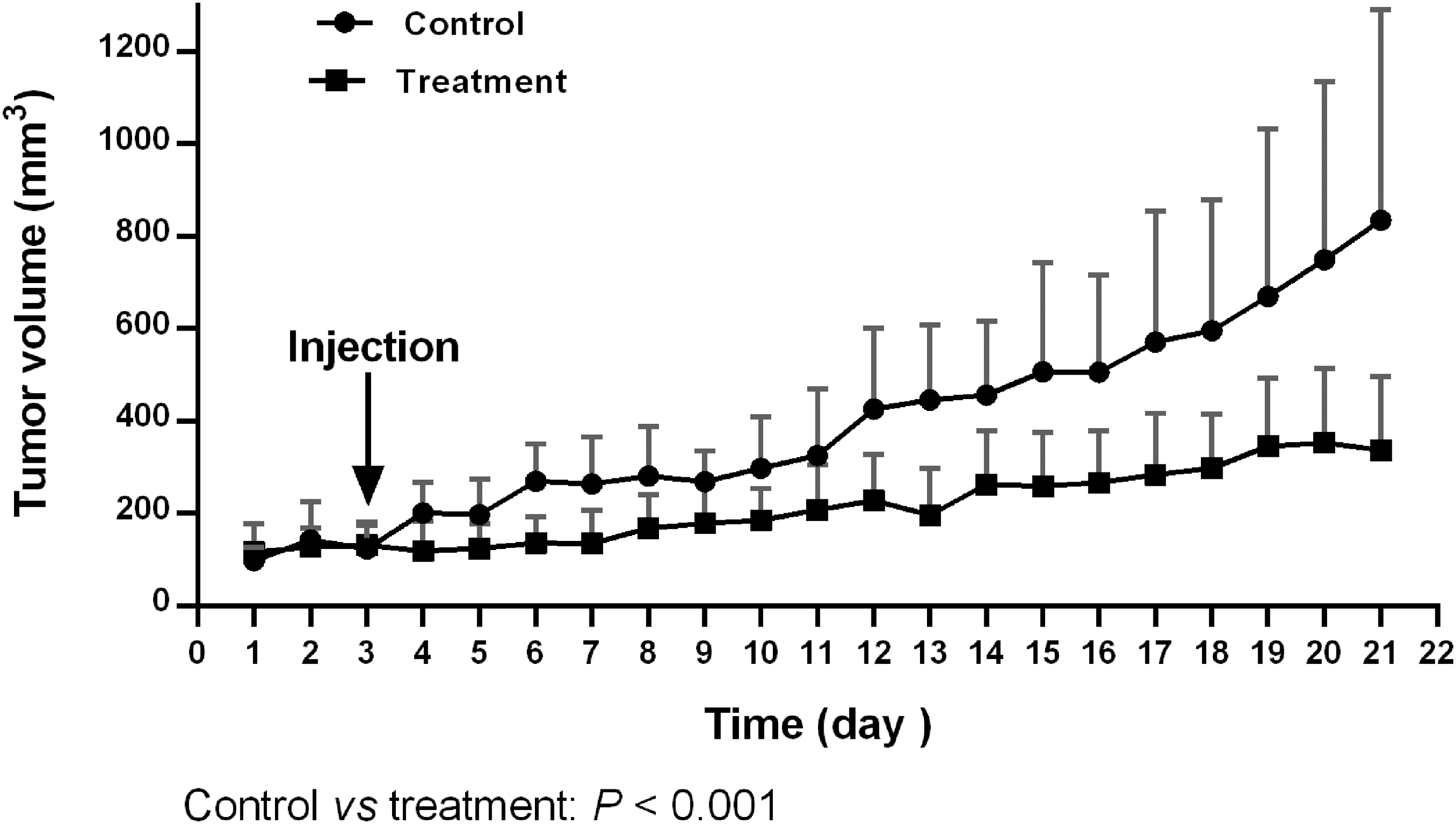
Tumor size change in nude mice with an ovarian tumor in control and paclitaxel treatment groups (n = 10 in each group). During the experiment, unfortunately, three mice were lost (control group: two and treatment group: one).
Biodistribution studies
Biodistribution data of radiolabeled peptide in ovarian tumor-bearing mice are shown in Table 1. The highest level of uptake was observed in the kidneys, in control and paclitaxel-treated groups to be 2.43% ± 0.72 and 2.94% ± 0.62, respectively. Furthermore, the major excretion route of the 99mTc-labeled peptide is renal system, however, a low level of radioactivity remains in muscle and bone. Tumor uptakes were 0.12% ± 0.02 and 0.11% ± 0.01 for control and paclitaxel-treated groups, respectively, which were statistically nonsignificant. In control and paclitaxel-treated groups, no remarkable differences between tumor/blood and tumor/muscle ratios were observed.
Biodistribution of 99mTc-HYNIC-(Ser)3-LTVPWY Peptide in Mice with Subcutaneous SKOV-3 Cancer Cells Each Mouse of Both Groups (n = 4) a
Nude mouse was intravenously administered 1 μg of 99mTc-HYNIC-(Ser)3-LTVPWY peptide (n = 4 each group) and sacrificed 4 h after injection.
Comparison of control with treatment groups; p-value = 0.14, insignificant.
ID/g, injection dose per gram; SD, standard deviation; S&T, salivary glands and thyroid.
Tumor imaging
The tumor imaging in female nude mice was gathered by dual-head γ-CAM at 4 h after radiolabeled peptide injection. The anesthetized mice were kept in the prone position on the bed and tumors were successfully seen in both groups. Radioactivity absorption was measured as a tumor-muscle ratio in control and paclitaxel-treated mice (Fig. 3).
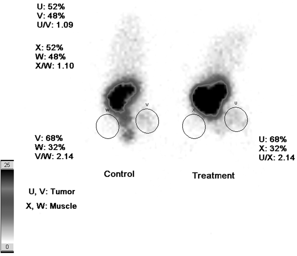
Images of ovarian tumor-bearing mice 4 h after injection of 99mTc-HYNIC-(Ser)3-LTVPWY in control and paclitaxel-treated (treatment) animals.
Histopathological findings
In the histological assay, a large number of cells showed cell division in the control group. In some of the control group samples, histological images confirmed the invasion of cancer cells into surrounding tissues, such as muscle tissue. Also, the effects of paclitaxel on inhibiting cell proliferation, increasing necrosis, fibrosis, and cell death in the tumoral mass were observed in the treatment group (Fig. 4A, B: control; Fig. 4C, D: treatment). Subsequently, paclitaxel could suppress neovascularization in the tumoral mass. Histopathologic findings revealed about 50% to 75% necrosis in the paclitaxel-treated group (Table 2).
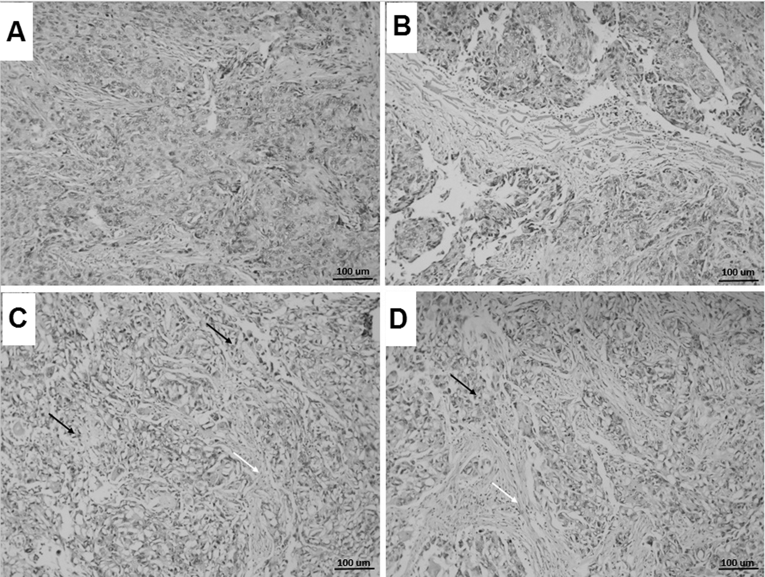
Histopathological findings of the effect of paclitaxel on ovarian cancer tissue (SKOV-3) are shown.
Semiquantitative Analysis of Histological Staining
Data were displayed as a percentage of total tissue area. +: 0%–25%, ++: 25%–50%, +++: 50%–75%, ++++: 75%–100%. The treatment group received paclitaxel.
Immunohistochemical findings
According to IHC, paclitaxel has an inconsequential effect on the HER2 receptor alteration. The intensity of colors represents the immunoreactivity of HER2 receptor, which is close in both control (Fig. 5A, B) and treatment group (Fig. 5C, D). Figure 6 presents the quantitative analysis of the immunohistochemical detection of HER2 expression in both groups. The high immunoreactivities of HER2 were confirmed by quantitative analysis in control (36.8 ± 1) and paclitaxel-treated mice (34.2 ± 1.02). No significant difference in HER2 immunoreactivity level was found between control and paclitaxel-treated groups (p > 0.05).

Immunohistochemical findings of the paclitaxel effect on HER2 overexpression of ovarian cancer mass in tumor xenografted mice.
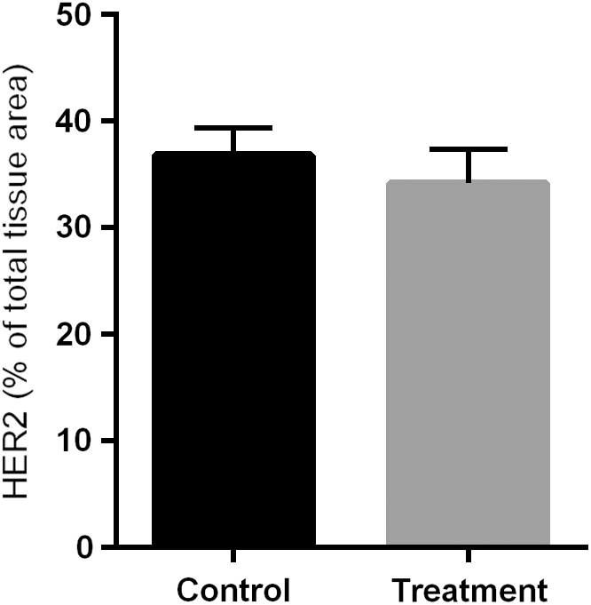
Densitometry analysis of immunohistochemical staining for HER2. Data are presented as a percentage of total tissue area. HER2 expressions in the control and treatment groups were not particular to mention. It was a negligible difference between the two groups in HER2 staining. Data are presented as mean ± standard deviation. Two mice were analyzed in each group.
Discussion
In this study, paclitaxel markedly decreased the size of ovarian tumor compared to control group. Histopathological findings showed the ability of paclitaxel to decrease tumor invasion. The biodistribution results showed that the tumor uptake of radiolabeled peptide were nonsignificantly different between control and paclitaxel groups. Also, the imaging experiment showed the same tumor visualization at both control and treatment groups with the same tumor size. Besides, the imaging findings completely confirmed the biodistribution data. In IHC study, HER2-expression was the same for results in both groups. It is demonstrated that paclitaxel reduced tumor size, whereas it did not alter HER2 status in the tumor of the mice.
HER2 is overexpressed in ovarian cancer patients, that is playing a crucial role in invasive and metastasis. 30,31 Evidently, HER2 expression is associated to poor prognosis and it can be used as a predicting biomarker for prognosis in ovarian cancer patients. 9 Currently, HER2 status is examined by IHC and/or FISH through tumor biopsy specimens in patients. The heterogeneous expression of HER2 was observed in patients that results in failure trastuzumab response and survival outcome in patient. 32,33 A noninvasive diagnostic tool for evaluation of HER2 expression can help physicians to follow-up HER2 status in patients for stratification, which leads to impressive therapy. The status of HER2 greatly impacts on choosing an effective treatment protocol in patient with cancer.
Also, chemotherapy might alter HER2 status in tumor tissue, and it is necessary assay the tumor tissue after chemotherapy for optimizing therapy. 34,35 Altered HER2 expression levels occurred in 7.1% of patients after neoadjuvant chemotherapy, broadly as a switch from positive to negative HER2 status. While a minimal alteration in the HER2 status was found in the control patients, 34 in another study, a notable difference in HER2 status was observed between core needle biopsies and surgical resection specimens in patients receiving neoadjuvant chemotherapy. After neoadjuvant chemotherapy, 23.4% of tumors were determined to have downregulated HER2 expression as evaluated by IHC. HER2 protein overexpression level was accompanied by promising pathologic response to anthracycline and taxane-based chemotherapy regimens. 24 Vincent-Salomon et al. 36 reported that HER2 overexpression level was unaltered after chemotherapy in metastatic sites. HER2 negative primary tumors did not convert to HER2 positive in patients after receiving chemotherapy. A decreased HER2 expression level was observed after chemotherapy or in secondary tumors in few patients. 36 Radiopharmaceutical molecular imaging techniques such as positron emission tomography (PET) and SPECT are emerging noninvasive techniques for tumor imaging. While PET has better resolution, sensitivity, and more accurate quantification of radioactivity concentrations in vivo, SPECT is cheaper and more widely available than PET. Both tools have been used for diagnosing of ovarian cancer. 37 18 F-FDG as a PET radiopharmaceutical has been applied for gaining imperative prognostic information for diagnosis and relapse of ovarian cancer in patients. The accumulation of 18 F-FDG in the tumor is based on glucose metabolism and does not predict the HER2 status on the cell surface. 38 This study was to investigate whether paclitaxel causes a selective alteration in ovarian cancer with HER2 overexpression, whereas previous researches have reported controversial results on the impact of anticancer therapy on HER2 receptors, however, the effects of paclitaxel on HER2 status in ovarian cancer is indiscernible. Next, the authors compared the results of HER2 status between 99mTc-HYNIC-(Ser)3-LTVPWY and IHC in tumor-bearing mice with/without paclitaxel therapy. They have demonstrated an agreement in HER2 expression assessment with IHC, biodistribution data, and imaging considering injection of 99mTc-labeled LTVPWY peptide into ovarian tumor-bearing mice. Results of these three methods demonstrated that HER2 status in ovarian tumor was unaltered after paclitaxel therapy in mice. The authors' previous study illustrated that 99mTc-HYNIC-(Ser)3-LTVPWY peptide was efficiently accumulated in the tumor through the specific binding on HER2. The specific binding of this radiolabeled peptide was approved with in vitro and in vivo studies with blocking experiments. 14 This radiolabeled peptide has fast washout from nontarget tissues and accumulated reasonably in the tumor for imaging at 4 h after injection. 14 111In-DTPA-pertuzumab as a HER2-targeted radiolabeled monoclonal antibody has been used for its capability to detect and monitor tumor response to trastuzumab therapy in MDA-MB-361 tumor-bearing mice with HER2 overexpression. HER2-targeted antibody therapy resulted in 4.5-fold reduction in 111In-labeled pertuzumab accumulation in the tumor.
HER2 status in tumors was tested with IHC in trastuzumab-treated mice, which revealed an impressive decrease in HER2-positive tumor cells, in comparison with phosphate-buffered saline-treated mice. 39 Affibody molecule ZHER2:342 binds to HER2 noncompetitively with trastuzumab, however, the 99mTc-Affibody molecule was performed for HER2 status trastuzumab-treated mice bearing SKOV-3 tumors. The accumulation of 99mTc-Affibody molecule represented notably reduced in the tumors of mice that were treated with trastuzumab. The reduction in HER2 status in the trastuzumab-treated tumor was confirmed by IHC. Zhang et al. 40 reported that high abdominal accumulation of 99mTc-Affibody molecule was hampering the use of this radiolabeled Affibody in the detection of tumors with HER2 expression in abdominal region. 99mTc-Affibody molecule has faster blood clearance and higher tumor to nontarget tissue ratios and presents advantages compared to radiolabeled trastuzumab, pertuzumab, or its fragments in the detection of HER2 downregulation. 40
Trastuzumab therapy resulted in downregulated HER2 status in the tumor, which was detected and monitored with HER2-targeted radiolabeled antibody and Affibody molecule. The current study showed that paclitaxel did not influence the HER2 status, while it had markedly antitumor effect on ovarian cancer in mice.99mTc-HYNIC-(Ser)3-LTVPWY was useful for detecting HER2 status after chemotherapy of ovarian tumor xenograft in nude mice. 99mTc-HYNIC-(Ser)3-LTVPWY has advantages over radiolabeled trastuzumab and Affibody molecule to detect HER2 status in tumors, including small size, rapid blood washout through kidneys, and negligible abdominal accumulation. Low tumor uptake is disadvantage of 99mTc-HYNIC-(Ser)3-LTVPWY in comparison with radiolabeled trastuzumab and Affibody molecule. Animal SPECT is unavailable in the university and that is a limitation of this study, it impacted on quality of animal image.
Conclusions
The authors conclude that paclitaxel does not alter the HER2 status in ovarian tumor-bearing mice, while HER2 status was evaluated with IHC, biodistribution, and gamma imaging. This is a substantial issue to address since 99mTc-HYNIC-(Ser)3-LTVPWY peptide is a useful tool for predicting the HER2 status in the tumor after chemotherapy regarding being less expensive and noninvasive tool compared to IHC.
Footnotes
Disclosure Statement
There are no existing financial conflicts.
Funding Information
This study was supported by a grant from Mazandaran University of Medical Sciences, Sari, Iran (ID # 3604).