
Abstract
Background:
Long noncoding RNA (lncRNA) MORT is silenced in many malignancies, but its role in cancer remains hardly known.
Methods:
The expression of MORT and NOTCH1 was determined by real-time quantitative polymerase chain reaction and enzyme-linked immunosorbent assay, respectively. Correlation between MORT and NOTCH1 was analyzed by Pearson's correlation analysis. To further investigate the interaction between MORT and NOTCH1, overexpression experiments were performed.
Results:
In our study, MORT expression was downregulated in hepatocellular carcinoma (HCC), while NOTCH1 expression was upregulated in HCC patients. Hepatitis B virus and hepatitis C virus infection and tumor size did not significantly affect MORT expression, but MORT expression was lower in metastatic HCC patients compared with nonmetastatic HCC patients. MORT and NOTCH1 were inversely correlated across HCC tissues. MORT overexpression decreased NOTCH1 expression, while NOTCH1 overexpression did not significantly affect MORT. MORT overexpression inhibited the migration and invasion of HCC cells, while NOTCH1 overexpression promoted the migration and invasion of HCC cells. In addition, NOTCH1 overexpression attenuated the effects of MORT overexpression on cell migration and invasion.
Conclusion:
Therefore, MORT overexpression may inhibit HCC by downregulating NOTCH1.
Introduction
Hepatocellular carcinoma (HCC) is one of the most common types of malignancies for both incidence and mortality. 1 Although the incidence of HCC did not change significantly during the past several decades in the United States, obvious increase was observed in mortality rate. 2,3 In China, HCC has been a major public health burden for decades, mainly owing to the high infection rates of hepatitis B virus (HBV) and hepatitis C virus (HCV). 4 The application of surgery, which is the only radical treatment, is limited by the fact that a considerable portion of patients with HCC are diagnosed with the existence of tumor metastasis. 5
NOTCH1 is a key regulator of cancer cell behaviors, 6 such as cancer cell invasion and migration. 7,8 In effect, NOTCH1 inhibition suppresses HCC. 7 NOTCH1 may play its roles by interacting with long noncoding RNAs (lncRNAs), 9,10 which are a subgroup of noncoding RNAs composed of 200 nucleotides and play essential roles in cancer biology. 11 MORT is silenced in many malignancies, but the functionality in cancer biology is hardly known. 12 Our preliminary transcriptome analysis showed that MORT and NOTCH1 mRNA were inversely correlated in HCC tumor tissues. In this study, we proved that MORT overexpression could inhibit HCC possibly by downregulating NOTCH1.
Methods
Human specimens and HCC cell lines
Our study included 82 HCC patients (42 males and 40 females; 32–68 years; 46.8 ± 4.4 years) at Xinhua Hospital affiliated to Shanghai Jiao Tong University School of Medicine from March 2015 to March 2018. Patients' inclusion criteria were as follows: (1) confirmed by pathological examinations; (2) patients fully understood the experimental procedure. Exclusion criteria were as follows: (1) patients with other diseases; (2) patients with initiated therapy. Healthy controls were enrolled to match the age and gender distributions of HCC patient group. Among the 82 patients, there were 39 HBV-positive cases, 21 HCV-positive cases, and 22 cases who were negative for both HBV and HCV. There were 12 cases of American Joint Committee on Cancer (AJCC) stage I, 18 cases of AJCC stage II, 20 cases of AJCC stage III, and 32 cases of AJCC stage IV. Biopsies were performed to collect paired tumor and adjacent healthy tissue. Blood was transferred to an anticoagulant BD Vacutainer plastic EDTA tube, followed by centrifugation at 1200 g for 10 min to collect the supernatant (plasma). SNU-398 and SNU-182 (ATCC) HCC cell lines were included. Cell culture was performed following instructions from ATCC.
Enzyme-linked immunosorbent assay
NOTCH1 enzyme-linked immunosorbent assay (ELISA) Kit (MyBioSource, San Diego, CA) was used for the measurement of NOTCH1 protein. All operations were performed following the recommendations from MyBioSource. Plasma levels of NOTCH1 were expressed as ng/g.
Real-time quantitative polymerase chain reaction
MPure™ Total RNA Extraction Kit (117022160; MP Biomedicals) was used for RNA isolation. After reverse transcriptions, quantitative polymerase chain reactions (qPCRs) were performed to measure the mRNA levels of MORT and NOTCH1 with β-actin as internal control. PCR conditions were 40 cycles with 95°C for 65 s, 95°C for 11 s, and 58.5°C for 45 s. Expression level of MORT was normalized to endogenous control β-actin using 2−ΔΔCT method.
Transfection
Vectors expressing MORT and NOTCH1 were designed and synthesized by Sangon (Shanghai, China). Cells of both HCC cell lines were cultivated overnight to reach 70%–80% confluence. Lipofectamine 3000 reagent (Thermo Fisher Scientific) was used to transfect vectors at a dose of 10 nM. Empty vector-transfected cells were negative control (NC) cells. Control (C) cells were untransfected cells.
In vitro cell migration and invasion assay
The mRNA expression of MORT and NOTCH1 was determined by real-time quantitative PCR (RT-qPCR) at 24 h after transfection. Cell migration and invasion were detected by Transwell assay when MORT and NOTCH1 overexpression rates were >200%. In brief, 3000 cells in 0.1 mL nonserum medium were transferred to the upper chamber. To induce cell invasion and migration, medium containing 20% fetal bovine serum was used to fill the lower chamber. Matrigel-coated chambers (356234; Millipore) and uncoated chambers were used for invasion and migration assay, respectively. Cells were cultivated under normal conditions for 12 h, followed by staining with 0.5% crystal violet (Sigma-Aldrich) for 20 min at room temperature.
Western blot
After total protein isolation (radioimmunoprecipitation assay buffer solution; Sigma-Aldrich) and protein quantification (bicinchoninic acid assay, Sigma-Aldrich), protein samples were denatured (10 min in boiling water) and separated (8% sodium dodecyl sulfate polyacrylamide gel electrophoresis). After that, polyvinylidene fluoride membranes were used to transfer proteins. Phosphate-buffered saline (5% nonfat milk) was used to block the membranes. NOTCH1 (ab65297; Abcam) and GAPDH (ab9485; Abcam) primary antibodies were used to incubate the membranes for 15 h at 4°C, followed by further incubation for 2 h with secondary antibody (1:1400, MBS435036; MyBioSource) at room temperature. Signals were produced using ECL (Sigma-Aldrich). Expression level of NOTCH1 was normalized to GAPDH using ImageJ v1.46 software.
Statistical analysis
Correlation was analyzed by Pearson's correlation analysis. Paired tissues were compared by paired t test. Two independent groups were compared by unpaired test. One-way analysis of variance and Tukey's test were used to compare multiple groups. The 82 HCC patients were divided into high and low MORT level groups (n = 41, cutoff value = median level of MORT expression in tumor tissues). p < 0.05 was considered statistically significant.
Results
MORT and NOTCH1 expressions were dysregulated in HCC patients
The expression of MORT and NOTCH1 in paired tissues from 82 HCC patients was measured by RT-qPCR and ELISA. The expression of MORT was downregulated (Fig. 1A), while NOTCH1 expression was upregulated (Fig. 1B) in tumor tissues compared with adjacent healthy tissues of HCC patients (p < 0.05). The expression of MORT in tumor tissues was not closely correlated with patients' gender, age, AJCC stages, tumor multiplicity, smoking, and drinking habit (Table 1).

MORT and NOTCH1 were dysregulated in tumor tissues of HCC patients. RT-qPCR and ELISA results showed that MORT was downregulated
Correlations Between Expression Levels of MORT in Tumor Tissues and Patients' Clinicopathological Data
AJCC, American Joint Committee on Cancer.
MORT and NOTCH1 were inversely correlated across tumor tissues, but not across adjacent healthy tissues
As shown in Figure 2A, correlation analysis revealed that expression levels of MORT and NOTCH1 were significantly and inversely correlated with each other across tumor tissues. However, MORT and NOTCH1 were not significantly correlated across adjacent healthy tissues (Fig. 2B).
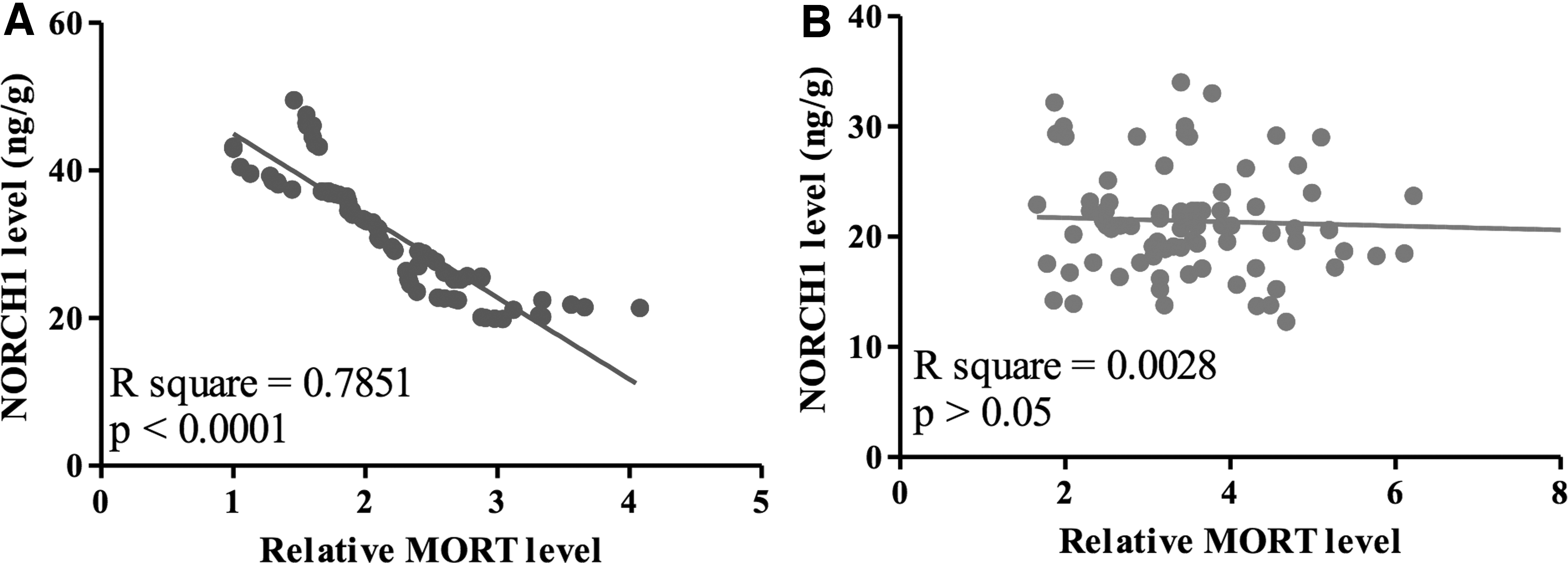
MORT and NOTCH1 were inversely correlated in tumor tissues, but not in adjacent healthy tissues. Pearson's correlation coefficient analysis showed that expression levels of MORT and NOTCH1 were inversely correlated across tumor tissues
MORT overexpression led to NOTCH1 inhibition in HCC cells
The overexpression of MORT and NOTCH1 was achieved and revealed by RT-qPCR (Fig. 3A). MORT overexpression decreased NOTCH1 expression in both SNU-398 and SNU-182 cells (Fig. 3B, p < 0.05). In contrast, NOTCH1 overexpression did not significantly affect MORT expression in those cells (Fig. 3C).
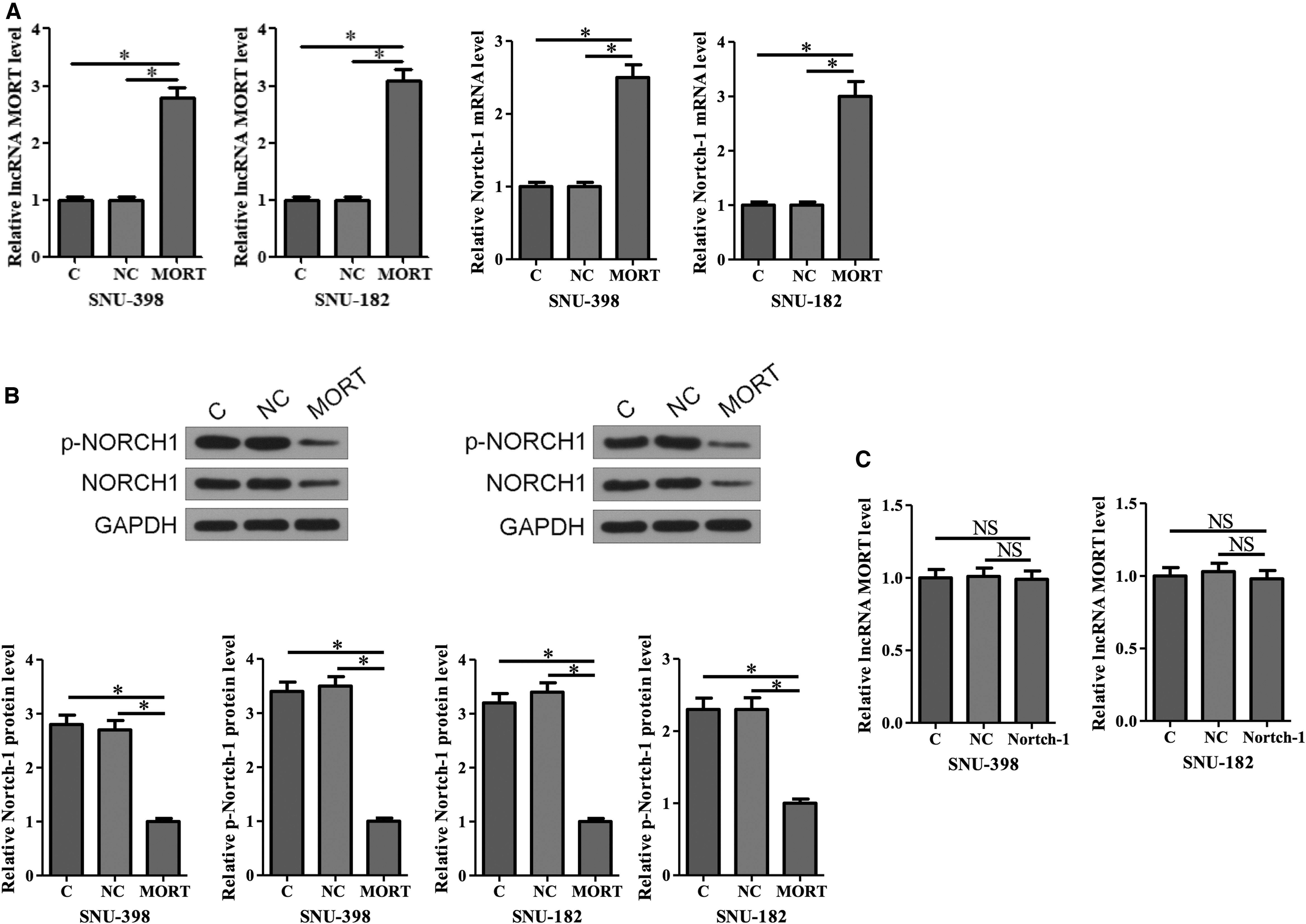
MORT overexpression decreased NOTCH1 expression in HCC cells. Overexpression of MORT and NOTCH1 was reached after transfection
Expression of MORT was regulated by tumor metastasis
Among 82 HCC patients, there were 39 HBV-positive cases (HBV), 21 HCV-positive cases (HCV), and 22 cases who were negative for both HBV and HCV (NCs). As shown in Figure 4A, MORT expression was not significantly different among HBV, HCV, and NCs. Based on the diameter of primary tumors, patients were divided into 0–2 cm group (n = 20), 2–5 cm group (n = 34), and >5 cm group (n = 28). As shown in Figure 4B, tumor size did not significantly affect MORT expression. There were 50 cases of nonmetastatic HCC and 32 cases of metastatic HCC. Compared with nonmetastatic HCC patients, the expression levels of MORT were significantly lower in tumor tissues of metastatic HCC patients (Fig. 4C, p < 0.05).
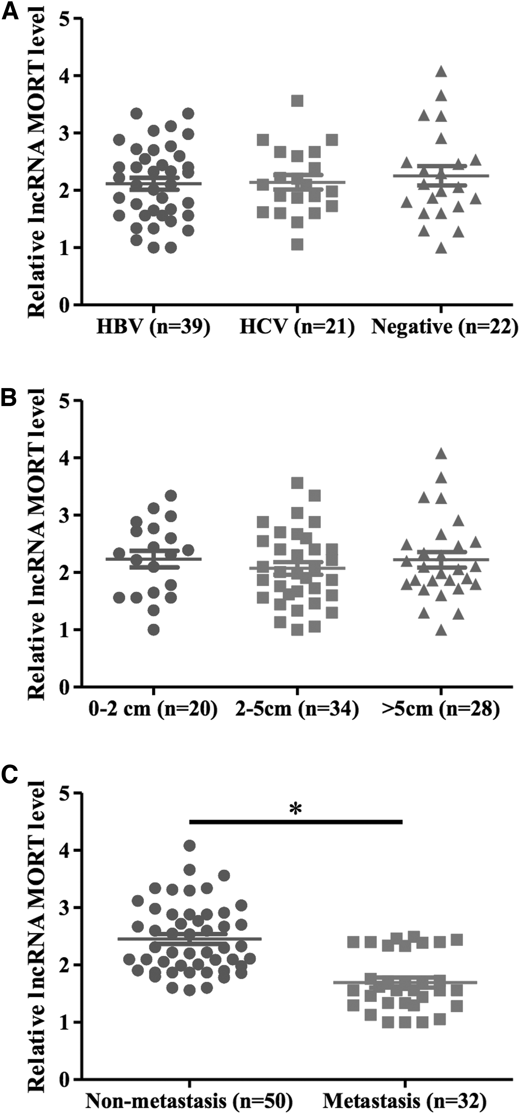
Expression of MORT was regulated by tumor metastasis. Expression of MORT in tumor tissues was not regulated by HBC or HCC infection
MORT overexpression inhibited HCC cell migration and invasion through NOTCH1 inhibition
The above-mentioned data indicated the involvement of MORT in tumor metastasis of HCC. Therefore, we further investigated the involvement of MORT and NOTCH1 in cancer cell migration and invasion. We found that MORT overexpression decreased cell migration and invasion, while NOTCH1 overexpression significantly increased cell migration (Fig. 5A) and invasion (Fig. 5B) of both SNU-398 and SNU-182 cells (p < 0.05). In addition, NOTCH1 overexpression significantly attenuated the inhibitory effects of MORT overexpression (p < 0.05).

MORT overexpression inhibited HCC cell migration and invasion through NOTCH1 inhibition. MORT overexpression significantly increased cell migration and invasion, while NOTCH1 overexpression significantly decreased cell migration
Discussion
As a recently identified lncRNA, MORT was proven to be downregulated in many types of cancers. 12 However, the role of MORT in cancer development is unknown. We first reported that MORT was downregulated in HCC, and it could inhibit HCC cell migration and invasion by inhibiting the expression of NOTCH1.
Infections of HBV and HCV have been proved to be major causes of the occurrence of HCC. 13 More and more studies have revealed that HBV and HCV may regulate the expression of lncRNAs to participate in the pathogenesis of HCC. 14,15 In this study, we found that MORT was downregulated in HCC and not significantly affected by HBV and HCV infection. Therefore, MORT may participate in HCC through HBV- and HCV-independent pathways.
NOTCH1 was upregulated in different types of cancers including HCC, and inhibition of NOTCH1 expression might inhibit the progression of HCC. 16,17 Consistently, our study observed the significantly upregulated NOTCH1 expression in HCC. Interestingly, our study showed that MORT could serve as an upstream activator of NOTCH1 in HCC cells. However, the regulation of NOTCH1 by MORT might be indirect because the expression levels of MORT and NOTCH1 were negatively correlated only across tumor tissues. It has been reported that MORT may downregulate ROCK1, 18 which can affect the expression of NOTCH1. 19 It is known that ROCK1 is upregulated in HCC. 20 Therefore, the upregulated ROCK1 in HCC may mediate the interaction between MORT and NOTCH1. We will explore this possibility in our future studies. NOTCH1 is a critical player in cancer metastasis. 7 In this study, we showed that the inhibition of NOTCH1 by MORT was likely involved in the regulation of metastasis of HCC cells. These data suggest that overexpression of MORT may serve as a therapeutic target for the treatment of HCC by inhibiting NOTCH1.
Conclusions
In summary, MORT is downregulated in HCC, and NOTCH1 is upregulated in HCC. MORT overexpression may inhibit cancer cell migration and invasion in HCC by downregulating NOTCH1.
Ethics Approval and Consent to Participate
This study was approved by the Ethics Committee of Xinhua Hospital affiliated to Shanghai Jiao Tong University School of Medicine. The research has been carried out in accordance with the World Medical Association Declaration of Helsinki. All patients provided written informed consent before their inclusion in the study.
Availability of Data and Materials
The analyzed data sets generated during the study are available from the corresponding author on reasonable request.
Footnotes
Authors' Contributions
X.Q.L. and J.W. contributed to study design; X.Q.L., G.H.G., and F.J. performed analysis and investigation; X.Q.L. and J.W. contributed to article preparation.
Disclosure Statement
There are no existing financial conflicts.
Funding Information
No funding was received for this article.