
Abstract
Background:
Targeted α particle therapy using long-lived in vivo α particle generators is cytotoxic to target tissues. However, the redistribution of released radioactive daughters through the circulation should be considered. A mathematical model was developed to describe the physicochemical kinetics of 212Pb-labeled pharmaceuticals and its radioactive daughters.
Materials and Methods:
A bolus of 212Pb-labeled pharmaceuticals injected in a developed compartmental model was simulated. The contributions of chelated and free radionuclides to the total released energy were investigated for different dissociation fractions of 212Bi for different chelators, for example, 36% for DOTA. The compartmental model was applied to describe a 212Bi retention study and to assess the stability of the 212Bi-1,4,7,10-tetrakis(carbamoylmethyl)-1,4,7,10-tetraazacyclododecane (212Bi-DOTAM) complex after β− decay of 212Pb.
Results:
The simulation of the injection showed that α emissions contribute 75% to the total released energy, mostly from 212Po (72%). The simulation of the 212Bi retention study showed that (16 ± 5)% of 212Bi atoms dissociate from the 212Bi-DOTAM complexes. The fractions of energies released by free radionuclides were 21% and 38% for DOTAM and DOTA chelators, respectively.
Conclusion:
The developed α particle generator model allows for simulating the radioactive kinetics of labeled and unlabeled pharmaceuticals being released from the chelating system due to a preceding disintegration.
Introduction
An increasing number of preclinical and clinical studies in targeted α particle therapy (TAT) reveal the great potential of α-emitting radionuclides in targeted radionuclide therapy. 1 –4 The short path length (50–80 μm) and the high linear energy transfer (LET) of α particles of around 100 keV/μm result in high efficacy in killing specific tumor cells, while sparing surrounding normal tissues. 5 The choice of the radionuclide must be based on the type of disease, the half-life and the decay scheme of the radionuclide, the associated chemical properties, and the availability of the appropriate radionuclide. 6 In TAT, the efficacy of the chelating system to retain the daughter nuclei at the target location must be considered. 7
The application of short-lived α emitters, such as 212Bi (t 1/2 = 60.6 min) and 213Bi (t 1/2 = 45.6 min), is most promising in clinical trials when rapid accumulation at target sites is ensured. For example, short-lived α emitters are used for targeting cell clusters or micrometastases such as metastatic melanoma and acute myeloid leukemia. 8,9 The advantage of using short-lived α emitters is the possibility to reduce toxicity to nontarget tissues by reducing the systemic distribution of the radioactivity. However, the short half-life of these radionuclides is a major obstacle in targeting tumors that are difficult to access. Using an atomic generator system, which consists of a pharmaceutical labeled with a longer lived radioactive parent, such as 212Pb (t 1/2 = 10.6 h) or 225Ac (t 1/2 = 10.0 d), to produce short-lived α-emitting daughters will overcome this limitation. 10,11
In vivo α particle generators overcome the limitation of radiopharmaceuticals with slow pharmacokinetics, thus increasing accumulation in the tumors and deliver a higher dose per administered activity. 12,13 However, the transitions between multiple radioactive daughters in the decay scheme of the parent have to be considered. Mirzadeh et al. reported that the 212Pb decay in the 212Pb-DOTA complex results in 36% dissociated 212Bi atoms due to the electronic excitation of the produced 212Bi daughter atoms. The energy of the emitted γ following 88% of the β− decays of the parent are transferred to the inner orbital electrons in around 36% of the decays. The reorganization of valence electrons, induced by the cascade of conversion electrons, emits characteristic X-rays and Auger electrons. Subsequently, these electronic excitations generate a highly ionized Bi5+ cation, which dissociates from its DOTA-chelator. 14,15
The released 212Bi atoms (36%) distribute to normal tissues causing toxicity to nontarget tissues. 16 The toxicity to nontarget tissues is further increased due to the distribution of radionuclides set free after α particle emissions. Therefore, using α particle generator systems and developing stable bifunctional chelation agents in TAT necessitates the assessment of the radiochemical stability and the pharmacokinetics of these generator systems. Also, the pharmacokinetics of α particle generator systems and the subsequent localization of the released radionuclides in normal tissues require further investigations.
In this work, a mathematical model was developed to simulate the complex physical decay scheme and the chemical fate of 212Pb-labeled pharmaceuticals. The developed model describes the kinetics of the 212Pb-labeled pharmaceutical and its decay products. The contributions of α and β− emissions of each chelated and free radionuclide to the total released energy are determined. The basic 212Pb model is applied to assess the stability of the 212Bi-DOTAM complex after β− decay of 212Pb by simulating an in vitro retention study. The effect of the kinetic stability of the 212Bi-chelator complex after β− decay of 212Pb on the production of free radionuclides was investigated.
Materials and Methods
Compartmental model
212Pb as part of the decay chain of 228Th decays to 212Bi. The α emitter 212Bi decays via a branched pathway to 208Tl and 212Po and both, in turn, decay to stable 208Pb (Fig. 1). 17

A linear compartmental model for chelated 212Pb and its decay products (Fig. 2) was developed and implemented using the modeling software SAAM II version 2.3 (The Epsilon Group, TEG, USA). 18 The model represents the decay scheme of 212Pb attached to a pharmaceutical, including physical and chemical transfer rates. Starting with 212Pb, each radionuclide in the decay scheme in Figure 1 was modeled by a single compartment. All compartments were connected via the physical decay rates of the radionuclides (Supplementary Table S1 in Supplementary Data A). 17
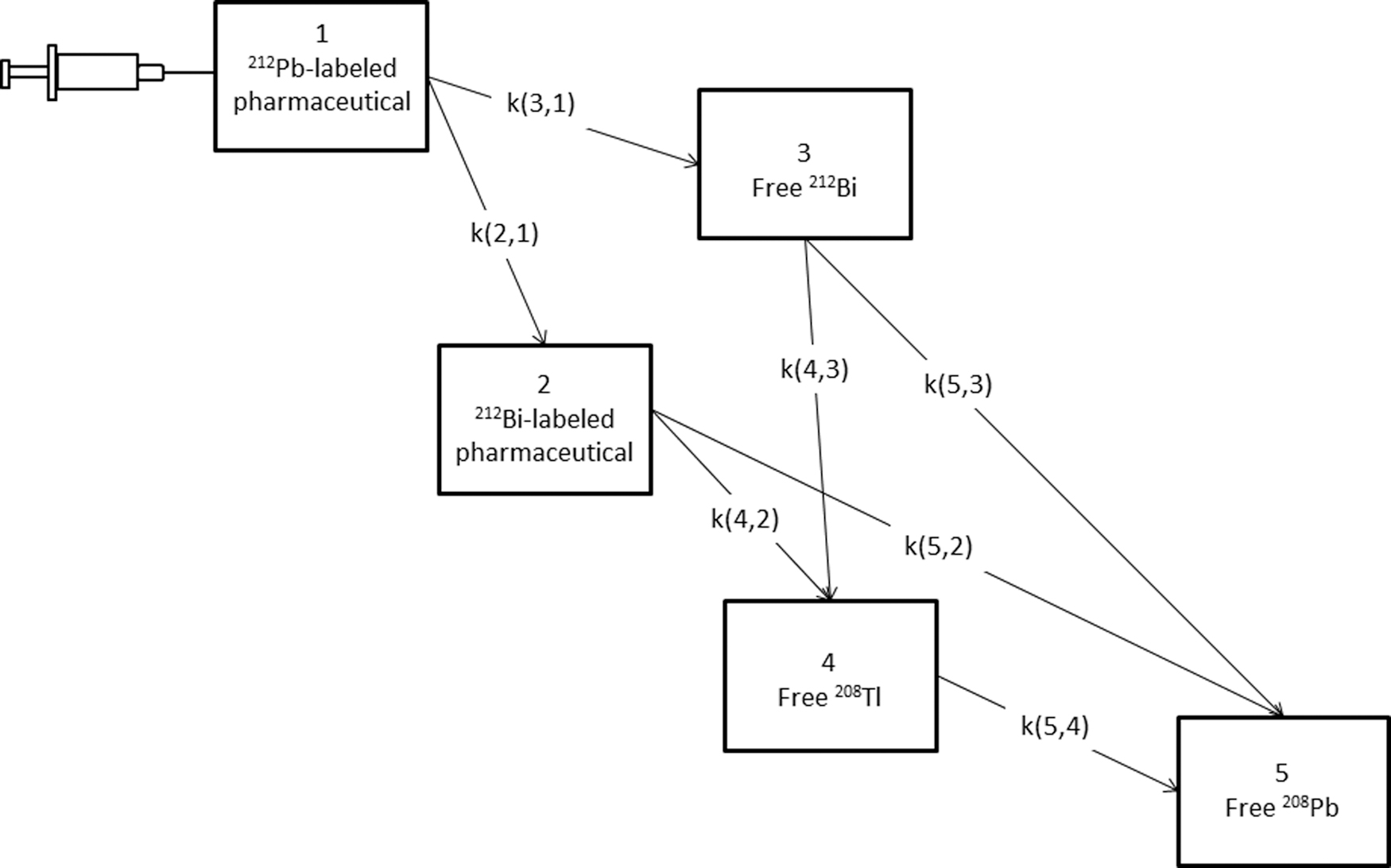
Compartmental model developed to simulate the kinetics of 212Pb-labeled pharmaceuticals and its radioactive progeny, including physical and chemical transfer rates. k(a, b) indicates the transfer rate from compartment b to compartment a. Compartments 1 and 2 represent chelated 212Pb and 212Bi radionuclides and the other three represent the free 212Bi, 208Tl, and 208Pb nuclides. The free 212Bi compartment (no. 3) results from the dissociation of a fraction (ƒ) of Bi-chelator complexes after the 212Pb decay. 12 The compartment representing 212Po is not included in the model because the short half-life of 212Po (t 1/2 = 0.3 μs) prevents its redistribution in the blood circulation. Instead, it is integrated in the calculations after its production using the transfer rates k(5,2) and k(5,3). 212Pb-labeled pharmaceuticals are injected into compartment 1.
The β− decay of chelated 212Pb was modeled based on the decay scheme shown in Figure 3A. All energy levels of 212Bi are presented as a single excited-state compartment of chelated 212Bi (Fig. 3B). Around 12% of β− decays of chelated 212Pb result in chelated 212Bi in the ground state. The rest of 212Pb decays results in excited-state chelated 212Bi. Depending on the chelating system used, a fraction of 212Bi in the excited state will be released from the chelator after internal conversion as described above. The other fraction goes to the 212Bi ground state after γ emissions and remains bound in the chelator. However, due to the short lifetime of the excited state of the 212Bi (10−12 s–10−13 s), 14 the final structure representing the decay of 212Pb was reduced as shown in Figure 3C.

The branched decay of 212Bi (Fig. 2) was modeled by representing the free daughter in each branch by a single compartment (Supplementary Fig. S1 in Supplementary Data B).
Kinetics of 212Pb-labeled radiopharmaceuticals
To study the kinetics of 212Pb and its progeny, a 212Pb-labeled radiopharmaceutical was injected into compartment 1 (Fig. 2) and the time course in each compartment was simulated. The activity and the energy release rate for each radionuclide were calculated. In addition, the contributions of chelated and free radionuclides to the total released energy were calculated for 212Bi-chelator complexes with dissociation fractions ƒ (Fig. 3) of 16% (the percentage will be referred to later in the in vitro retention study results), 30%, and 36%.
In vitro retention study of 212Bi
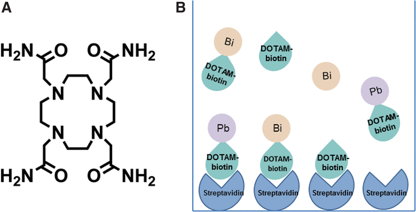
DOTAM-biotin (2-(4-amino-N-((N-biotinyl)-aminohexyl-oyl)-butyl)-1,4,7,10-Tetrakis (carbamoylmethyl)-1,4,7,10-tetraazacyclododecane) is a ligand with a linking moiety stemming out of the macrocycle ring, conjugated to biotin through an aminocaproic acid linker (Fig. 3A).
The retention experiment performed at Orano Med LLC was carried out in 8 wells of a streptavidin-coated clear strip plate (Pierce) with bovine serum albumin after triplicate rinsing with Dulbecco's phosphate-buffered saline (DPBS) (GE). The binding of 212Pb-DOTAM-biotin on the receptors of streptavidin molecules was prepared by adding 100 μL of diluted 212Pb-DOTAM-biotin (40 μCi/mL) with DPBS to each well and incubating the system for 30 min at room temperature while shaking.
After the incubation period, the wells were rinsed five times with 250 μL of DPBS per well so that only radiolabeled DOTAM-biotin-streptavidin complexes were found in the system. One hundred microliters of phosphate-buffered saline was then added to each well containing the bound biotin and incubated at room temperature till the transient equilibrium was reached (6 h) between 212Pb and 212Bi to have a measurable amount of dissociated 212Bi in the supernatant. The supernatants were removed from each well after the completion of the incubation. The amounts of 212Pb and 212Bi were quantified in all separated supernatant samples and respective wells by using a Biodex Atomlab 500 wipe test counter (Supplementary Table S2 in Supplementary Data C). The kinetic stability of the 212Pb-DOTAM complex was confirmed by conducting a Radio-HPLC experiment (Supplementary Data D).
Simulation approach of the in vitro retention study
A scheme of the in vitro retention study is depicted in Figure 4B. At time zero of the simulation, all the 212Pb-DOTAM-biotin molecules and their physical decay products (212Bi-DOTAM-biotin molecules) are assumed to be bound on the receptors of streptavidin molecules and none of the species is present in the supernatant. Then, the bound radiolabeled and unlabeled biotin molecules will dissociate and associate with rate constants (8.80 ± 0.06) × 10−5 s−1 and (5.50 ± 0.08) × 108 M−1 s−1, respectively. 19 Therefore, at later time points, the unbound radiolabeled biotin molecules (212Pb- and 212Bi-DOTAM-biotin molecules), the unlabeled biotin molecules, and the free radioactive daughters 212Bi and 208Tl are found in the supernatant (Fig. 2).
A compartmental model was developed to simulate the experiment. The model consists of the basic 212Pb generator model (Fig. 2) that represents the unbound radiolabeled α particle generators (212Pb-DOTAM-biotin) and the physical decay products (212Bi-DOTAM-biotin and free 212Bi, 208Tl and stable 208Pb) in the supernatant. In addition, two compartments 6 and 7 are added to represent the 212Pb- and 212Bi-DOTAM-biotin molecules bound to streptavidin molecules (Fig. 5). The two compartments are connected with compartments 1 and 2 by the processes of the association and dissociation.
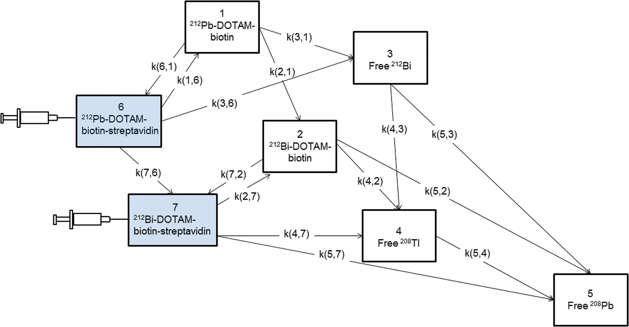
212Pb generator model (compartments 1–5) plus the two compartments (6 and 7) for the streptavidin-bound species to simulate and fit the 212Bi retention study. The highlighted compartments 6 and 7 are on the streptavidin-coated plates and the others are in the supernatant. The initial amounts of bound 212Pb-DOTAM-biotin and 212Bi-DOTAM-biotin are injected into compartments 6 and 7. The total amount of streptavidin binding sites is constant. The compartments representing the bound and free unlabeled biotin (Fig. 4B) are not shown. The free binding sites on streptavidin molecules are integrated in the differential equations. 208Tl and 208Pb are not measured but included in the model. Color images are available online.
The physical decay of bound 212Pb-DOTAM-biotin molecules in compartment 6 produces the bound 212Bi-DOTAM-biotin (compartment 7) on the streptavidin-coated plate and the released Bi in the supernatant. All 212Pb atoms found in the well are associated to biotin molecules because of the used kinetically stable 212Pb-DOTAM complex as confirmed by Radio-HPLC data (Supplementary Fig. S2 in Supplementary Data D). The decay products of compartment 7, bound 212Bi-DOTAM-biotin, are transferred by physical decay rates into the corresponding compartments in the supernatant. The values of the transfer rates in Figure 5 are given in Supplementary Tables S1 and S3 in Supplementary Data A and E, respectively. The initial amount of 212Pb-DOTAM-biotin molecules, the amount of binding sites on streptavidin molecules, and the dissociation fraction ƒ of the Bi-DOTAM complex (Fig. 3) were estimated by fitting the compartmental model to the experimental data using SAAM II.
Results
Simulation of the kinetics of 212Pb-labeled pharmaceuticals
The energy release rates due to α and β− emissions of each radionuclide are presented in Figure 6. The α emitter 212Po has the highest energy release rate among these radionuclides. α emissions contribute 75% to the total released energy (Table 1) and 72% of the emitted energy originates from the 212Po radionuclide. The kinetics of the activities of radiolabeled pharmaceuticals and free radionuclides over time are shown in Supplementary Figure S3 in Supplementary Data F.

Energy release rates of α or β− emissions of each radionuclide. Pure 212Pb is assumed at the start time of the simulation. The values of the energy release rates are normalized to the maximum energy emitted at 3.8 h. Color images are available online.
Contribution of Each Chelated and Free Radionuclide to the Total Released Energy for Different Dissociation Fractions (16%, 30%, and 36%) of Bi-chelator Complexes
Radionuclides are bound to the pharmaceutical.
Radionuclides are detached from the pharmaceutical.
Figure 7 shows the dependence of the released energy on the dissociation fraction ƒ. For a chelator system that releases only about half as much Bi as the DOTA chelator, that is, a dissociation fraction ƒ of 16% instead of 36%, the percentage of the released energy by the free radionuclides (16%) is reduced to half of that produced in the case of the DOTA chelator (34%).

Contributions of α and β− emissions to the total released energy for different dissociation fractions (ƒ) of Bi-chelator complexes. The presented data points are for dissociation fractions of Bi-chelator complexes of 16%, 30%, and 36% (Table 1). All dissociated 212Bi are assumed to be redistributed in nontarget tissues and chelated 212Bi are found at the target sites. 212Po produced from the decay of chelated 212Bi are included in α-Bound Radionuclides. On the contrary, 212Po produced from the decay of dissociated 212Bi is included in α-Free Radionuclides. More detailed descriptions are given in Table 1. Color images are available online.
In vitro retention study and fitting results
The average counts of 212Pb and 212Bi after 6 h incubation on the streptavidin-coated plates were (43,595 ± 5858) and (9521 ± 1062) cpm and in the supernatants of the 8 wells (9190 ± 1171) and (4195 ± 542) cpm, respectively.
The fit of the parameter model to the experimental data was good based on visual inspection (Supplementary Fig. S4 in Supplementary Data G). The fitted initial amount of 212Pb-DOTAM-biotin and the amount of binding sites on streptavidin molecules in a well were (0.12 ± 0.01) and (0.43 ± 0.13) fmol, respectively. The dissociation fraction ƒ of the Bi-DOTAM complex was 0.16 ± 0.05.
Discussion and Conclusion
Using in vivo α particle generators of long half-life in TAT, such as 212Pb-labeled pharmaceutical, allows for delivering high doses to tumor tissues. However, the redistribution of the radioactive daughters dissociated from the pharmaceuticals results in the irradiation of nontarget tissues. Posttreatment pharmacokinetic studies and dosimetry calculations were based on blood samples, urine excretion, planar images, and portable detectors measuring the redistribution effect of the released radioactive daughters. 20 –22 However, some of the reported dosimetry measurements of these in vivo α particle generators did not consider the possibility of interim translocation of the released radioactive daughters. 13 Monte Carlo simulations were performed to calculate dose distributions and evaluate the therapeutic potential of long-lived radioactive parents in TAT. 7,23
In the current study, a mathematical compartmental model of the complete decay chain of 212Pb-labeled pharmaceuticals was developed and implemented. This model represents the production of chelated and released radionuclides through the complete decay chain of the atomic generator system. The importance of modeling this generator approach is that it allows quantification of the redistribution of radioactive daughter nuclei, thereby addressing concerns about exposure of normal tissue. 24 This model is used for calculating the contribution of each radionuclide to the total released energy based on its physicochemical properties. 212Pb is assumed to be chelated with a stable chelator, such as 2-(4-isothiocyanatobenzyl-1,4,7,10-tetraaza-1,4,7,10-tetra-(2-carbamonylmetyl)-cyclododecane (TCMC) (pH >2), which has a similar behavior as DOTAM. 6,25,26 Therefore, free 212Pb atoms are not included in the model.
Although pure 212Pb was assumed at the start of the simulation in Figure 6, the model allows also for predicting the release rate of each radionuclide for other starting values such as in transient equilibrium, which is attained after preparation and quality control testing. When the biological effectiveness of radiation is taken into account (in this study the α weighting factor was 5), the results will be similar (93.7%) to the reported 93.4% contribution of high LET α emissions obtained by Yong et al. 6 The time-activity curves (Supplementary Fig. S3) demonstrate the transient equilibrium of 212Pb and 212Bi, which is in agreement with previously published data. 15
The 212Pb generator model (Fig. 2) can be used as a building block for broad applications. For example, the presented mathematical model in Figure 5 was successfully implemented to describe the 212Bi retention study and to fit its experimental data (Supplementary Fig. S4). Therefore, the model can be used for evaluating different bifunctional chelating agents for sequestering radioactive metal ions, as shown for the 212Pb-DOTAM. Table 1 shows that the fraction of the energy released by 212Po, either bound to a pharmaceutical or free, is the highest among other 212Pb radioactive daughters. For example, the use of the bifunctional chelator DOTA (ƒ = 0.36) in 212Pb-based therapy results in two times more energy released due to α emissions by free 212Po compared to the energy released when the DOTAM chelator (ƒ = 0.16) is used. For an optimal chelator (with ƒ = 0), 6% of the total released energy comes from the free β−-emitter 208Tl (Fig. 7). At least this fraction thus contributes to the nontarget toxicity.
Because of the physicochemical properties of the structure of the generator model, the in vivo stability of the generator system depends only on the type of the chelator used. Therefore, this generator system can be applied to simulate the pharmacokinetics of different radiopharmaceuticals, such as radiolabeled antibodies and radiolabeled peptides.
For in vivo modeling, the developed physicochemical generator model is combined with a respective physiologically based pharmacokinetic (PBPK) model, that is, the patient. 27 As a result, the full PBPK model will allow for simulating the pharmacokinetics of bound and unbound radiopharmaceuticals as well as free radionuclides in the whole body. 28 Specifically, such a 212Pb-PBPK model will allow for studying the biodistribution of the free 212Bi and the cytotoxic effects of its 212Po daughters on organs at risk, for example, kidneys. As a consequence, the potential of performing dosimetry calculations through such a 212Pb-PBPK model is a prerequisite for predicting the optimal dosing regimens for different prescribed doses. 29
Footnotes
Authors' Contributions
N.R.R.Z. designed the study, developed, implemented, and tested the model, performed the simulations, and wrote the article. P.K. evaluated the model, wrote, and edited the article. A.J.B. wrote and edited the article. T.A.R.S. and J.J.T. performed the experiment, wrote, and edited the article. G.G. designed the study, developed the model, and evaluated the implementation of the model and the simulations, wrote, and edited the article. All coauthors have reviewed and approved the article before submission.
Disclosure Statement
J.J.T. and T.A.R.S. are employees of Orano Med. No competing financial interests exist.
Funding Information
The authors gratefully acknowledge the funding from the DAAD (German Academic Exchange Service, Research Grants, Doctoral Programs in Germany 2018/19-57381412) and the Research Campus M2OLIE (German Federal Ministry of Education and Research (BMBF) within the Framework “Forschungscampus—public-private partnership for Innovations” funding code 13GW0388A).
Supplementary Material
Supplementary Data
References
Supplementary Material
Please find the following supplemental material available below.
For Open Access articles published under a Creative Commons License, all supplemental material carries the same license as the article it is associated with.
For non-Open Access articles published, all supplemental material carries a non-exclusive license, and permission requests for re-use of supplemental material or any part of supplemental material shall be sent directly to the copyright owner as specified in the copyright notice associated with the article.