
Abstract
Objective:
This study aimed to explore the role and regulatory mechanism of hsa-miR-147b in lung squamous cell carcinoma (LUSC) through The Cancer Genome Atlas (TCGA) database.
Methods:
The expression and clinical value of miR-147b in LUSC were analyzed in the TCGA database. The target genes of miR-147b were screened via miRWalk 2.0 and verified in TCGA database. Gene ontology (GO) annotation and Kyoto Encyclopedia of Genes and Genomes (KEGG) were performed to analyzed the differential target genes of miR-147b. Kaplan-Meier survival analysis and Cox regression were used to screen the prognosis-related target genes.
Results:
The expression of miR-147b in LUSC tissues increased, and was associated with poor prognosis, gender, and stage of LUSC patients. The area under the curve (AUC) of miR-147b was 0.8478 by the receiver-operating characteristic curve. There were 428 differentially expressed genes of miR-147b that played a critical role in drug transport, DNA binding, calcium signaling pathway, and Ras signaling pathway through GO and KEGG. PTGIS, SUSD4, ARC, HTR2C, SHISA9, and PLA2G4D were independent risk factors for poor prognosis in LUSC patients. LUSC patients in the high-risk group had a higher risk of death. The time-dependent AUC was 0.673.
Conclusions:
MiR-147b might be a potential molecular marker for poor prognosis in patients with LUSC.
Introduction
Lung cancer is one of the malignant tumors with the highest morbidity and mortality in the world. As the main type of lung cancer, non-small cell lung cancer (NSCLC) accounts for 85% of lung cancer. Lung squamous cell carcinoma (LUSC) is one of the main types of NSCLC, accounting for 20%–30% of lung cancer. 1 –3 Due to the metastatic nature of LUSC and the lack of biomarkers for diagnosis and prognosis, the 5-year overall survival (OS) of LUSC patients is not more than 15%. 4 Improving the prognosis of patients with LUSC has been the focus of attention of many researchers. Therefore, it is a significant need to look for new biomarkers related to the prognosis of LUSC, increase the early diagnosis and recognition of LUSC patients, and improve the prognosis of LUSC patients.
MicroRNA (miRNA) is a small, adjustable, and noncoding short-stranded RNA, which can specifically bind to the target messenger ribonucleic acid (mRNA) and inhibit post-transcriptional gene expression, thereby playing an important role in regulating tumor gene expression, cell cycle, apoptosis, invasion, and migration. 5,6 MiR-411 targeting SETD6 can inhibit gastric cancer cell proliferation, colony formation, and migration, while SETD6 overexpression promotes cell proliferation, colony formation, and migration. 7 MiR-199 targeting RGS17 can significantly inhibit the proliferation, migration, and invasion of hepatocellular carcinoma (HCC) cells. The overexpression of RGS17 promotes the proliferation, migration, and invasion of HCC cells. 8 MiR-487a promotes the proliferation and metastasis of hepatoma (HCC) cells by targeting SPRED2 or PIK3R1. In addition, the expression of miR-487a in HCC tissues is upregulated, and the high expression of miR-487a is significantly correlated to the poor prognosis of HCC patients, suggesting that it may be a new target for the treatment of HCC. 9 With the in-depth study of miRNA, significant progress has been made in understanding of role and molecular mechanism of miRNA in the occurrence and development of NSCLC. For example, the expression of miRNA-221 in tissues and cells of NSCLC is significantly higher than that in normal tissues and cells. The high expression of miRNA-221 is closely correlated to short OS in patients with NSCLC. The univariate and multivariate analysis reveal that the high expression of miRNA-221 may be a risk factor for poor prognosis in patients with NSCLC. 10 MiRNA-221 targeting TIMP2 can promote NSCLC cell proliferation, cell cycle, and cell migration, and invasion. 11 However, the mechanism of some miRNAs in this process remains unclear.
According to existing research, the expression of miR-147b is abnormal in many kinds of tumors. The expression level of miR-147b in the tissue of colorectal cancer (CRC) was significantly lower than that in normal tissue. MiR-147b targeting RAP2B inhibited the proliferation of CRC cells and the tumorigenicity of nude mice. 12 In tumor tissues of HCC, the expression of miR-147b was abnormally elevated, which interfered with the expression of miR-147b and inversely regulated the expression of UBE2N, thereby inhibiting the proliferation of HCC cells and the tumorigenesis ability of nude mice. 13 In addition, it was found that the expression of miR-147b in tissues of esophageal carcinoma (ESCC) significantly increased, and this high expression was significantly correlated with clinical stage, invasion, histological type, and lymph node metastasis. In addition, miR-147b downregulation could significantly inhibit the cell proliferation and cell cycle, and decrease the invasive ability of ESCC cells. 14 At present, the value and regulatory mechanism of miR-147b in LUSC remain unknown. Thus, this study aims to evaluate the potential value of miR-147b in the occurrence and development of LUSC by mining and analyzing the LUSC transcripts group sequencing data of The Cancer Genome Atlas (TCGA), which is the largest public tumor database in the world.
Materials and Methods
TCGA data processing and visualization analysis
The count data for TCGA-LUSC miRNA and mRNA on the TCGA website (
Target genes of miR-147b
MiRwalk 2.0 (
Gene ontology, Kyoto Encyclopedia of Genes and Genomes, and protein–protein interactions analysis
The Venn diagram was used to show the overlapping genes between differentially expressed genes in the TCGA database and miR-147b target genes. The overlapping genes were analyzed by gene ontology (GO) and Kyoto Encyclopedia of Genes and Genomes (KEGG) with the Metascape online software, to explore the biological functions and signaling pathways involved in miR-147b.
16
–18
In addition, the Search Tool for the Retrieval of Interacting Genes (STRING): the functional protein association networks database (
Construction of risk assessment model
Kaplan-Meier survival analysis was applied to screen miR-147b target genes with prognostic significance (p < 0.05), and univariate Cox regression was used to analyze the prognostic significance of the target genes. In addition, the multivariate Cox regression model was employed to screen independent risk factors for poor prognosis. Independent risk factors that influenced the prognosis of LUSC patients were grouped into risk groups. LUSC patients were divided into high- and low-risk groups to evaluate the risk of death of LUSC patients. 19 Furthermore, the time-dependent receiver-operating characteristic (ROC) curve analysis model was used to evaluate the sensitivity and specificity of the diagnosis of LUSC.
Statistical analysis
The data processing and statistical analysis were processed by Perl and R (v.4.0.1). The miR-147b expression and its relationship with the clinicopathological features of LUSC patients were analyzed by Wilcoxon signed-rank test and logistic regression analysis. The relationship between miR-147b and its target gene expression, and the OS of LUSC patients was analyzed by Cox regression and Kaplan-Meier analysis. Our performed the ROC analysis on the miR-147b expression and the risk model to determine whether this had clinical value for LUSC.
Results
Increased expression of miR-147b in LUSC and its potential clinical significance
The expression of miR-147b in LUSC tissues significantly increased (Fig. 1A, B). Compared to the 45 normal lung tissues, the expression of miR-147b in unmatched (N = 478) and matched (N = 45) LUSC tissues significantly increased. The elevated expression of miR-147b was associated to the gender and stage of LUSC patients (Fig. 1C, D). The area under the curve (AUC) of miR-147b was 0.8478 by the ROC curve (Fig. 1E). The Kaplan-Meier survival analysis revealed that the prognosis of LUSC patients with high miR-147b expression was worse compared with LUSC patients with low miR-147b expression (Fig. 1F).
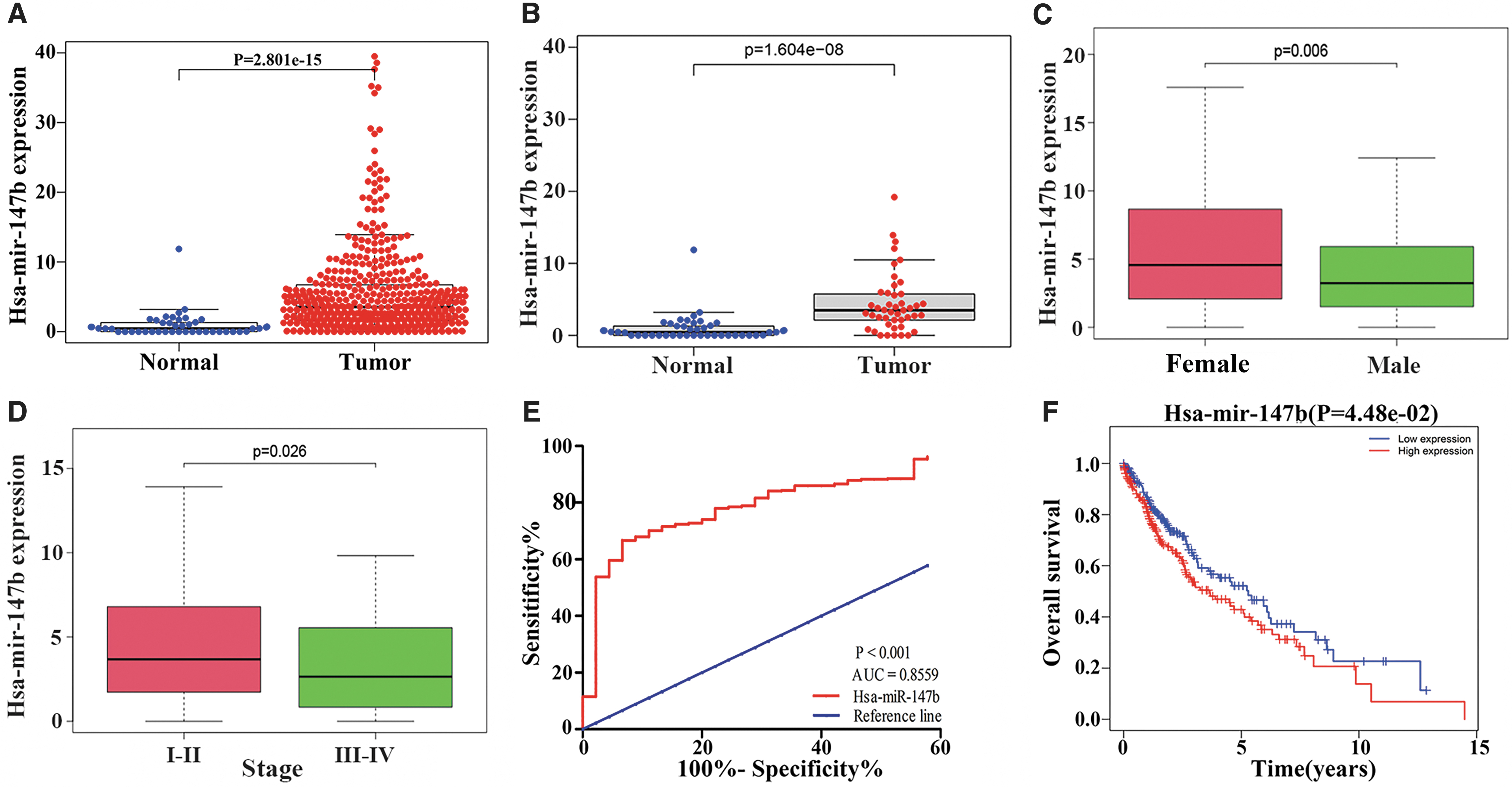
The clinical significance of the increase in miR-147b expression in LUSC tissues.
Bioinformatics analysis of miR-147b target genes
Using the miRwalk 2.0 software to predict miR-147b target genes, it was found that there were 2704 target genes with a targeting relationship. Furthermore, there were 428 differentially expressed genes in the TCGA database (fold change >2, p < 0.05) (Fig. 2 and Table 1). Among these, our visualized the top 15 differentially expressed genes in the form of a heatmap (Fig. 3). To further determine the potential function and molecular mechanism of miR-147b in the development of LUSC, our analyzed the GO, KEGG, and PPI of 428 differential target genes. The GO and KEGG analysis revealed that specific DNA binding, drug transport, cell differentiation, the calcium signaling pathway, and the Ras signaling pathway were of great significance (Fig. 4A, B). In addition, our introduced 428 differential genes into the STRING database to explore the interaction between miR-147b target genes, and Cytoscape 3.6.1 was used for the visualization (Fig. 5).
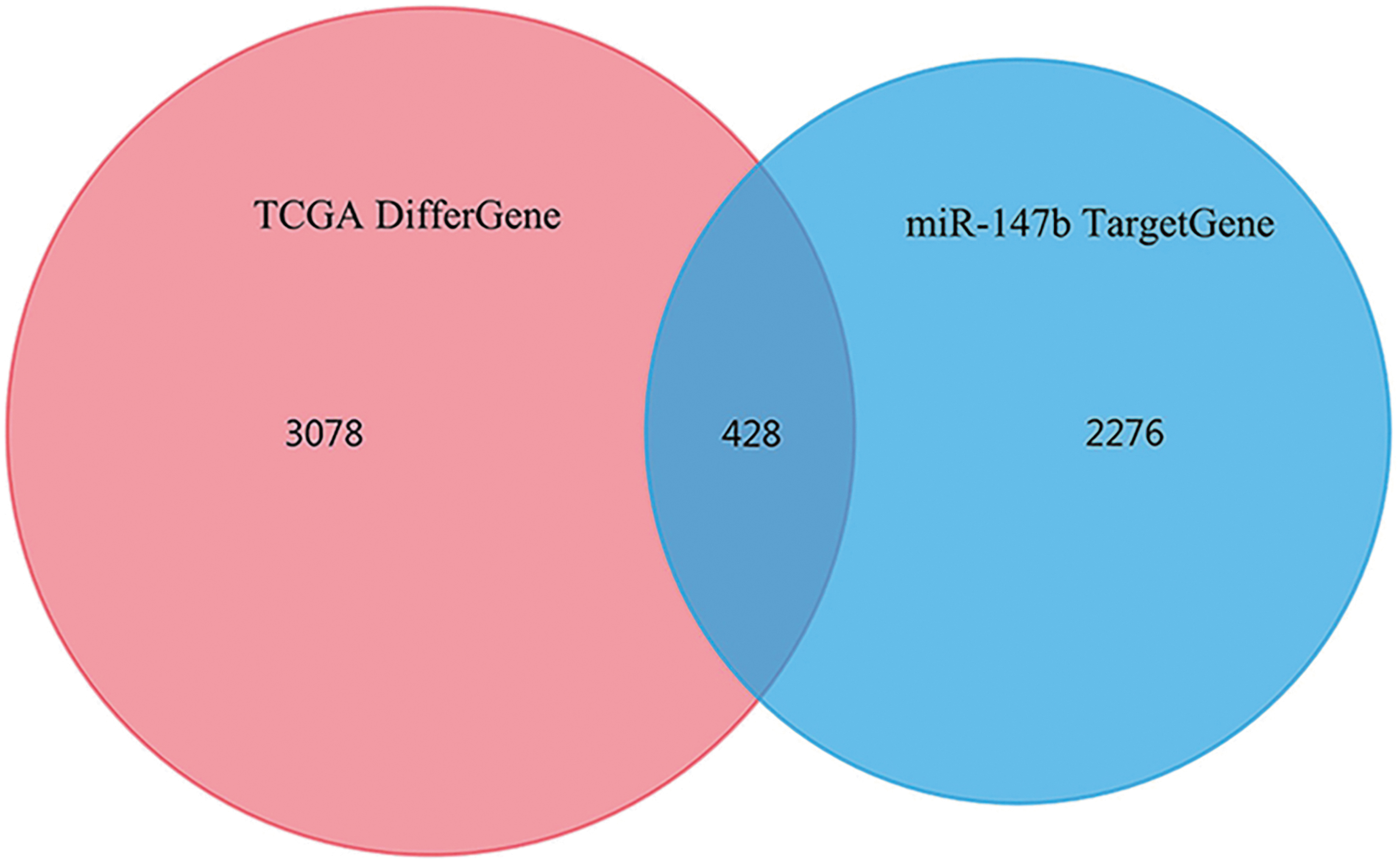
The Venn diagram of differentially expressed genes of LUSC from the TCGA database and the miR-147b target genes.
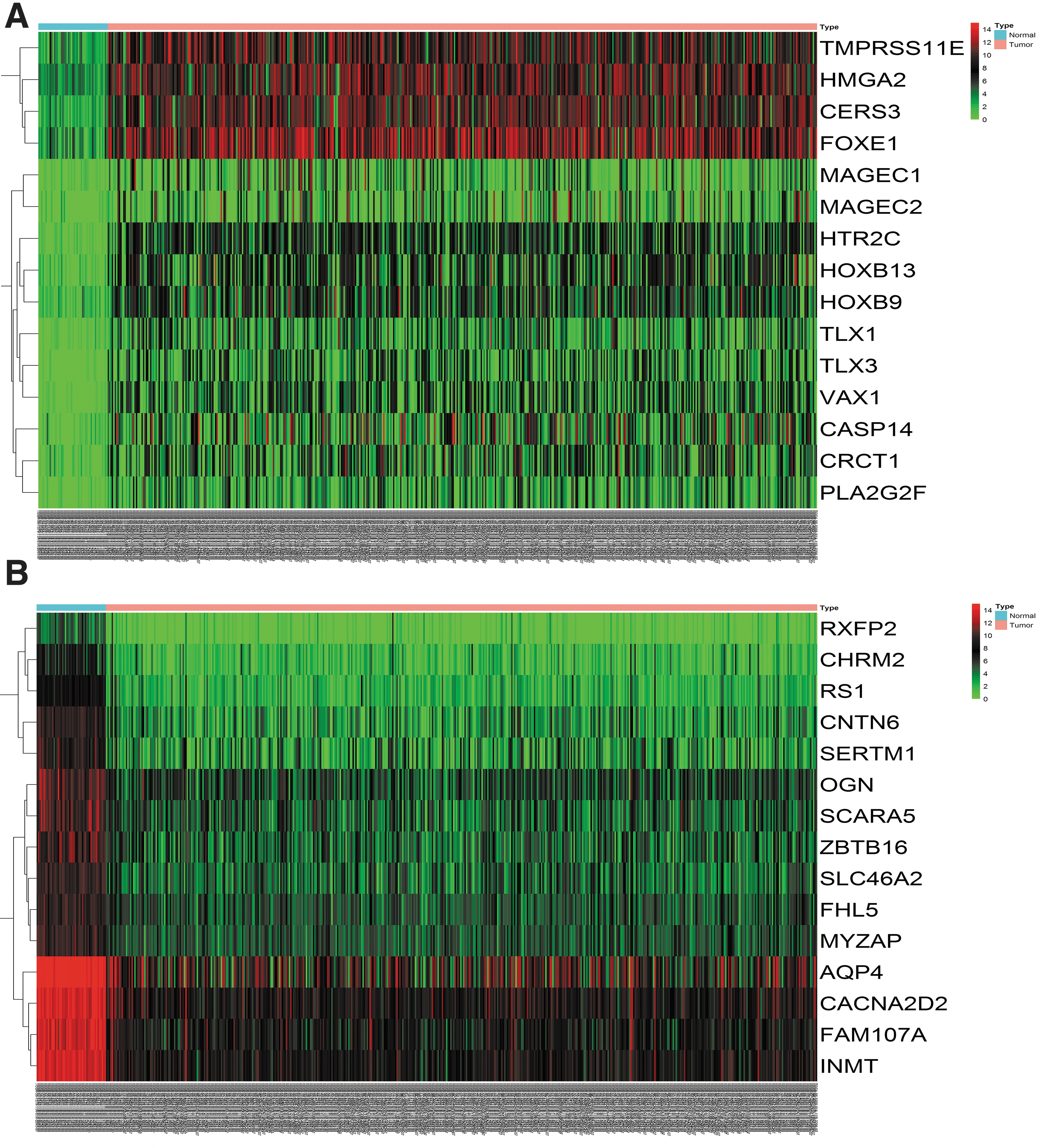
The heatmap shows the top 15 differentially expressed target genes in the TCGA database.

Biological functions of the miR-147b target genes.

The construction of the PPI network for differential target genes (combined score >0.7). PPI, protein–protein interaction.
Overlapping Genes of Differentially Expressed Genes in the TCGA Database and miR-147b Target Genes
TCGA, The Cancer Genome Atlas.
Kaplan-Meier survival analysis of the miR-147b target genes
Through the Kaplan-Meier survival analysis of 428 differential target genes, it was found that 70 target genes were significantly correlated with OS in patients with LUSC, such as CITED2, SLC35G1, CCDC68, FHL5, CHODL, ANO5, RBBP8NL, HES7, GREM2, HES6, and CYP2S1 (Fig. 6 and Table 2). The differential expressed genes were shown in Table 2.
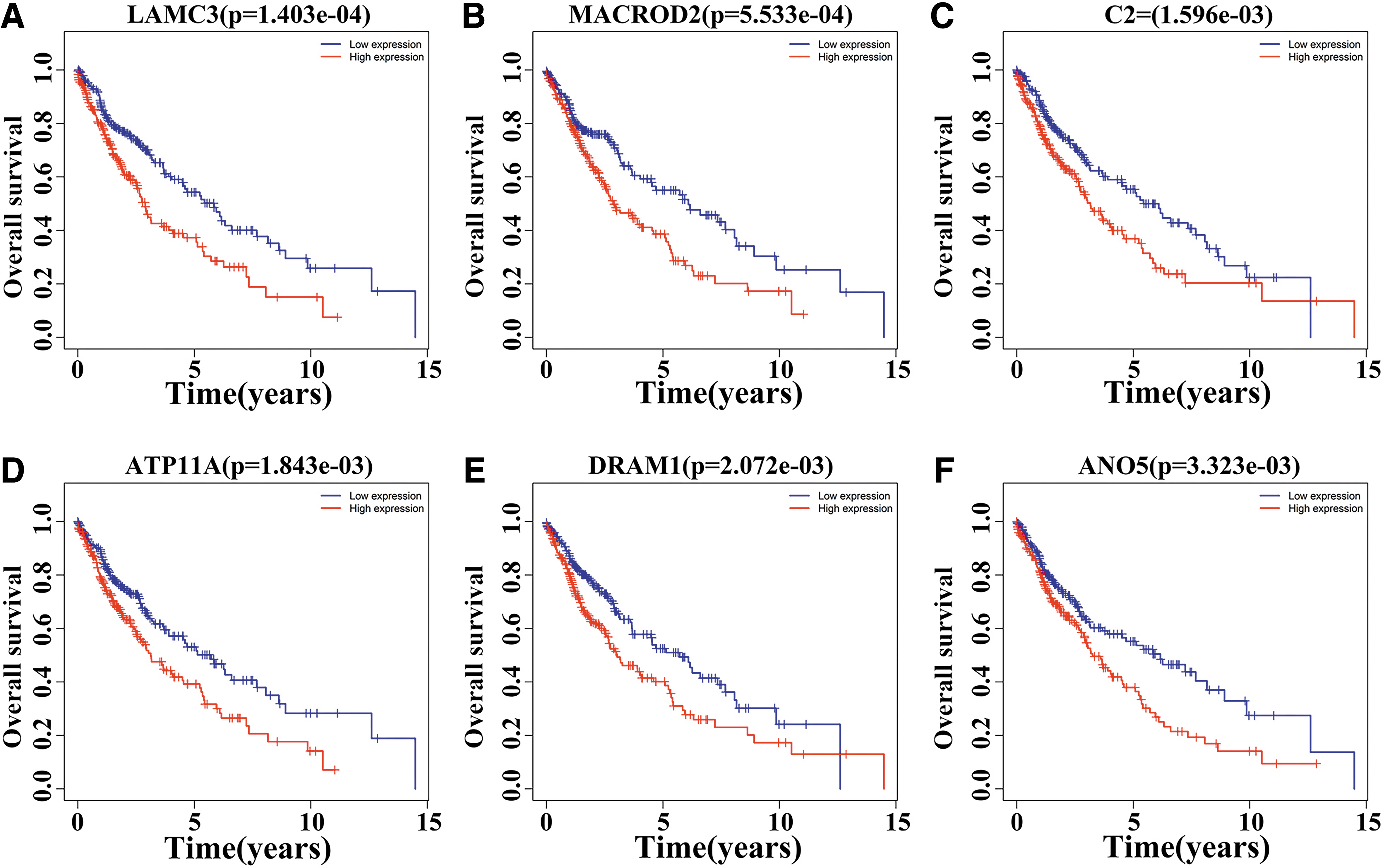
The top 6 miR-147b target genes associated with OS in LUSC patients.
The Expression and Prognosis of 70 Target Genes in Lung Squamous Cell Carcinoma Patients
FDR, false discovery rate; logFC, logFoldchange; LUSC, lung squamous cell carcinomas; OS, overall survival.
Construction of Cox risk proportion model
The univariate Cox regression analysis revealed that there were 48 target genes (PKNOX2, KLF6, CCDC68, PTGIS, SLC35G1 SUSD4, MYZAP, AKAP2, RGMA, TIMP3, DRAM1, MACROD2, FLT4, ZBTB16, FHL5, KCNK3, ATP11A, SLC46A2, CYP2S1, LAMC3, CITED2, ABCC5, MRAP2, ARC, DCDC2, OTX1, PLA2G4D, TMEM204, C2, SHISA9, CD93, ESCO2, INMT, NTRK3, FAM84A, ANO5, IQSEC3, PTGDS, PFN2, CHODL, HTR2C, FOXD3, AK1, SORBS1, SHROOM4, MFAP4, SLC8A3, and GREM2) correlated with OS in LUSC, which were statistically significant (Table 3). The multivariate Cox regression analysis revealed that six mRNAs were risk factors for prognosis in patients with LUSC. Risk score = (PTGIS*2.805) + (PLA2G4D*2.288) + (SUSD4* −1.991) + (ARC*1.527) + (HTR2C* −1.753) + (SHISA9* −1.499). 20
The Univariate Cox Regression Analysis of Target Genes Related to the Prognosis of Lung Squamous Cell Carcinoma Patients
HR, hazard ratio.
The patients were divided into two groups according to the risk score. The Kaplan-Meier survival analysis revealed that the survival rate in the low-risk group was significantly higher than that in the high-risk group (p < 0.05, Fig. 7A). Patients in the low-risk group might have higher levels of SUSD4, SHISA9, and HTR2C. In contrast, patients in the high-risk groups tended to have higher levels of PTGIS, PLA2G4D and ARC (Fig. 7B). The time-dependent AUC was 0.673 (p < 0.05, Fig. 7C).

The Cox risk proportional model.
Discussion
Previous studies have found that the occurrence of LUSC is correlated with the activation of proto-oncogenes and the inactivation of various miRNA-regulated tumor suppressor genes, which would lead to the progression and metastasis of LUSC. A study found that the expression level of miRNA-223 in serum of patients with NSCLC significantly decreased. In addition, miRNA-223 inhibited NSCLC cell proliferation and induced apoptosis by regulating the EGFR/PI3K/AKT signaling pathway. 21 The expression of miR-218 was significantly downregulated in lung cancer. MiR-218 inhibited the NSCLC cell proliferation, invasion, colony formation, and tumorigenesis in nude mice by targeting the IL-6/JAK/STAT3 signaling pathway. 22 The expression of miRNA-383 was significantly downregulated in lung cancer tissues and cells. MiR-383 could directly target endothelial Pas domain protein 1 (EPAS1) to inhibit the migration, invasion, and tumorigenesis of human lung cancer cells in nude mice, and exert the function of tumor suppressor. 23 In addition, the expression level of miR-577 in NSCLC was significantly downregulated, which could inhibit the proliferation, migration, invasion, and epithelial-stromal transformation of NSCLC cells through the Wnt/β-catenin signaling pathway, and this might play an important role in NSCLC. 24
Studies have found that miR-147b can inhibit and promote the occurrence and development of cancer. For example, Xu et al. reported that miR-147b was elevated in thyroid cancer tissues and cells. The interference with the miR-147b expression could reduce the proliferation, colony formation, and invasion ability of thyroid cancer cells. 25 Yi et al. reported that the expression of miR-147b decreased in CRC tissues, and that this was an independent risk factor for the poor prognosis of CRC patients. The elevated expression of miR-147b inhibited CRC cell proliferation in vitro and tumor growth in vivo. 12,26 Surprisingly, Ning et al. reported that the expression of miR-147b was significantly downregulated in NSCLC tissues and cells. Increasing the expression of miR-147b could reduce the proliferation, colony formation, and invasion ability of NSCLC cells. 27 However, Feng et al. reported that the expression of miR-147b was significantly elevated in lung adenocarcinoma (LUAD) tissues and cells, and that the prognosis of LUAD patients with an elevated miR-147b expression was poor. The elevated miR-147b expression promoted the LUAD cell proliferation, clone formation, migration, and invasion. 28 The results of these two were not consistent. This indicates that the expression of miR-147b in LUSC tissues and the value of miR-147b in the diagnosis and prognosis of LUSC need to be further studied. Therefore, our study used the TCGA database to analyze the role and prognostic value of miR-147b and its target gene regulatory network in the progression of LUSC, find new biomarkers, and provide new ideas and methods for the diagnosis and treatment of LUSC patients.
It was found that the expression of miR-147b in LUSC tissues was significantly elevated, and was associated with the gender, stage, and diagnosis of LUSC patients by Wilcoxon signed-rank test, logistic regression analysis, and ROC analysis. The Kaplan-Meier survival analysis revealed that the prognosis of LUSC patients with high miR-147b expression was worse than those with low miR-147b expression. This would provide a new field of vision and new direction for the targeted therapy of patients with LUSC.
In this study, our applied the GO and KEGG analysis to understand the biological function of miR-147b. The GO and KEGG analysis revealed that miR-147b target genes were involved in drug transport, specific DNA binding, calcium signaling pathway, Ras signaling pathway, and so on. The calcium signaling pathway played an essential role in cell senescence, cell proliferation, embryonic development, wound healing, cancer, and other pathophysiological conditions. 29 For example, the expression of Rap2B increased in breast cancer cell lines and normal breast cell lines. Furthermore, Rap2B could increase the level of intracellular calcium, promote the phosphorylation of extracellular signal-related kinase (ERK)-1, and finally promote the ability of cell proliferation, migration, and invasion. 30 In addition, Ras signaling pathway was closely correlated with the occurrence and development of lung cancer. Interfering with the AMPH-1 gene expression could activate the Ras-Raf-MEK-ERK signaling pathway, which could significantly increase cell proliferation, reduce apoptosis, and promote cell cycle progression. 31 Nogo-B receptor promoted the epithelial-stromal transformation of NSCLC cells through the Ras/ERK/SNAIL1 pathway. 32 Therefore, it is reasonable to speculate that the miR-147b-mRNA regulatory network plays a very critical role in the occurrence and development of lung cancer.
This study deepens the authors' understanding of the treatment and prognosis of LUSC at the molecular level, improves the understanding of the pathogenesis and potential molecular regulation of LUSC, and contributes to the timely diagnosis and prognosis of patients with LUSC. Furthermore, this lays a foundation for future clinical research. However, it is worth emphasizing that the mechanism of miRNA and the regulatory network of miRNA-mRNA interaction is particularly complex. Our provided theoretical knowledge and the analysis of clinical results. However, more scientific research is needed to confirm these findings, and investigate the potential value for clinical application, to improve the survival and prognosis of patients with LUSC.
Conclusions
In summary, the high expression of miR-147b is associated with poor prognosis in patients with LUSC, which may be a potential molecular marker for poor prognosis in patients with LUSC.
Footnotes
Availability of Data and Materials
The datasets generated for this study are available on request to the corresponding author.
Authors' Contributions
X.X. and T.-Y.F. designed this study and checked the data processing. D.K. drafted the article and analyzed the data. Q.G. explained the results and critically revised the important academic content in the article. All authors read and approved the final article.
Disclosure Statement
The patient samples were obtained from the TCGA database, and did not need to be approved by the ethics committee.
Funding Information
This study was supported by the Science and Technology Bureau of Zunyi city, Guizhou province [No. (2018) 59].