
Abstract
Background:
The goal of this research was to investigate the feasibility of 64Cu labeling in prostate-specific membrane antigen imaging and therapy (PSMA I&T) for PSMA positron emission tomography (PET) imaging and biodistribution evaluation.
Materials and Methods:
PSMA I&T was labeled with 64Cu, and stability in human and mouse sera was evaluated. Prostate cancer cell lines were used for specific uptake assays (22RV1 for PSMA-positive, PC-3 for -negative). Both PC-3 and 22RV1 cells were transplanted into the left and right thighs in a mouse for PET/computed tomography (CT) imaging. Biodistribution was performed using 22RV1 tumor models.
Results:
Labeling yield (decay corrected) of 64Cu-PSMA I&T was more than 95% compared to the free 64Cu peak. The serum stability of 64Cu-PSMA I&T was maintained at more than 90% until 60 h. Regarding the specific binding of 64Cu-PSMA I&T was 7.5-fold higher to 22RV1 cells than PC-3 cells (p < 0.001). On PET/CT imaging, more specific 64Cu-PSMA I&T uptake was observed to 22RV1 tumors than to PC-3 tumors. In the PSMA blocking study using 2-phosphonomethoxypropyl adenine (2-PMPA), the 64Cu-PSMA I&T signal significantly decreased in the 22RV1 tumor region. In the biodistribution study, the kidney uptake was the highest among all organs at 2 h (52.6 ± 20.8%ID/g) but sharply decreased at 24 and 48 h. Also, the liver showed similar uptake over time (range, 10–12%ID/g). On the contrary, 64Cu-PSMA I&T uptake of the tumors increased with time and peaked at 48 h (5.6 ± 0.1%ID/g).
Conclusions:
PSMA I&T labeled with 64Cu showed the feasibility of the PSMA specific PET imaging through in vitro and in vivo studies. Furthermore, 64Cu-PSMA I&T might be considered as the candidate of future clinical trial.
Introduction
Prostate-specific membrane antigen (PSMA, also known as glutamate carboxypeptidase II) is a transmembrane protein that has been reported to be overexpressed in castration-resistant prostate cancer (PC). 1,2 Consequently, PSMA is a potential imaging and therapeutic biomarker for the evaluation of cancer progression and targeted radioligand therapy. 3,4
Since the development of PSMA inhibitors in targeting PCs, various radioligands for imaging and therapy have been applied in cancer patients. 5 –12 Among these PSMA inhibitors, PSMA imaging and therapy (PSMA I&T), which was developed by Weineisen et al. based on 1,4,7,10-tetraazacyclodocecane,1-(glutaric acid)-4,7,10-triacetic acid (DOTAGA) structure, has been studied in prostate cancer imaging and therapy from preclinical to clinical research using gallium (68Ga) and lutetium (177Lu). 13 –16 Positron emission tomography (PET) imaging using 64Cu is promising rather than 68Ga because of its relatively long half-life (12.7 h) and economic production by cyclotron. 17 In addition, 64Cu revealed higher image resolution than 68Ga due to lower energy at emission. For these reasons, it could be applicable at delayed time points and has a better image quality.
Regarding 64Cu labeled PSMA agents, some researchers revealed the feasibility of 64Cu-PSMA-617 through clinical trials. 18 –21 They demonstrated that 64Cu-PSMA-617 can show high diagnostic accuracy 20 and perform better than 18 F-choline in the case of low PSA values. 19 However, 64Cu-PSMA-617 is not readily available. Therefore, the authors searched for other PSMA inhibitors for 64Cu labeling and they found out that PSMA I&T can be a good candidate for 64Cu labeling, although a 64Cu-labeled PSMA I&T study has not been conducted yet. In terms of chemical structure of PSMA I&T, DOTAGA chelator has been reported to improve ligand pharmacokinetics and play a crucial role in high affinity on the basis of high tumor accumulation. 13,22,23 Also, this structure showed much lower clearance than other chelators such as NOTA and NODAGA. 24 This means that PSMA I&T may be more suitable for labeling with a relatively long half-life, such as 64Cu, rather than 68Ga. 25
In this study, the authors performed 64Cu labeling with PSMA I&T for targeted imaging using PET. Specifically, they observed the serum stability of 64Cu-PSMA I&T and evaluated its specific binding in prostate cancer cells in proportion to PSMA expression. In addition, they established mouse xenograft models to perform specific targeted imaging with PET/computed tomography (CT), and evaluated the organ distribution patterns of 64Cu-PSMA I&T in tumor models.
Materials and Methods
Cell culture
Human prostate cancer cell lines (22RV1; PSMA-positive, and PC-3; PSMA-negative) were used. Both cells were obtained from the American Type Culture Collection (ATCC) and maintained in RPMI 1640 with 10% fetal bovine serum (Gibco) containing 1% antibiotics (penicillin G, 100 U/mL, and streptomycin 10 μg/mL; Gibco). Cells were incubated at 37°C in a 5% CO2 atmosphere.
Radiolabeling of 64Cu-PSMA I&T
PSMA I&T, purchased from the Technical University of Munich, was synthesized as described previously. 13 For PSMA I&T labeling, the 64Cu eluate 26 (481 kBq) was added to a labeling vial containing 1 mM sodium acetate buffer (pH 5.5) and 1 nmol PSMA I&T. Final volume was ranged from 150 to 200 μL. The reaction mixture was heated for 5 min at 42°C. Incorporation yield was determined by instant thin-layer chromatography (iTLC; Agilent Technologies) using 0.1 M citric acid (pH 5.5). The incorporation yield always exceeded 95%. For the in vivo study, 64Cu-PSMA I&T was labeled according to 481 kBq of 64Cu eluate per 1 nmol ratio at pH 5.5 and diluted with sodium chloride solution (0.9%).
Stability test
The stability of 64Cu-PSMA I&T was analyzed through iTLC after incubation in human and mouse sera, and phosphate-buffered saline (PBS) at 37°C for various times (1, 2, 4, 24, 48, and 60 h).
In vitro uptake assay of 64Cu-PSMA I&T
Uptake was examined using 3.75 nM of 64Cu-PSMA I&T in an assay medium (serum-free medium) with or without 100 μM 2-phosphonomethoxypropyl adenine (2-PMPA) (Sigma-Aldrich) for blocking studies. Incubation was performed for 2 h at 37°C. Assay protocol was described in a previous study. 27 In brief, the cells were washed twice with cold PBS containing 1% bovine serum albumin and lysed with 1% sodium dodecyl sulfate (SDS). The radioactivity of the cell lysates was measured using a γ-counter. Radioactivity was normalized to the amount of total protein at the time of the assay. All experiments were performed at least in triplicates.
Animal experiments
Six-week-old male BALB/C nude mice were obtained from NARA Bio, Inc., All animal experiments were approved by the Institutional Animal Care and Use Committee of KIRAMS (2019-0032). For establishment of prostate cancer xenograft mouse models, 22RV1 cells (5 × 10⁶/100 μL mixed cultured medium with Matrigel [Corning, Inc.]) were transplanted subcutaneously into the right thigh. For comparison between negative and positive tumors based on 64Cu-PSMA I&T targeting, PC-3 and 22RV1 cells were transplanted into the left and right thighs of a mouse, respectively. Small animal PET imaging and biodistribution studies were performed when tumor sizes were more than 0.5 cm in diameter.
Small-animal PET/CT imaging
PET/CT images of tumor-bearing mice were acquired by PET/CT (INVEON scanner; Siemens Healthcare). At 2, 24, and 48 h after intravenous injection (i.v.) of 8.1–8.9 MBq of 64Cu-PSMA I&T per head of 22RV1 xenograft model only group (n = 4), images were obtained for 20 min under inhalation anesthesia (isoflurane, 1.5%). In addition, both PC-3 and 22RV1 xenograft models (n = 2) were obtained at 24 and 48 h after i.v. of 18 F-FDG (5.55 MBq) as well as 64Cu-PSMA I&T (7.2–7.4 MBq) with or without 800 μM 2-PMPA to assess the specific binding of PSMA. Images were reconstructed and analyzed using the INVEON software and AMIDE algorithm (A Medical Image Data Examiner).
Biodistribution study
Biodistribution studies were performed to evaluate the uptake of 64Cu-PSMA I&T in tumor-bearing mice (n = 3, at each time point). All mice were intravenously injected with 2.0 MBq of 64Cu-PSMA I&T. Each group was sacrificed at 2, 24, and 48 h after i.v. Various organs, including tumor and blood samples, were weighed and radioactivity was measured. The γ-counter data were represented by the percentage of injected dose per gram of tissue (%ID/g).
Statistical analyses
All statistical analyses were performed using GraphPad Prism software. All data were evaluated as means ± standard deviation and are representative of at least independent two-times biological experiments performed as quadruple per one set. Statistical significance between the groups was compared using one-way analysis of variance and unpaired Student's t-test. p < 0.05 was considered statistically significant.
Results
Preparation of 64Cu-PSMA I&T and in vitro uptake assay
To evaluate the radiolabeling efficiency of PSMA I&T with 64Cu, the authors measured 64Cu-PSMA I&T products by iTLC. In Figure 1A, 64Cu-PSMA I&T was successfully labeled (more than 95%) compared to the free 64Cu peak. They also evaluated serum stability in human and mouse sera, as well as PBS (pH 7.4). The stability of 64Cu-PSMA I&T was maintained at more than 90% until 60 h (Fig. 1B). To confirm the 64Cu-PSMA I&T binding (uptake rate) in proportion to PSMA expression, they treated 3.75 nM of 64Cu-PSMA I&T into PC-3 and 22RV1 cells with or without 100 μM of 2-PMPA. This result showed that 22RV1 cells were 7.5-fold higher than PC-3 cells (p < 0.001). With respect to the blocking study, 22RV1 cells with the 2-PMPA application were 10.6-fold lower than those without 2-PMPA application (p < 0.01) (Fig. 1C). On the contrary, there was no difference in PC-3 cells between the PMPA application group and the PMPA nonapplication group [p = 0.265]). These results demonstrated that 64Cu-PSMA I&T was stably labeled and specifically targeted in proportion to PSMA expression.

Preparation of 64Cu-PSMA I&T
In vivo PET/CT imaging of 64Cu-PSMA I&T
To evaluate the PSMA-specific targeted imaging using 64Cu-PSMA I&T in in vivo, they established tumor xenograft models using PC-3 and 22RV1 cells into both thighs from a mouse, and confirmed both tumors using 18 F-FDG PET/CT for evaluation of tumor prognosis. This result showed 18 F-FDG uptake in both tumors in the xenograft model (Supplementary Fig. S1A–C). Next, they evaluated the targeted imaging using 64Cu-PSMA I&T between the tumors. Through the maximum intensity projection image as well as static images, 64Cu-PSMA I&T-specific uptake was higher in the 22RV1 tumor region than in the PC-3 tumor (Fig. 2). On the contrary, in the coinjection groups with 800 μM of 2-PMPA, 64Cu-PSMA I&T uptake was significantly decreased in the 22RV1 tumor region (Supplementary Fig. S2). These results demonstrated that tumors with high PSMA expression could be clearly visualized by 64Cu-PSMA I&T PET/CT.
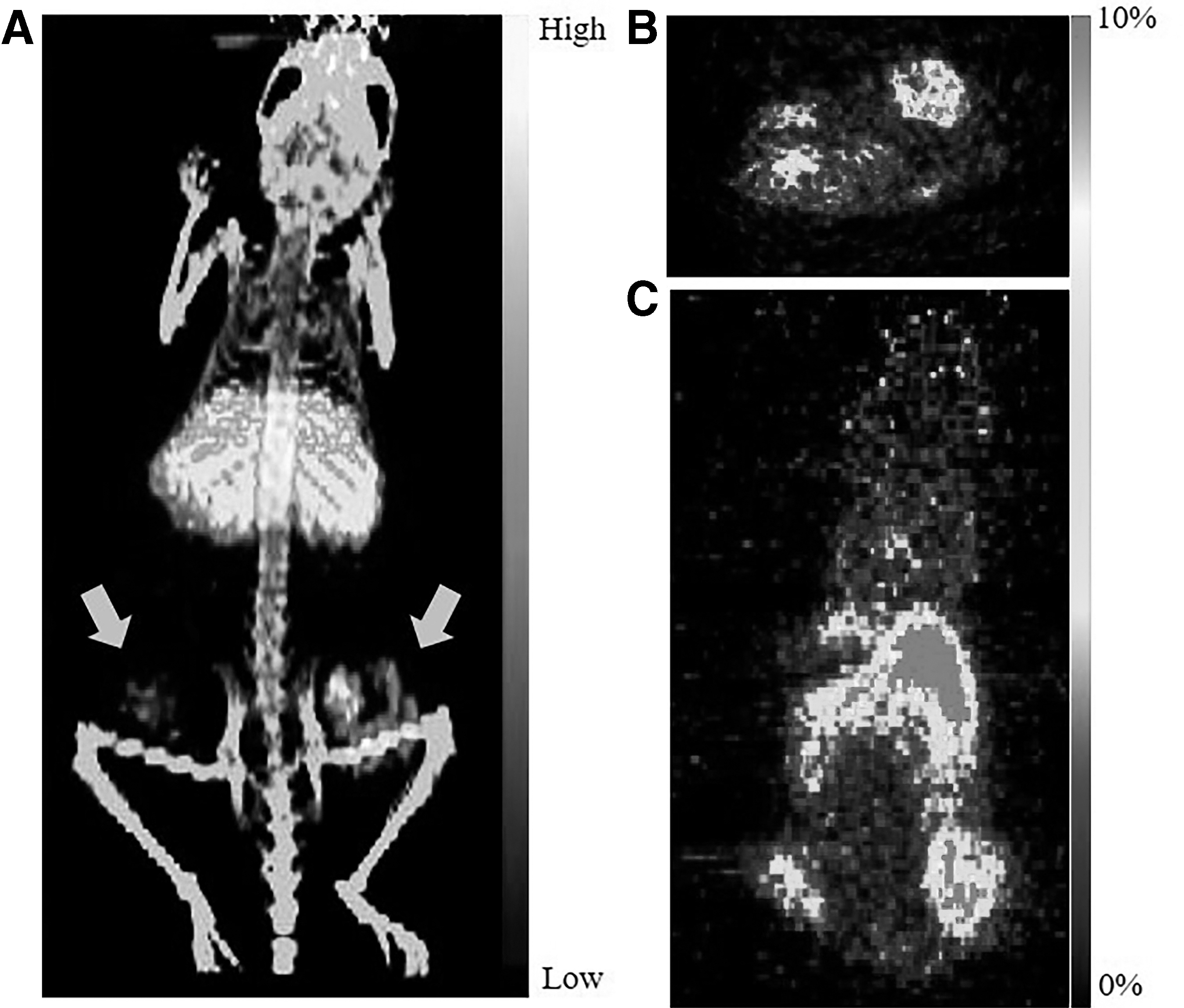
64Cu-PSMA I&T PET/CT imaging at 24 h after the intravenous injection (∼7.4 MBq)
In vivo biodistribution of 64Cu-PSMA I&T
The authors established tumor xenograft models using only 22RV1 cells to confirm the in vivo distribution patterns of 64Cu-PSMA I&T at various time points. In the PET/CT imaging, the authors observed not only specific tumor imaging targeted PSMA but also a decreasing pattern of 64Cu-PSMA I&T uptake, except for the liver (Fig. 3A). Based on the PET/CT imaging results, they evaluated the in vivo distribution patterns of 64Cu-PSMA I&T (%ID/g) in tumor-bearing mice. Table 1 and Figure 3B show that kidney uptake at 2 h after i.v. was highest (52.6 ± 20.8), but sharply decreased at 24 h (15.6 ± 3.9) and 48 h (9.2 ± 2.5), and liver uptake was similar at all three time points (ranges from 10 to 12). On the contrary, 64Cu-PSMA I&T uptake in the tumors increased with time and peaked at 48 h (5.6 ± 0.1). In addition, the tumor-to-liver ratio was similar at 24 and 48 h (Fig. 3C). However, the tumor-to-kidney ratio at 48 h (0.61% ± 0.05%) was the highest among those times (p < 0.05) (Fig. 3D). Consequently, these results showed the feasibility of PSMA imaging using 64Cu-PSMA I&T PET/CT.
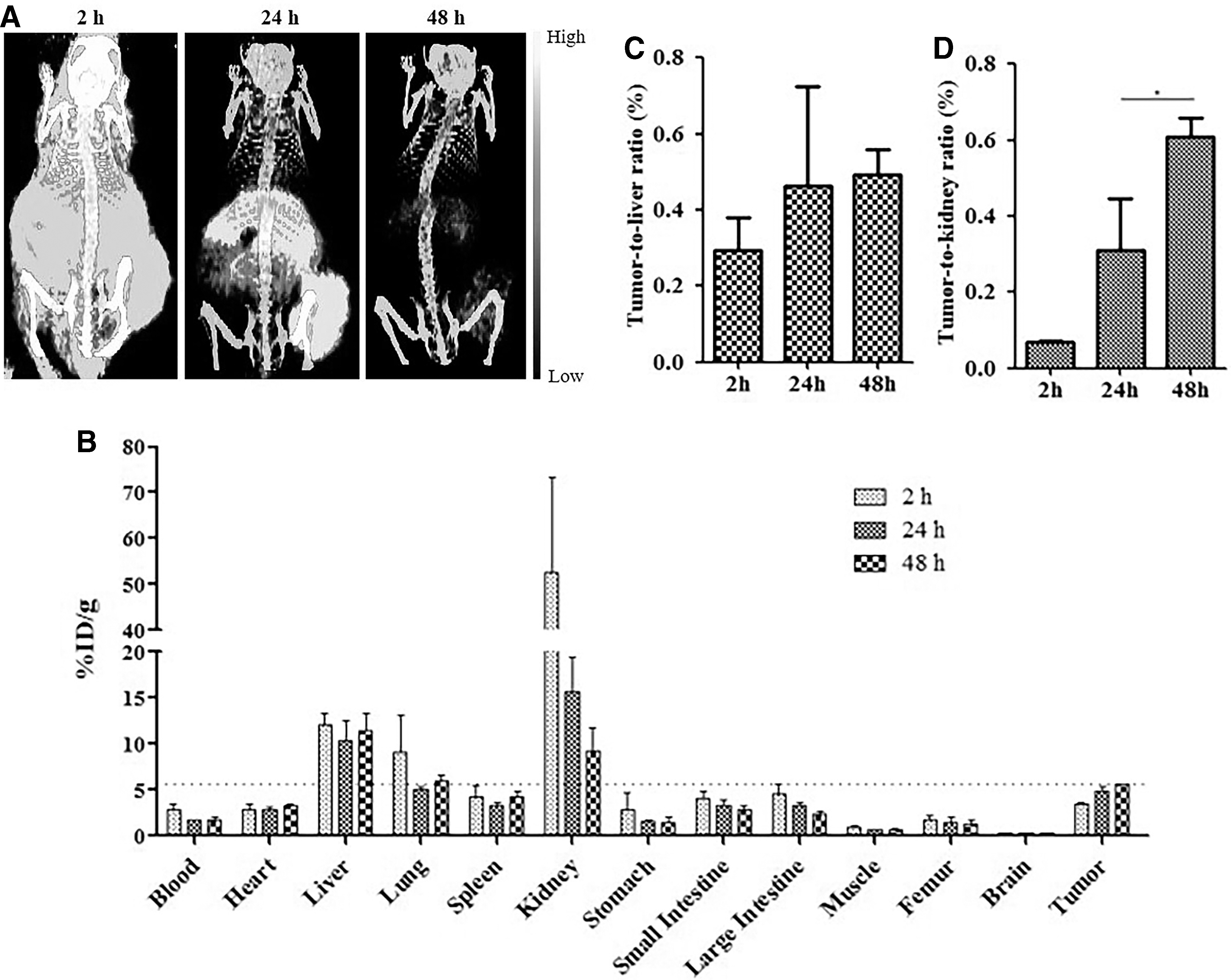
In vivo evaluation of 64Cu-PSMA I&T in 22RV1 tumor models.
Biodistribution Results of 22RV1 Tumor-Bearing Mice After Intravenous Injection of 64Cu-PSMA I&T (%ID/g)
Discussion
The aim of this study was to evaluate 64Cu-labeled PSMA I&T in optimizing in vivo PET imaging. The authors successfully confirmed the serum stability of 64Cu-PSMA I&T using in vitro-specific binding and in vivo experiments such as PSMA PET imaging and biodistribution studies.
The development of efficient and applicable radiopharmaceuticals using radiometal isotopes has played an important role in diagnostic imaging and therapy. PSMA I&T was first introduced as a radiopharmaceutical of PSMA ligand using 68Ga for diagnosis. However, this radioisotope showed limitations of short half-life and relatively poor image quality. 13,14 To overcome these limitations, the authors searched for methods of using 64Cu, which is made especially useful because of the possibility of imaging and therapeutic surrogate radiometal isotope, 28 –30 and successfully established a technique for labeling 64Cu-PSMA I&T (Fig. 1A, B) and confirmed specific targeting of 64Cu-PSMA I&T to PSMA using in vitro assessment (Fig. 1C).
As mentioned above, 68Ga-labeled PSMA I&T have been used as representative radio-tracers for PET imaging. These studies showed that tumor uptake 1 h after i.v. was 4.95 ± 1.6%ID/g in the LNCaP (PSMA-positive prostate adenocarcinoma cell line) xenograft model. 13,14 On the contrary, tumor uptake of 64Cu-PSMA I&T at 48 h after i.v. (5.6 ± 0.1%ID/g) was slightly higher than that of 68Ga-labeled PSMA I&T, although at different time points (Fig. 3B and Table 1). This result means that 64Cu-PSMA I&T is comparable to 68Ga-PSMA I&T for PET imaging, and 64Cu-PSMA I&T can be utilized for PSMA PET imaging. 31
PSMA-targeting ligands have been labeled with 64Cu, which is a more favorable radioisotope for PET imaging. Among these radiopharmaceuticals, 64Cu-PSMA-617 has been reported as the most promising radiopharmaceutical in targeting PSMA-positive prostate cancer as well as gastric adenocarcinoma. 17,32 –35 In addition, Cantiello et al. showed the high diagnostic accuracy of 64Cu-PSMA-617 PET/CT for primary lymph node staging. 20 This means that 64Cu-labeled PSMA targeting agents play a crucial role in PET imaging, although the high uptake in the liver and gallbladder has been described as a main problem. 21 The results also showed that uptake by the kidney and all the organs decreased with time, while liver uptake remained (Fig. 3B and Table 1). High uptake in the liver is due to the hepatic metabolism related to 64Cu uptake and excretion. 36,37 In addition, 64Cu has been known to show high-quality images and spatial resolution. 38,39 Therefore, they suggest that 64Cu-PSMA I&T can be used for specific PSMA PET imaging.
In addition to prostate cancer, PSMA expression associated with neovasculature has been reported in the 68Ga/64Cu PSMA uptake region. 35,40,41 With respect to neovasculature uptake, Han et al. described that 64Cu-PSMA-617 uptake was observed in PC-3 tumor xenograft models (3.47 ± 0.48%ID/g at 24 h) as known for the PSMA-negative cell line using PET imaging. 35 Accordingly, it is possible to explain one of the reasons why 64Cu-PSMA I&T showed uptake in the PC-3 tumor (Fig. 2).
Regarding targeted therapy using PSMA I&T, some studies reported that 177Lu-PSMA I&T treatment in patients had mild toxicity and effective antitumor activity, 16 and 213Bi-PSMA I&T treatment as an alpha therapy showed DNA double-strand breaks in PSMA-positive tumor xenograft models. 42 It is possible to use 64Cu-PSMA I&T as a surrogate for 67Cu-PSMA I&T because of its analogous characteristics. 28 –30 67Cu is an attractive candidate for therapeutic radioisotopes because it emits particles with a maximum energy ranging from 0.4 to 0.6 MeV, with a good half-life (2.6 d). Similarly, 64Cu-PSMA I&T could be used to evaluate the dosimetry of 67Cu-PSMA I&T. Furthermore, 64Cu-PSMA I&T could be applied to simulate the distribution of 225Ac-PSMA I&T because 64Cu showed similar characteristics to 225Ac as a metal. Therefore, the authors expect to use 64Cu-PSMA I&T to calculate the dosimetry of 225Ac-PSMA I&T, although its characteristics are not exactly the same as those of 225Ac-PSMA I&T. Currently, they have confirmed the cell-binding assay and radio-labeling of 225Ac-PSMA I&T (data not shown) and hope to perform clinical trials using 225Ac-PSMA I&T in the near future.
Conclusions
PSMA I&T labeled with 64Cu showed the feasibility of the PSMA-specific PET imaging through in vitro and in vivo studies. Furthermore, 64Cu-PSMA I&T might be considered as a candidate of future clinical trial.
Editorial
This study was performed in accordance with the ethical standards of the authors' institutions and with the Declaration of Helsinki 1964 and its later amendments or comparable ethical standards.
Footnotes
Authors' Contributions
This study was designed by C.-H.L., I.L., S.-K.W., K.I.K., K.C.L., K.S., C.W.C., and S.M.L. Data acquisition and analysis were mainly performed by C.-H.L. C.-H.L. and I.L. wrote the article draft. All authors reviewed and edited this final version of the article.
Disclosure Statement
No competing financial interests exist.
Funding Information
This project has been funded, in part, with a grant from the Korea Institute of Radiological and Medical Sciences (KIRAMS), funded by the Ministry of Science, ICT (MSIT), Republic of Korea (no. 50547-2020) and, in part, with grants from the National Research Foundation of Korea (NRF) funded by the Ministry of Science, ICT (MSIT), Republic of Korea (No. 2019M2D2A1A02057204, 2020R1A2C2102492).
Supplementary Material
Supplementary Figure S1
Supplementary Figure S2
References
Supplementary Material
Please find the following supplemental material available below.
For Open Access articles published under a Creative Commons License, all supplemental material carries the same license as the article it is associated with.
For non-Open Access articles published, all supplemental material carries a non-exclusive license, and permission requests for re-use of supplemental material or any part of supplemental material shall be sent directly to the copyright owner as specified in the copyright notice associated with the article.