
Abstract
Objective:
Nuclear casein kinase and cyclin-dependent kinase substrate 1 (NUCKS1) is highly expressed in some tumors, including hepatocellular carcinoma (HCC). However, its clinical significance in HCC prognosis is still unclear. The aim of this study was to explore the expression and prognostic value of NUCKS1 in HCC.
Materials and Methods:
Quantitative real-time polymerase chain reaction was used to detect relative expression of NUCKS1 mRNA in HCC tissues and corresponding adjacent normal tissues. The relationship between NUCKS1 expression and clinical characteristics of patients was analyzed by χ2 test. Kaplan-Meier method and Cox regression analysis were applied to estimate prognostic value of NUCKS1 in HCC.
Results:
Compared with normal ones, the expression of NUCKS1 mRNA was significantly upregulated in HCC tissues (p < 0.001). Besides, NUCKS1 expression was closely associated with tumor differentiation, tumor node metastasis stage, vascular invasion, and metastasis (p < 0.05). Kaplan-Meier analysis revealed that overall survival was obviously longer in HCC patients with low expression of NUCKS1 than those with high NUCKS1 expression (log rank test, p = 0.001). NUCKS1 might be an independent prognostic factor for HCC patients (HR = 1.905, 95% CI = 1.106–3.283, p = 0.020).
Conclusions:
NUCKS1 may be correlated with the progression of HCC and serve as a potential predictive factor for the prognosis of this disease.
Introduction
Hepatocellular carcinoma (HCC) is one of the most aggressive fatal malignant tumors with high incidence and mortality worldwide. 1,2 The tumorigenesis and development of HCC include multiple aspects, such as liver cirrhosis, and hepatitis B virus and hepatitis C virus infection. 3 Surgical resection is a major therapeutic method for HCC patients, but it is not effective for patients at late stage. 4 Although varied treatments and perioperative management have been improved, the prognosis of HCC is still poor due to delayed diagnosis, recurrence, and metastasis. 5,6 Therefore, it is urgent to look for novel and effective biomarkers for the diagnosis and prognosis of HCC.
Nuclear casein kinase and cyclin-dependent kinase substrate 1 (NUCKS1, also named NUCKS) encoding gene NUCKS1 is located on human chromosome 1q32.1 and encodes a highly phosphorylated DNA binding protein. Moreover, it plays different roles through various modifications, such as methylation, ubiquitylation, acetylation, and formylation. 7 The expression of NUCKS1 is observed in almost all mammalian tissues, and NUCKS1 protein is a substrate of some protein kinases in vitro. 8,9 It has been proven to be involved in several cellular processes, including cell proliferation, cell cycle regulation, and the activation of various genes during rapid cell growth. 10,11 A large number of studies also demonstrate that aberrant expression of NUCKS1 emerges in a variety of human cancers and its overexpression is associated with tumor growth and metastasis. 12,13 Cheong et al. mentioned that the expression of NUCKS1 was significantly increased in HCC tissues compared with adjacent noncancerous tissues at the protein level. 14 However, few researches ever discussed prognostic value of NUCKS1 in HCC previously.
In this study, we investigated clinical significance of NUCKS1 in HCC patients through detecting its expression level in malignant and corresponding adjacent normal tissues, and analyzed the correlation between its expression and clinical features of the patients. Moreover, we also explored whether elevated NUCKS1 expression could be a predictive factor for poor prognosis of HCC patients.
Materials and Methods
Patients and samples
In this study, we enrolled 126 HCC patients who underwent surgery in Harrison international Peace Hospital between September, 2013, and March, 2015. Their diagnosis was completed through pathological examinations. None of the patients had received any therapies before surgery. This study was approved by the Ethical Committee of the hospital and written informed consents were signed by all participators in advance.
HCC tissues and corresponding adjacent normal ones (3 cm away from tumor capsule) were collected from these 126 patients. Tissue samples were immediately frozen in liquid nitrogen and stored at −80°C for use. Detailed clinicopathological characteristics of HCC patients, including age, gender, liver cirrhosis, tumor size, alpha-fetoprotein (AFP) level, tumor differentiation, tumor node metastasis (TNM) stage, vascular invasion, and metastasis, are shown in Table 1. Follow-up lasted for 5 years through telephone calls or questionnaires. Patients who died from other diseases or unexpected events were excluded from our study.
Relationship Between NUCKS1 Expression and Clinical Features of Hepatocellular Carcinoma Patients
AFP, alpha-fetoprotein; NUCKS1, nuclear casein kinase and cyclin-dependent kinase substrate 1; TNM, tumor node metastasis.
RNA extraction and quantitative real-time polymerase chain reaction
Total RNA was severally isolated from tissue samples using TRIzol reagent (Invitrogen, Carlsbad, CA). Reverse transcription was performed with PrimeScript™ RT reagent kit (Takara, Dalian, China). SYBR Premix Ex Taq (Takara) was used to detect relative expression of NUCKS1 mRNA in specimens, on Applied Biosystems 7900 Fast Real-Time PCR system (Applied Biosystems, Foster City, CA). β-Actin served as the internal reference and 2−ΔΔCt method was applied to calculate relative NUCKS1 expression. Each sample was examined in triplicate.
Statistical analyses
All statistical analyses were carried out in SPSS 18.0 software (SPSS, Inc., Chicago, IL) and GraphPad Prism 5.0 (GraphPad Software, Inc., San Diego, CA). Data were expressed as mean ± standard deviation. Difference in NUCKS1 expression between two groups was evaluated by Student's t-test. χ 2 test was applied to analyze the relationship between NUCKS1 expression and clinical features of patients. Survival curve was established through Kaplan-Meier method with log rank test. Univariate and multivariate Cox regression analyses were utilized to estimate prognostic value of NUCKS1 in HCC patients. p < 0.05 was considered a statistically significant level.
Results
Clinical characteristics of HCC patients
As shown in Table 1, HCC patients contained 44 females and 82 males, with a mean age of 57.26 ± 15.25 years (ranging from 34 to 77 years). Of 126 patients, 87 had liver cirrhosis. Tumor sizes were less than 5 cm in 71 patients and more than 5 cm in 55 cases. AFP levels were less than 20 ng/mL in 47 patients and more than 20 ng/mL in 79 cases. Tumor differentiation was well or moderate in 68 cases and poor in 58 cases. Among these patients, 66 were at stage I–II and 60 at stage III–IV. Fifty-two patients developed vascular invasion and 45 showed metastasis.
Relative expression of NUCKS1 was upregulated in HCC patients
Quantitative real-time polymerase chain reaction (qRT-PCR) was used to detect relative expression levels of NUCKS1 mRNA in HCC and adjacent normal tissues. Obtained result showed that NUCKS1 expression was significantly increased in HCC tissues compared with corresponding adjacent normal ones (p < 0.001, Fig. 1).
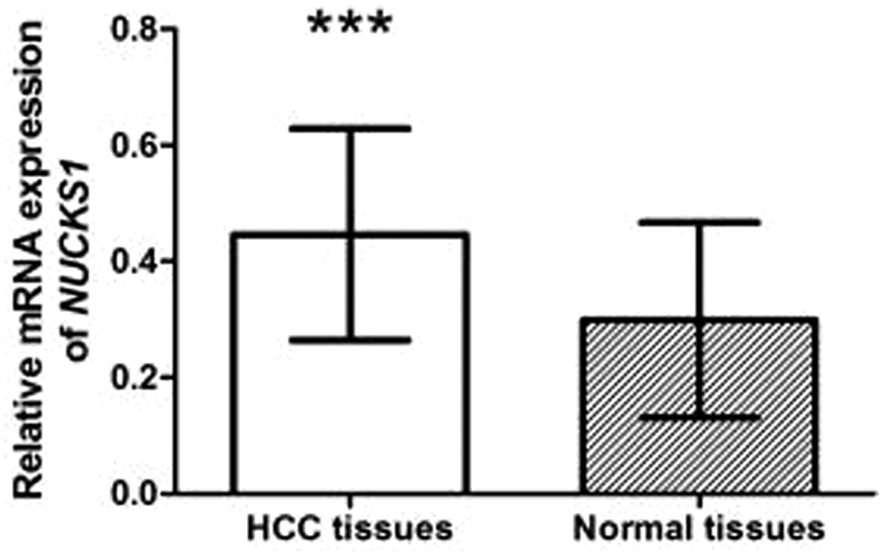
Relative expression of NUCKS1 mRNA in HCC and corresponding adjacent normal tissues. Compared with normal ones, NUCKS1 mRNA expression was obviously upregulated in HCC tissues. *** Indicated p < 0.001. HCC, hepatocellular carcinoma; NUCKS1, nuclear casein kinase and cyclin-dependent kinase substrate 1.
Association between NUCKS1 expression and clinical characteristics of HCC patients
To explore the role of NUCKS1 in HCC, 126 patients were divided into high and low expression groups according to mean expression of NUCKS1 in HCC tissues (0.446 ± 0.016). Then the relationship between NUCKS1 expression and clinical features of the patients was analyzed. NUCKS1 expression was closely associated with tumor differentiation (p = 0.039), TNM stage (p = 0.014), vascular invasion (p = 0.046), and metastasis (p = 0.035) (Table 1). However, there was no obvious correlation between NUCKS1 expression and age, gender, liver cirrhosis, tumor size, or AFP level (p > 0.05, Table 1). Moreover, we found that in patients with I–II stage, but without vascular invasion and metastasis, the expression of NUCKS1 was also significantly higher in diseased tissues than in adjacent normal tissues (p < 0.001, Fig. 2), and so was for those at III–IV stage and with vascular invasion and positive metastasis.
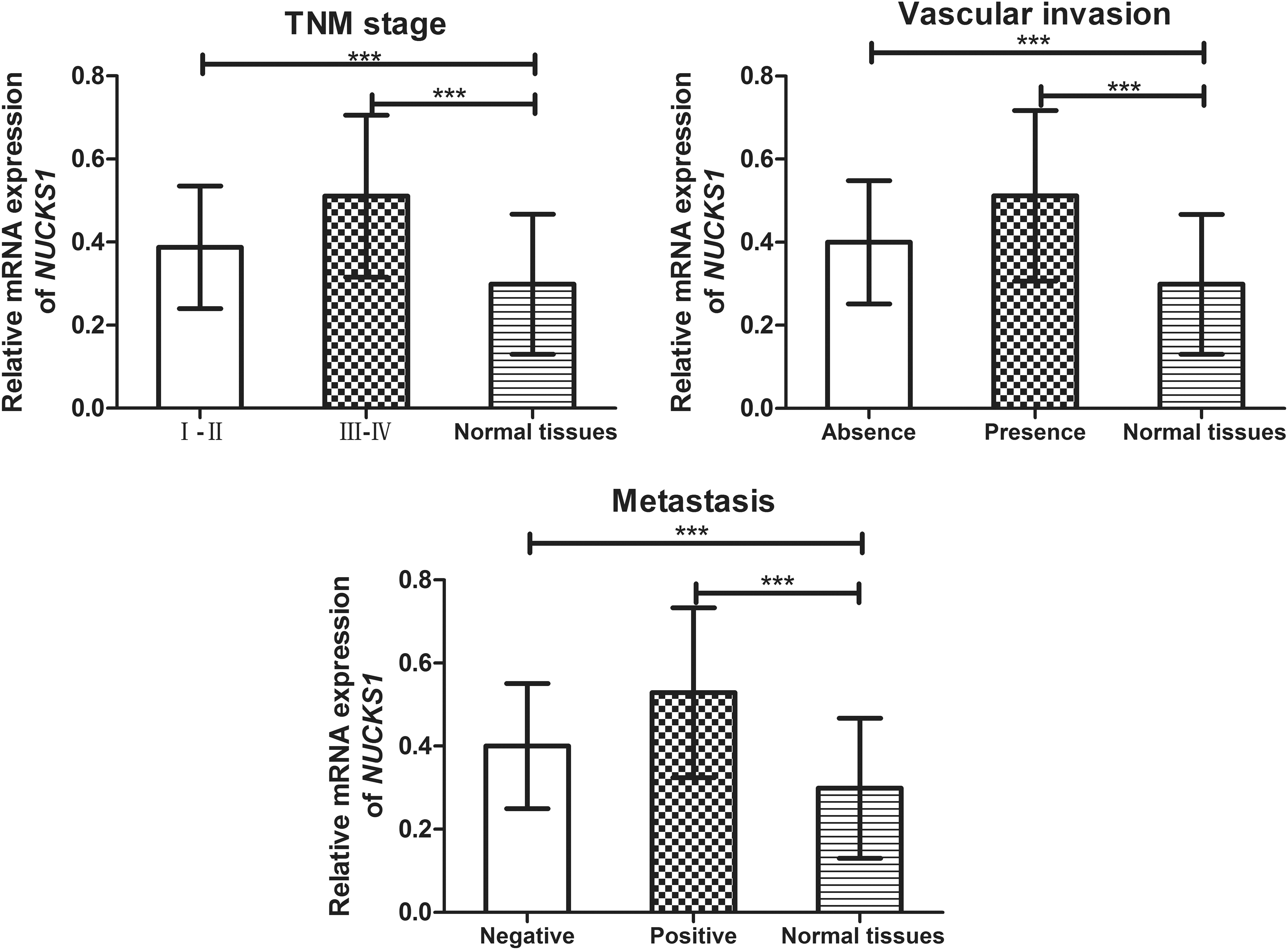
The comparison of NUCKS1 expression between different TNM stages, vascular invasion statuses, metastasis statuses, and adjacent normal tissues. *** Indicated p < 0.001. NUCKS1, nuclear casein kinase and cyclin-dependent kinase substrate 1; TNM, tumor node metastasis.
Increased NUCKS1 expression was correlated with poor prognosis of HCC patients
To estimate prognostic value of NUCKS1 in HCC patients, follow-up examination was conducted for 5 years. Treatments for patients after surgery followed doctor's advice. According to Kaplan-Meier analysis, patients with high expression of NUCKS1 had obviously shorter overall survival than those with low NUCKS1 expression (log rank test, p = 0.001, Fig. 3). Univariate Cox regression analysis demonstrated that NUCKS1 expression (p = 0.001), tumor size (p = 0.022), AFP level (p = 0.024), tumor differentiation (p = 0.045), TNM stage (p = 0.008), vascular invasion (p = 0.014), and metastasis (p = 0.005) were all associated with overall survival of HCC patients (Table 2). Besides, NUCKS1 expression (HR = 1.905, 95% CI = 1.106–3.283, p = 0.020), TNM stage (HR = 1.845, 95% CI = 1.074–3.170, p = 0.027), and metastasis (HR = 1.867, 95% CI = 1.100–3.167, p = 0.021) were all independent prognostic factors for HCC patients (Table 2).
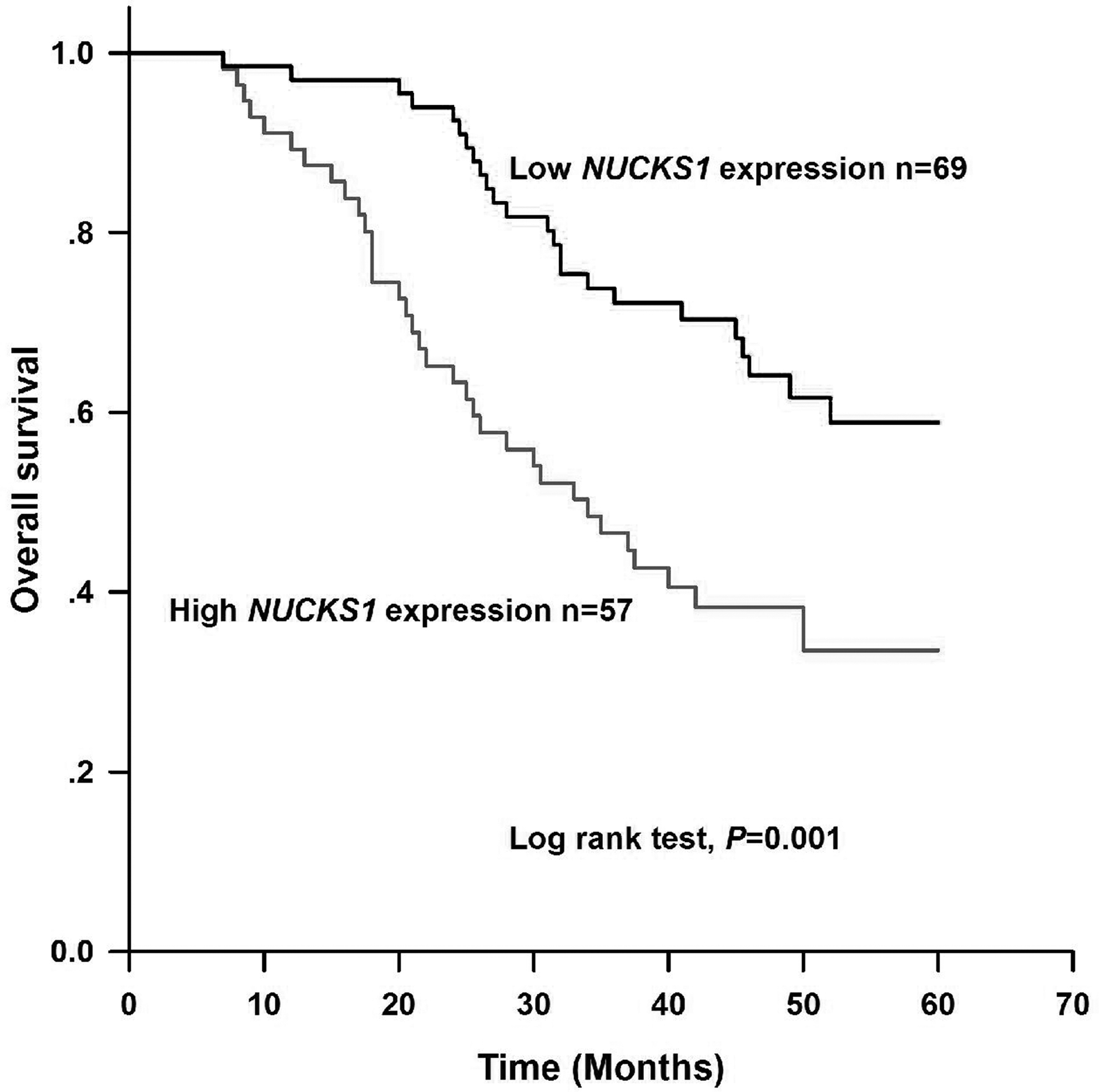
Kaplan-Meier curve analysis for overall survival of HCC patients. Overall survival was significantly longer in patients with low expression of NUCKS1 than those with high NUCKS1 expression (log rank test, p = 0.001). HCC, hepatocellular carcinoma; NUCKS1, nuclear casein kinase and cyclin-dependent kinase substrate 1.
Univariate and Multivariate Cox Regression Analyses of Prognostic Factors in Patients with Hepatocellular Carcinoma Patients
—, Indicated no data.
HR, hazard ratio.
Discussion
In this study, we detected relative expression levels of NUCKS1 mRNA in HCC tissues and adjacent normal ones. The result of qRT-PCR showed that NUCKS1 expression was significantly higher in HCC tissues than in normal controls. In addition, increased NUCKS1 expression was tightly associated with some clinical factors, including poor tumor differentiation, advanced TNM stage, the presence of vascular invasion, and positive metastasis in the patients. Such results revealed that NUCKS1 functioned as an oncogene in HCC, and was involved in carcinogenesis and tumor progression.
NUCKS1 is identified as one of cell cycle-related proteins and participates in responses to DNA damage induction. 8,15 Previous studies reported that NUCKS1 played a crucial role in the regulation of glucose homoeostasis and insulin signaling. 16,17 Besides, NUCKS1 was shown to be a key transcriptional regulator and chromatin modifier, and could be a potential biomarker for human diseases, including cancers and metabolic diseases. 18 Similar to high-mobility group proteins, NUCKS1 belongs to the class of the most modified proteins in the proteome of mammals and is associated with the progression of human cancer. 19 A study carried out by Kikuchi et al. demonstrated that the expression levels of NUCKS1 mRNA and protein were significantly upregulated in colorectal cancer (CRC) tissues in comparison to normal tissues. 20 A notable correlation was found between NUCKS1 overexpression and invasion, TNM stage, and metastasis in CRC patients. In addition, Liu et al. revealed that NUCKS1 expression was strongly correlated with tumor stage, histological grade, and metastasis in endometrial cancer. 21 Our results were consistent with those from previous studies.
Due to frequent recurrence and metastasis, the prognosis of HCC remains undesirable. Commonly, serum AFP level is regarded as a diagnostic and prognostic factor for HCC patients. 22 Moreover, TNM stage and metastasis are both associated with the prognosis of HCC patients. However, the efficiency of these factors is unsatisfactory. 23,24 Accumulated proofs have supported that increased NUCKS1 is linked to poor prognosis of human cancers. 20,25 However, its prognostic value in HCC remains poorly known. In this study, Kaplan-Meier survival analysis showed a negative relationship between NUCKS1 expression and overall survival of HCC patients. Moreover, Cox regression analysis further demonstrated that NUCKS1 expression level was an independent prognostic factor for HCC patients. Besides, TNM stage and metastasis both were independent factors to predict HCC prognosis.
There were still some limitations in our study. First, the expression of NUCKS1 was indicated to be associated with tumor size in patients with cervical squamous cell carcinoma, 26 which was inconsistent with our findings. The functions of NUCKS1 in different tumors might be varied. The sample size was relatively small, which might cause deviation in final result. Meanwhile, the study subjects were from the same region, and whether final results were suitable for individuals in other regions or other races needs to be further studied. Next, precise molecular mechanism of NUCKS1 in the regulation of HCC is still unclear. Thus, given these constraints, further studies are needed to gain insight into the functions of NUCKS1 on the progression of HCC.
Conclusions
In conclusion, the expression of NUCKS1 is remarkably upregulated in HCC and associated with the progression of the disease. Moreover, NUCKS1 may be a potential biomarker for the prognosis of HCC.
Footnotes
Authors' Contributions
Conceived and designed the experiments: X.Z. and X.L. Performed the experiments: X.Z. and H.B. Analyzed the data: G.L. and N.L. Contributed reagents/materials/analysis tools: H.L. and N.L. Wrote the article: J.D. and H.L.
Author Confirmation Statement
All coauthors have reviewed and approved of the article before submission.
Disclosure Statement
No competing financial interests exist.
Funding Information
The authors did not receive support from any organization for the submitted work.