




Abstract
Background:
Cutaneous squamous cell carcinoma (CSCC) is the most deadly disease among nonmelanoma skin cancers. LINC00641 plays a role in various cancers, but its role in CSCC has not been reported so far.
Methods and Materials:
The expression of LINC00641 and miR-424 in cells was detected by RT-qPCR. CCK-8 and colony formation assay were used to detect the proliferation of cells. Western blot was used to detect the expression levels of proliferation-, invasion-, and migration-related proteins. Wound Healing and Transwell experiments detected the ability of cell invasion and migration. In animal experiments, a tumor-bearing model was established in nude mice, and tumor volume was measured and photographed. The expression levels of proliferation-, invasion-, and migration-related proteins were detected by Western blot.
Results:
The expression of LINC00641 was significantly decreased in CSCC cell lines. The overexpression of LINC00641 at the cellular level inhibited the proliferation and migration of CSCC cell line A431 by downregulating the expression of miR-424. The overexpression of LINC00641 in animals inhibited the tumor volume of nude mice by downregulating the expression of miR-424 to inhibit the expression of proliferation- and migration-related proteins.
Conclusion:
LINC00641 inhibits the development of CSCC by downregulating miR-424.
Introduction
Cutaneous squamous cell carcinoma (CSCC) is a malignant tumor originating from keratinocytes of the epidermis or appendage. 1 CSCC can occur on the skin or mucous membrane, and it often forms the basis of precancerous lesion of some skin diseases. CSCC is second only to basal cell carcinoma and accounts for about 20% of skin cancers. Worldwide, there are 40,000 to 60,000 new cases of CSCC each year. 2 Ultraviolet radiation is a major driver of the disease. Other risk factors include exposure to carcinogenic chemicals, chronic skin ulcers, and immunosuppression, etc. 3 CSCC is the most deadly disease among nonmelanoma skin cancers. Therefore, it is important to find potential therapeutic targets for CSCC.
Long intergenic noncoding RNA 00641 (LINC00641) has been reported to play an anticancer role in a variety of cancers. The downregulation of LINC00641 in bladder cancer patients predicted poor prognosis, and the upregulation of LINC00641 significantly inhibited the proliferation, migration, and invasion of bladder cancer cells. Xenograft assay also confirmed that the overexpression of LINC00641 inhibited the growth of bladder cancer in vivo. 4 Study has shown that the expression level of LINC00641 in tissues of breast cancer was decreased, and the expression level of LINC00641 was negatively correlated with tumor size, lymph node metastasis, and clinical stage. Overexpression of LINC00641 inhibited cell proliferation, migration, and invasion and stimulated BC cell apoptosis. LINC00641 overexpression also significantly reduced the growth and metastasis of BC in vivo. 5 However, the role of LINC00641 in CSCC has not been reported.
Starbase predicted that LINC00641 could target miR-424. Research shows that the expression of miR-424 in CSCC is significantly upregulated. 2,6 In addition, the upregulation of miR-424-5p in laryngeal squamous cell carcinoma is closely related to the poor differentiation, advanced tumor stage, and cervical lymph node metastasis. Also, the overexpression of miR-424-5p promotes the proliferation, migration, invasion, and adhesion of laryngeal squamous cell carcinoma cells and affects the cell cycle process. 7 Therefore, the authors speculated that LINC00641 could target mir-424 and play an important role in the development and progression of CSCC.
In this article, the expression of LINC00641 and miR-424 in CSCC was detected and the mechanism was discussed by in vitro and in vivo experiments so as to provide scientific basis for targeted therapy of CSCC.
Materials and Methods
Cell culture
Cell lines of CSCC were purchased from the Type Culture Collection of the Chinese Academy of Science and incubated in DMEM (Gibco; Thermo Fisher Scientific) supplemented with 10% FBS (Gibco; Thermo Fisher Scientific) in a 37°C, 5% CO2 environment.
The model preparation of human skin squamous cell carcinoma xenograft in nude mice
Twenty healthy female BALB/C nude mice 6–8 weeks of age were selected and fed for adaption for 1 week. The nude mice were raised in a clean biological laminar flow rack, which had been UV disinfected on a regular basis and maintained at a constant temperature (25°C ± 2°C) and humidity (45% to 50%). A431 cells at logarithmic growth stage were inoculated subcutaneously at the right back near the upper limb of nude mice by 6 × 10⁷ U/mL. Weight and tumor volume were measured weekly. At week 5, the tumor was stripped and photographed. The mice were divided into Oe-NC (injected with A431 cells transfected with idle plasmid), Oe-Linc00641 (injected with A431 cells transfected with overexpressing Linc00641 plasmid), Oe-Linc00641+mimic-NC (injected with A431 cells transfected with overexpressing Linc00641 plasmid and idle plasmid), and Oe-Linc00641+miR-424 mimic (injected with A431 cells transfected with overexpressing Linc00641 plasmid and idle plasmid). The body weight and tumor volume of mice were recorded during 5 weeks of modeling. There were five mice in each group.
RT-quantitative polymerase chain reaction
Total RNA was extracted from cells using RNAzol RT (Sigma-Aldrich), following the manufacturer's protocol. Total mRNA concentration and purity were evaluated with the Thermo Fisher Scientific NanoDrop 2000C (cells). The SuperScript III Platinum SYBR Green One-Step qRT-PCR Kit (Invitrogen) was used according to the manufacturer's instructions. The following primers were used: Linc00641 forward, 5′-CAGCAGCTTGACACACGGTA-3′, and reverse, 5′-AAACACCAAAGTGGCATGTGA-3′; miR-424 forward, 5′-GCGGCGGCAGCAGCAATTCATG-3′, and reverse, 5′-ATCCAGTGCAGGGTCCGAGG-3′.
Transfection assay
The cells in a logarithmic phase were transfected with the overexpression of Linc00641 plasmids and miR-424 mimic using Lipofectamine™2000 transfection reagent (USA Invitrogen Corporation), according to the manufacturer's instructions. The transfected cells were cultured for 48 h and injected into the rats intravenously.
Cell counting kit-8 assay
Transfected cells (4 × 10³ cells/well) were seeded in 96-well plates for 48 h. Then, the cells was incubated with 10 μL of cell counting kit-8 (CCK8) for 4 h. Then, 150 μL DMSO was added into the medium. Absorbance at 450 nm was measured using a microplate reader (VersaMax).
Colony formation assay
Cells were seeded in six-well plates and cultured at 37°C for about 21 d until the visible clones appeared. Cells were fixed with methanol for 15 min and stained with 0.1% (w/v) Crystal Violet for the capture of pictures.
Western blot assay
Lysis buffer (Sigma) was added to the cells to isolate total protein. The concentration of protein was determined using the Bicinchoninic Acid Assay Protein Assay Kit. Proteins (25 μg/lane) were separated using sodium dodecyl sulfate/polyacrylamide gel electrophoresis and transferred to membranes of polyvinylidene fluoride. These membranes were then incubated with primary antibodies and after 12 h, incubated with goat anti-rabbit horseradish peroxidase-conjugated secondary antibodies (1:5000; cat. no. ab181658; Abcam) at room temperature for 2 h. Finally, the Chemiluminescence Kit and ImageJ software were used to detect the blots. Anti-ki67 (1:1000, cat. no. ab245113; Abcam), anti-PCNA (1:1000; cat. no. ab92552), anti-MMP7 (1:1000; cat. no. ab207299), anti-MMP9 (1:1000; cat. no. ab219372), and anti-GAPDH (1:1000; cat. no. ab181602) were purchased from Abcam.
Wound healing assay
The treated cells were seeded into six-well plates and starved overnight in serum-free DMEM medium. Artificial wounds were generated using a sterile 200 μL pipette tip, and the cells were washed with FBS to remove floating cells and debris. Images were obtained at 0 and 24 h using a microscope (Olympus Corp., Tokyo, Japan). Migration ability was assessed by measuring changes about the size of the wound areas.
Transwell assay
Cell invasion was detected by using Transwell assay with a pore size of 8 μm (Millipore, Bedford, MA). In brief, cell suspensions were added to 24-well Transwell chambers at a concentration of 1–1.5 million cells/mL. The lower chamber was filled with 0.5 mL of DMEM containing FBS. For the invasion experiment, the upper chamber with an 8-μm pore size was coated with Matrigel according to the manufacturer's instructions. Twenty-four hours later, cells were fixed with paraformaldehyde and stained with 0.1% Crystal Violet. Cells on the upper surface of the membrane were removed using cotton swabs. Finally, the cells were counted under a microscope ( × 200).
Dual-luciferase reporter assay
miR-424 mimic plasmids (or its empty vector control) were cotransfected with wild-type (or mutant) Linc00641-Luc plasmids in A431 cells, allowing for expression for 48 h. The cells were washed with PBS and lysed subsequently. The luciferase activity was measured using a plate reader (BD Bioscience) and normalized to the transfection efficiency by the Renilla Luciferase Activity Kit (pRL-TK). All procedures followed the manufacturer's instructions. All plasmids were constructed by Life Technologies Corporation (Carlsbad, CA).
Statistical analysis
All data were processed by SPSS 22.0 software (IBM Corp. Armonk, NY). Measurement data were expressed as mean ± standard deviation (SD). Comparison between two groups was conducted by t-test. Comparisons among multiple groups were assessed by one-way analysis of variance. p < 0.05 was considered to be statistically significant.
Results
Overexpression of LINC00641 inhibited the proliferation, migration, and invasion of A431 cells
First, the expression of LINC00641 in CSCC cell lines (HAC-5, A431, SCC-13, SCL-1) and normal human immortalized keratinocytes (HaCaT) was detected. They found that compared with normal cells, the expression of LINC00641 in CSCC cell lines was significantly decreased, among which its expression in A431 decreased most significantly (Fig. 1A). Therefore, A431 cells were selected for the subsequent experiments. Transfection efficiency was detected by RT-qPCR after LINC00641 overexpression by cell transfection technique (Fig. 1B). The cells were divided into control, overexpression-NC (Oe-NC), and overexpression-Linc00641 (Oe-Linc00641) groups. The results from CCK-8 (Fig. 1C) and colony formation assay (Fig. 1D) showed that compared with the Oe-NC group, the proliferation of cells in the Oe-Linc00641 group decreased, accompanied by decreased expression of proliferation-related proteins, Ki67 and PCNA (Fig. 1E). They then examined cell proliferation and invasion. They found that, compared with Oe-NC group, the migration (Fig. 2A) and invasion (Fig. 2B) of the Oe-Linc00641 group were decreased, accompanied by the decrease of invasion- and migration-related proteins MMP7 and MMP9 (Fig. 2C).

Overexpression of Linc00641 inhibited the proliferation of A431 cells.
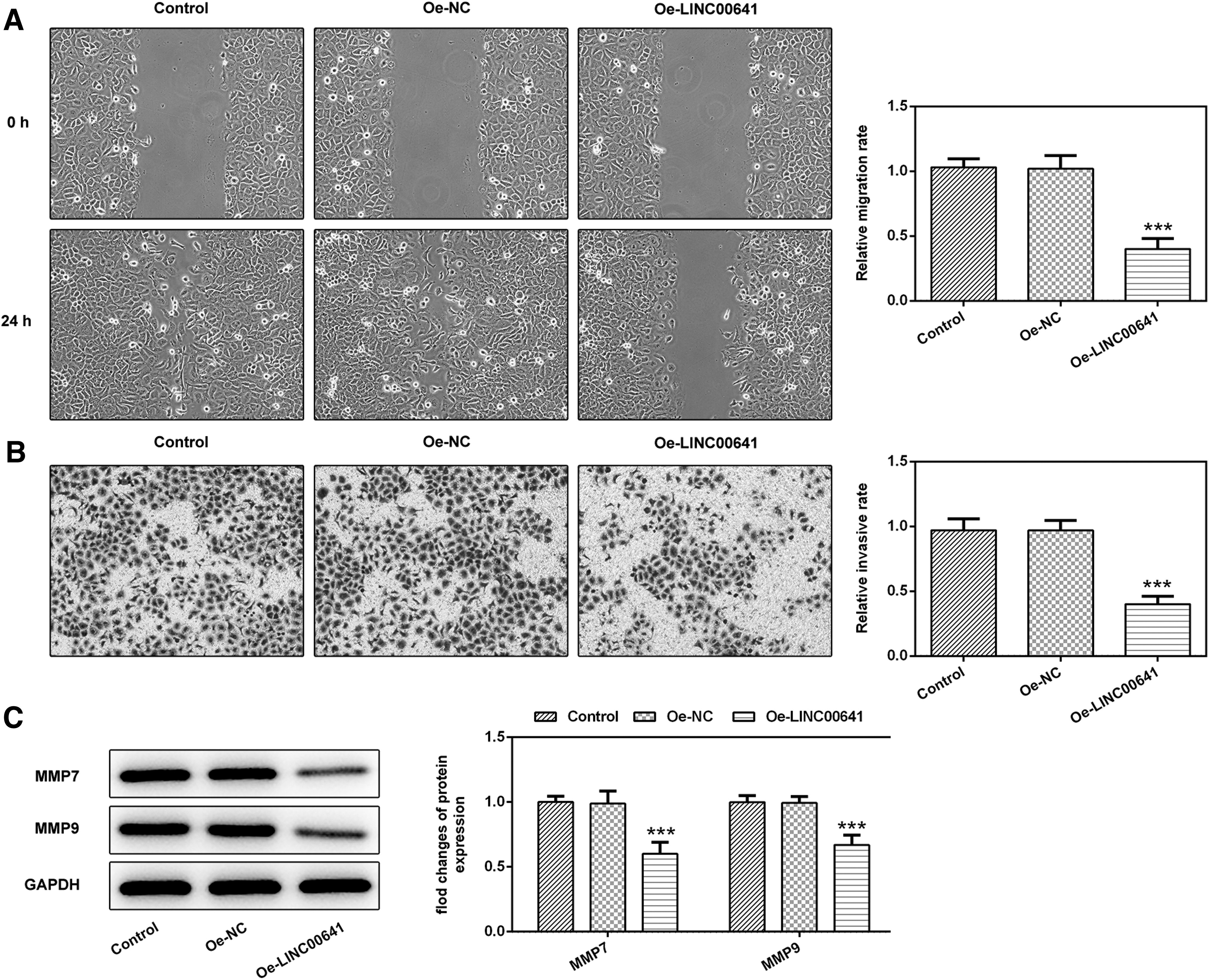
Overexpression of Linc00641 inhibited the migration and invasion of A431 cells.
Overexpression of LINC00641 inhibited the progression of CSCC in tumor-bearing nude mice
We divided the tumor-bearing mice into Oe-NC and Oe-Linc00641 and found that compared with the Oe-NC group, the body weight of the mice in the Oe-Linc00641 group had no significant differences (Fig. 3A), but the tumor volume of the mice was significantly decreased (Fig. 3B). Subsequently, Western blot results showed that compared with Oe-NC, Ki67, and PCNA expressions in tumor tissues in the Oe-Linc00641 group were decreased, as were the expressions of invasion-related proteins MMP7 and MMP9 (Fig. 3C). This indicated that overexpression of Linc00641 inhibited the development of CSCC in tumor-bearing nude mice.
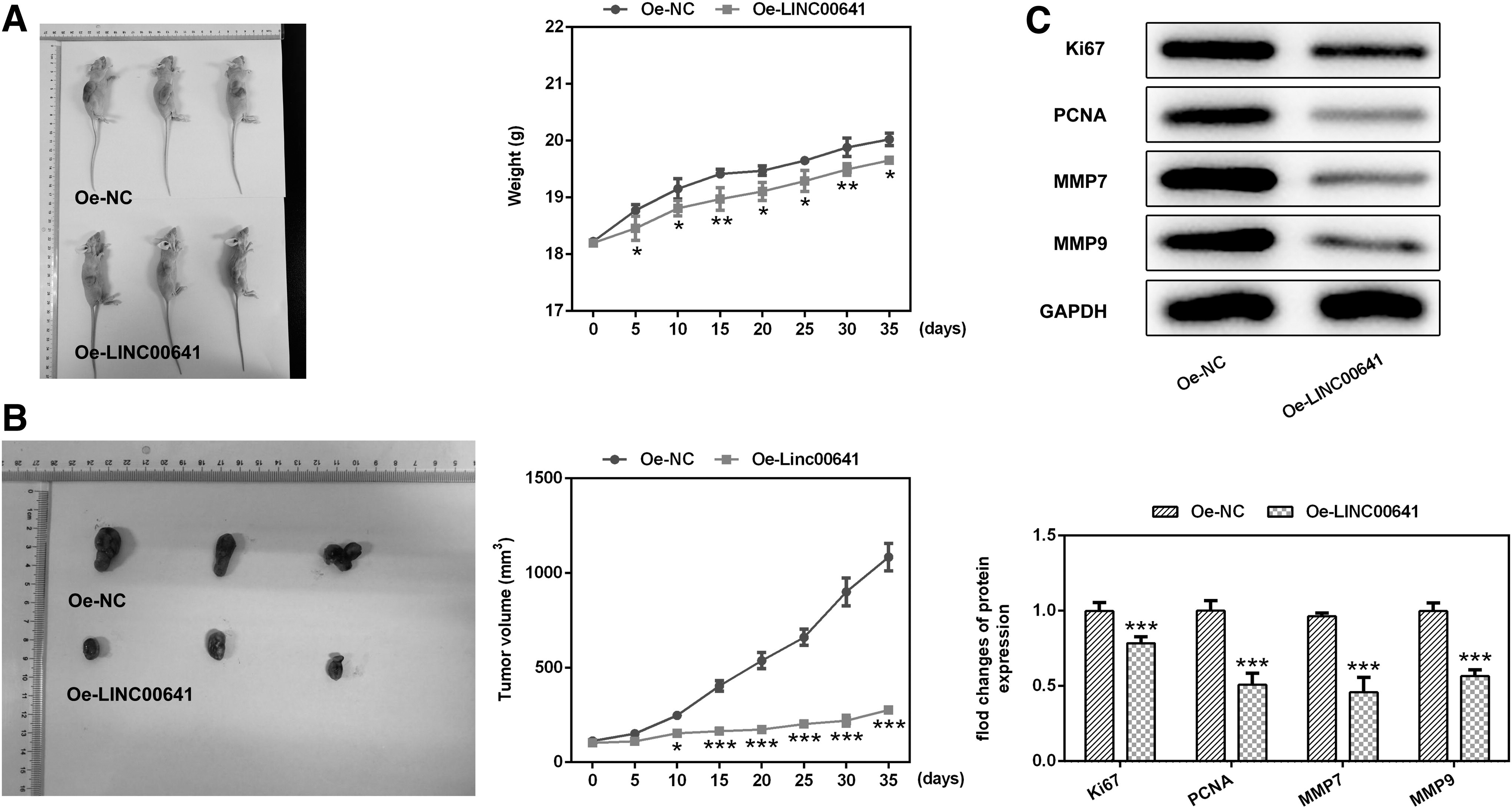
Overexpression of Linc00641 inhibited the progression of CSCC in tumor-bearing nude mice.
LINC00641 targeted and regulate miR-424
During the experiment, they found that the expression of miR-424 in CSCC cell lines was increased obviously (Fig. 4A). Starbase predicted that LINC00641 could target miR-424. In addition, the targeted binding of Linc00641 and miR-424 was also verified by the luciferase reporter gene (Fig. 4B). Subsequently, they overexpressed LINC00641 through cell transfection, and found that the expression of miR-424 in the Oe-LINc00641 group was decreased compared with Oe-NC group (Fig. 4C). This indicated that Linc00641 targeted and regulated miR-424.

Linc00641 can target and regulate miR-424.
LINC00641 targeted miR-424 to inhibit the proliferation, migration, and invasion of A431 cells
The expression of miR-424 in A431 cells was overexpressed by cell transfection (Fig. 5A). The cells were also divided into Control, Oe-Linc00641, Oe-Linc00641+mimic-NC, and Oe-Linc00641+miR-424 mimic groups. CCK-8 results showed that compared with Oe-Linc00641+mimic-NC, the cell proliferation level of Oe-Linc00641+miR-424 mimic significantly increased (Fig. 5B). The results from cell colony formation assay were consistent with those from CCK-8 (Fig. 5C). Compared with Oe-Linc00641+mimic-NC, the expressions of ki67 and PCNA in Oe-Linc00641+miR-424 mimic group increased (Fig. 5D). They also tested the ability of cell invasion and migration. The results are shown in Figure 6. Compared with Oe-Linc00641+mimic-NC, the migration (Fig. 6A) and invasion rate (Fig. 6B) of Oe-Linc00641+miR-424 mimic group increased, accompanied by increased protein expressions of MMP7 and MMP9 (Fig. 6C). These results indicated that overexpression of miR-424 reversed the inhibitory effects of overexpression of Linc00641 on proliferation, invasion, and migration of A431 cells. The results showed that Linc00641 targeted regulation miR-424 to inhibit the proliferation, migration, and invasion of A431 cells.

Linc00641 targeted miR-424 to inhibit the proliferation of A431 cells.

Linc00641 targeted miR-424 to inhibit the migration and invasion of A431 cells.
Linc00641 targeted miR-424 to inhibit the progression of CSCC in tumor-bearing nude mice
The tumor-bearing mice were divided into Oe-Linc00641+mimic-NC and Oe-Linc00641+miR-424 mimic groups. The experimental results showed that compared with Oe-Linc00641+mimic-NC, the body weight of mice in Oe-Linc00641+miR-424 mimic rose slightly (Fig. 7A), and the tumor tissue volume increased gradually with the time (Fig. 7B). Subsequently, western blot results showed that compared with Oe-Linc00641+mimic-NC, the expressions of Ki67 and PCNA in tumor tissues of mice with Oe-Linc00641+miR-424 mimic increased, and the expressions of migration and invasion-related proteins, MMP7 and MMP9, also significantly increased (Fig. 7C). The results showed that overexpression of miR424 in animals could reverse the inhibitory effect of overexpression of Linc00641 on the progression of CSCC in tumor-bearing mice.
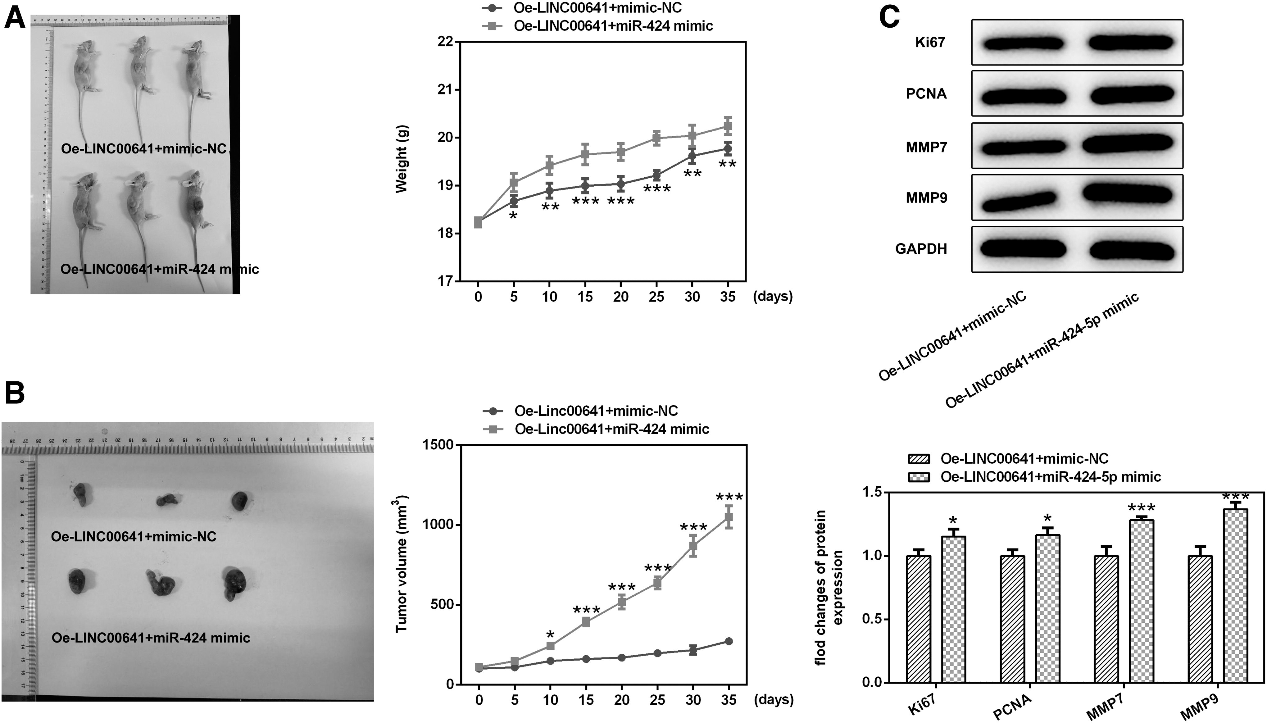
Linc00641 targeted miR-424 to inhibit the progression of CSCC in tumor-bearing nude mice.
Discussion
The high incidence and mortality of CSCC pose a serious threat to human health, and its development is a multistep process, involving many factors such as genetic change and epigenetic inheritance. 8,9 Therefore, understanding the pathogenesis of CSCC is of great significance for new therapeutic targets. The role of LncRNAs in the development of cancer has been a concern by many scholars, mainly because it can be used as signals and guide to regulate the pathophysiological processes in cells. 10,11
Linc00641 plays a role in many types of cancer. In glioma cells, LINC00641 limited the proliferation through regulating miR-4262/NRGN axis. 12 In cervical cancer, overexpression of LINC00641 inhibited the proliferation, migration, and invasion of cells and inhibited the characterization of epithelial mesenchymal transformation, suggesting that LINC00641 can inhibit the progression of cervical cancer. 13 However, there have been no reports on the study of Linc00641 in CSCC. They predicted through Starbase that the expression of Linc00641 was downregulated in CSCC patients. Subsequently, they observed that the expression of Linc00641 in CSCC cell lines was also significantly decreased. Through overexpression of Linc00641, they found that the proliferation, invasion, and mobility of CSCC cell line A431 cells were decreased. To further verify their experimental results, they constructed a tumor-bearing nude mouse model of CSCC, and found that the tumor volume of tumor-bearing nude mice decreased significantly by overexpression of Linc00641. In addition, the expression of proliferation- and invasion-associated proteins was also decreased significantly in tumor-bearing nude mice.
The expression of miR-424 in CSCC is upregulated, and it is predicted by bioinformatics website that LINC00641 can target bind to miR-424. 2,6 This targeting relationship was also verified by the luciferase reporter gene in their study. In addition, literature review found that in nonsmall-cell lung cancer, LINC00641 upregulated PLSCR4 by infiltrating miR-424-5p, so as to inhibit proliferation of nonsmall-cell lung cancer and induce apoptosis. 14 Therefore, they speculated LINC00641 could regulate cell proliferation, invasion, and migration in CSCC their miR-424. In their article, they found that overexpression of miR-424 can reverse the inhibition of proliferation, invasion, and migration of A431 cells after overexpression of LINC00641.
Conclusions
The author’s results indicate that LINC00641 was capable of inhibiting the development of CSCC by downregulating miR-424 in both cell and animal studies. Their study was of great significance for people to understand the pathogenesis of CSCC and to study new therapeutic targets of CSCC.
Ethics Approval and Consent to Participate
The study protocol was approved by the Shandong Provincial Hospital Affiliated to Shandong First Medical University, and all animal experiments comply with the ethical requirements of the animal council.
Footnotes
Disclosure Statement
The authors declare that they have no competing interests.
Funding Information
No funding was received for this article.
Supplementary Material
Western blot.pptx
References
Supplementary Material
Please find the following supplemental material available below.
For Open Access articles published under a Creative Commons License, all supplemental material carries the same license as the article it is associated with.
For non-Open Access articles published, all supplemental material carries a non-exclusive license, and permission requests for re-use of supplemental material or any part of supplemental material shall be sent directly to the copyright owner as specified in the copyright notice associated with the article.
