
Abstract
Objective:
The role and molecular mechanism of long-chain noncoding RNAs (lncRNAs) in lung cancer remain to be elucidated. The aim of this study was to investigate the association between a long coding RNA hypoxia-inducible factor-2α (HIF-2α) promoter upstream transcript (HIF2PUT) and clinical characteristics of non-small cell lung cancer (NSCLC) and its regulatory role in NSCLC.
Materials and Methods:
The correlation between HIF2PUT expression and pathological features of NSCLC was analyzed in NSCLC patient samples. Real-time polymerase chain reaction and Western blot were used to detect genes' mRNA and protein expression, respectively. Cell proliferation assay, invasion, and transwell assay were performed to determine the effects of HIF2PUT on NSCLC cells.
Results:
lncRNA HIF2PUT was downregulated in NSCLC tissues and cell lines. The authors found that HIF2PUT was mainly expressed in cytoplasm and overexpression of HIF2PUT attenuates cell proliferation and invasion in NSCLC cells. Moreover, low expression of HIF2PUT was significantly related to TNM stage (p = 0.045) and histological type (p = 0.025). Furthermore, HIF2PUT was found to play a role in cell proliferation and invasion in NSCLC through regulating HIF-2a.
Conclusion:
Based on this study, the inhibitory role of HIF2PUT on NSCLC proliferation, invasion could be blocked by HIF-2a silencing. In summary, this study suggests that HIF2PUT and HIF-2a may play an important role in the regulation of NSCLC progression, which provides new insights for clinical treatment.
Introduction
Lung cancer is the most common malignant tumor with the highest morbidity and mortality in the world, which seriously threatens human health and life. 1,2 At present, the molecular mechanism of the occurrence, development, and metastasis of lung cancer is still unclear. Moreover, with the lack of effective and sensitive early diagnostic indicators, patients are often in the middle and late stages of cancer, which is also a key factor for poor prognosis of patients. 3,4 In addition to surgery, the clinical application of targeted drugs has made considerable progress in the treatment of lung cancer. 5 However, how to improve the efficiency of treatment and reduce side-effects are still urgent problems to be solved in the treatment of lung cancer.
Non-small cell lung cancer (NSCLC) accounts for about 80% of all lung cancers; about 75% of the patients were diagnosed at the middle and advanced stages, and the 5-year survival rate was very low. 6 Compared with small cell carcinomas, NSCLC has faster growth and division, and earlier spread and metastasis. 7 Previous studies have shown that abnormal gene expression may be a key factor in the occurrence and development of malignant tumors. 8 It is the key research direction to study the mechanism of lung cancer and find the target genes that may be related to the biological activity of lung cancer.
Among the transcripts of human genome, only 2% of the genes can encode proteins. Ninety percent of the genes do not have this ability. 9 They are often transcribed into noncoding RNA (ncRNA) and these long-chain noncoding RNAs (lncRNAs) play an important role in the occurrence and development of many diseases. 10,11 According to the length of nucleotides, the authors named ncRNAs with >200nt as lncRNA. 8 With the in-depth study of lncRNA in recent years, many studies suggest that lncRNA plays a regulatory role in the occurrence and development of lung cancer. 8,12 Previous studies have shown that HIF2PUT was decreased significantly in osteosarcoma, and HIF2PUT overexpression inhibited proliferation and invasion after overexpression. 13 As a potential therapeutic agent, the expression and role of HIF2PUT in NSCLC are still unknown.
In this study, the authors aimed to study the relationship between clinical characteristics of NSCLC and HIF2PUT, and explore the role of HIF2PUT on proliferation and invasion of NSCLC and the potential mechanism underlying its' actions. The study may provide a new perspective for the treatment of NSCLC.
Materials and Methods
NSCLC tissue collection
All NSCLC tissues and adjacent normal tissues were collected in 45 patients from The Fifth People's Hospital of Shaanxi Province from December 2018 to December 2019. There were 38 males and 7 females. All the patient samples were histologically subtyped and graded according to the third WHO (World Health Organization) classification for lung cancer. Among them, there were 11 squamous cell carcinomas, 17 adenocarcinomas, and 14 other carcinomas. Twenty-three were poorly differentiated (G3–G4), and 22 were moderate or well differentiated (G1–G2). Twenty patients were staged 0–II and 25 were staged III–IV. The studies were approved by the Ethics Committee of The Fifth People's Hospital of Shaanxi Province and all patients were informed consents (approval number: SH20180049).
Cell culture and transfection
NSCLC cell lines of H1650, H1975, H358, and A549 used in this study were obtained from American Type Culture Collection. A549 cell line was cultured in DMEM (Hyclone; GE Healthcare) supplemented with 10% fetal bovine serum (Hyclone; GE Healthcare). Cells were maintained at 37°C in a humidified atmosphere with 5% CO2. The lncRNA HIF2PUT overexpression vector and HIF-2a siRNA vector were purchased from GenePharma (Shanghai, China). For the overexpression experiments, the transfection efficiency was verified in infected A549 by quantitative real-time polymerase chain reaction (qRT-PCR). Lipofectamine 3000 (Invitrogen, Carlsbad, CA) was used to transfect cells following the manufacturer's protocol. The siRNAs were purchased from GenePharma. The cells were subjected to the following experiments 24 h after transfection.
CCK-8 detection
NSCLC cell proliferation was examined using a CCK-8 assay kit (keyGEN Biotech, Nanjing, China). Cells were seeded (3 × 10⁴ cells per well) into 96-well plates. After transient overexpression of HIF2PUT or treatment with AKT inhibitor (Santa Cruz), 100 μL CCK-8 solution was added to each well at 0, 24, 48, 72, and 96 h and incubated for 2 h. The OD value at 450 nm was measured with a microplate reader (Thermo).
Transwell assay
2 × 10⁴ A549 cells were resuspended in 100 μL serum-free medium and were plated in the top chamber of each insert (8-μm pore size; Corning) with a Matrigel-coated membrane (BD Bioscience, San Jose) for the transwell assay. Lower chambers of the inserts were filled with DMEM medium with 10% fetal bovine serum. After 24 h of incubation, cells invaded to the lower surface of the insert were fixed, stained, and counted under a light microscope. After removal of the nonmotile cells at the top of the membranes with cotton swabs, 5 visual fields of 200 × magnification of each membrane were randomly selected and counted.
qRT-PCR analysis
Total RNA of NSCLC tissues, adjacent normal tissues, and NSCLC cell lines were extracted using Trizol according to the manufacturer's instructions. The purity of RNA was measured by detecting the absorbance ratio of 260/280 nm using a NanoDrop ND-1000 spectrophotometer (Thermo Scientific). Reverse transcription of RNA was performed by a PrimeScript RT Kit (RR047A; Takara, Tokyo, Japan), and qRT-PCR was using SYBR Premix kit (RR420A; Takara). The data were normalized to the levels of 18s rRNA (GeneBank ID) and further analyzed using the 2−ΔΔCT method. The primers used in the study were listed as follows: HIF2PUT, forward primer 5′-TTGGCAGACTACAAATATAC-3′, reverse primer 5′-AGCTTACTGGGGCTAATGA-3′; and HIF-2a, forward primer 5′-CGGCTCCGTTGGAACCCCA-3′, reverse primer 5′-GACTTGGGCGCCGTTGTGC-3′. The thermocycling conditions were as follows: incubation at 50°C for 2 min, followed by an incubation at 95°C for 2 min, followed by 40 cycles of 95°C for 15 s and 60°C for 32 s. Each sample was tested in triplicate and expression of each target was normalized to that of the human 18s.
Western blotting
The cells washed with PBS were lysed with RIPA buffer (20 mM HEPES, pH 7.5, 150 mM NaCl, 10% glycerol, 50 mM EDTA, and 1% Triton X-100), and the supernatant was collected by centrifugation. The extracted proteins were then subjected to 12% SDS-PAGE electrophoresis and transferred onto the PVDF membranes. The membranes were blocked with 5% nonfat milk and incubated with primary antibodies overnight at 4°C. Membranes were incubated with HRP-conjugated secondary antibodies for 2 h at room temperature. Immunoreactive proteins were visualized by using the chemiluminescence detection kit (Donghuan Biotech, Shanghai) and analyzed by Image J software. The following primary antibodies were used: anti-p-AKT (Abcam); anti-AKT (Abcam); anti-HIF-2a (Abcam); and anti-GAPDH (Abcam).
Statistical analysis
All the graph plotting and data analysis were performed by using the SPSS software package (version 17.0). All the data were shown as mean ± standard deviation. t Test was used to compare qRT-PCR, Western blotting, Transwell assay, and Transwell assay results. χ 2 -test or one-way ANOVA (comparison for more than two groups) and χ 2 test were used to examine the association of HIF2PUT expression level with various clinicopathologic parameters. p < 0.05 was considered to be statistically significant.
Results
lncRNA HIF2PUT is downregulated in NSCLC tissues and NSCLC cell lines
To investigate the potential role of lncRNA HIF2PUT in NSCLC, the authors first performed qRT-PCR analysis to determine the expression of HIF2PUT in NSCLC and normal adjacent tissues, and in NSCLC cell lines (H1650, H1975, H358, and A549). The results indicated that the expression of HIF2PUT was significantly decreased in NSCLC tissues compared with normal tissues. Similarly, HIF2PUT expression in NSCLC cell lines is also markedly significantly lower than in normal pulmonary endothelial bronchial cell line of BEAS-2B (Fig. 1A, B). In view of the lowest expression of HIF2PUT in A549 cell line, the authors chose A549 cells as NSCLC cell model in this study. In addition, nuclear separation study was performed to determine the location of HIF2PUT in A549 cells; they confirmed that HIF2PUT mainly expresses in the cytoplasm (Fig. 1C).
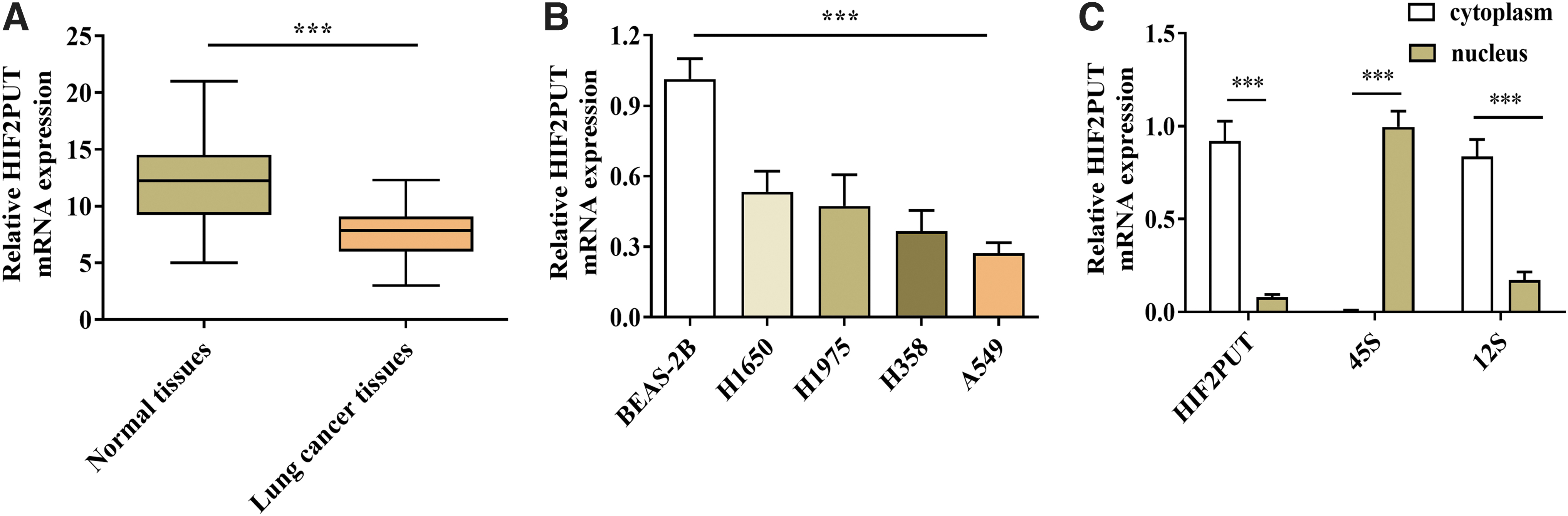
lncRNA HIF2PUT is downregulated in NSCLC tissues and NSCLC cell lines.
lncRNA HIF2PUT expression is related to NSCLC patients' clinical progression
To explore the clinical significance of HIF2PUT expression in NSCLC patients, a total of 45 NSCLC and normal adjacent tissue samples were detected for mRNA levels of HIF2PUT. Relative fold change of HIF2PUT was analyzed by qRT-PCR and the median expression of HIF2PUT was //////, all tissues' expression levels were divided into “high-expression” (n = 24) and “low-expression” (n = 21) HIF2PUT groups based on the median expression of HIF2PUT. Then, the relationship between HIF2PUT expression and pathological features of NSCLC was analyzed. As shown in Table 1, expression of HIF2PUT was significantly associated with TNM Stage (p = 0.045) and histological type (p = 0.025). Nevertheless, there was no significant association between HIF2PUT expression and age, gender, smoking, distant metastasis, and histological grade statistically.
Association Between HIF2PUT Expression and Clinicopathological Characteristics in 45 Non-Small Cell Lung Cancer Patients
Bold values signify p value was < 0.05, and were considered significant statistically.
lncRNA, long-chain noncoding RNA.
lncRNA HIF2PUT overexpression inhibits cell proliferation, invasion in NSCLC cells
To further investigate the role of HIF2PUT in NSCLC, HIF2PUT was overexpressed with adenovirus in A549 cells (Fig. 2A). HIF2PUT overexpression markedly suppressed the NSCLC cell proliferation by CCK-8 assay (Fig. 2B). Moreover, Transwell analysis results showed that overexpression of HIF2PUT significantly decreased NSCLC cell invasion (Fig. 2C).

HIF2PUT overexpression inhibits cell proliferation, invasion.
Overexpression of lncRNA HIF2PUT suppresses NSCLC cell proliferation, invasion through HIF-2a signaling
In addition to being proved to be an important factor in the occurrence and development of various tumors, a previous study also showed that HIF-2 gene can be regulated by lncRNA HIF2PUT in osteosarcoma. 13 In this study, Western blot and PCR analysis showed that HIF-2a can be significantly positively regulated by HIF2PUT (Fig. 3A, B). Moreover, the authors also indicated that HIF2PUT expression is positively correlated with HIF-2a in NSCLC tissues (Fig. 3C). Given that HIF2PUT expression was positively correlated with HIF-2a, the authors wonder whether HIF2PUT plays its function in NSCLC by regulating the HIF-2a expression. They overexpressed HIF2PUT in NSCLC cells with or without HIF-2a silencing. CCK-8 assay results showed that the proliferation of NSCLC cell inhibited by HIF2PUT overexpression was blocked after HIF-2a downregulation (Fig. 3D). Consistent with the CCK-8 results, transwell assay demonstrated that HIF2PUT overexpression significantly reduced NSCLC cell invasion, but was partially reversed by silencing of HIF-2a (Fig. 3E).
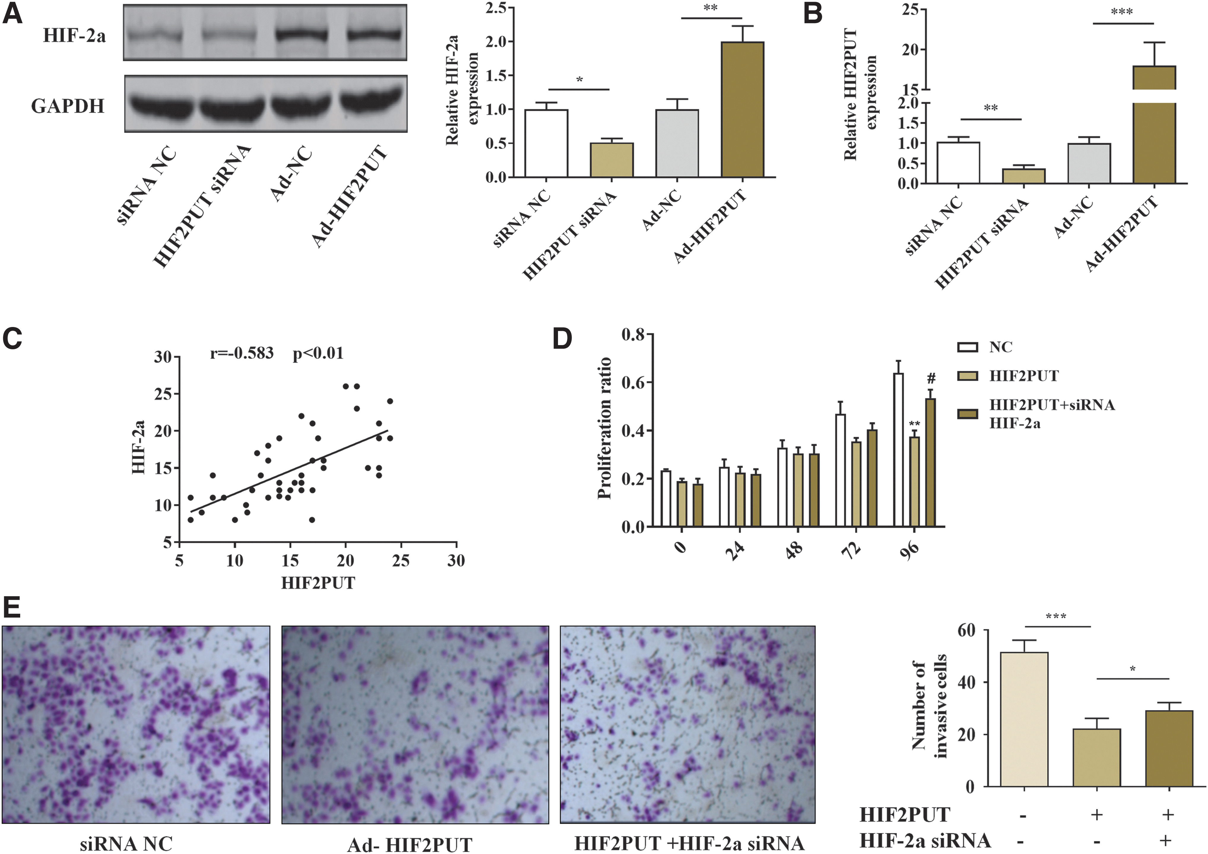
HIF2PUT suppresses NSCLC cell proliferation, invasion by HIF-2a.
Overexpression of lncRNA HIF2PUT suppresses NSCLC cell proliferation, invasion by AKT pathway
To further verify the pathway mechanism of HIF2PUT in NSCLC cells, the authors selected AKT and MAPK pathways, which related to HIF-2a gene in other studies. Western blot results showed that HIF2PUT could significantly increase the expression of phosphorylated AKT in NSCLC cells, but had no effect on the expression of MAPK (p-38; p-ERK and p-Jnk) (Fig. 4A, B). To determine whether HIF2PUT inhibits the proliferation and invasion of lung cancer cells through AKT pathway, they used AKT inhibitors to inhibit the expression of phosphorylated AKT (Fig. 5A). The authors found that the proliferation and invasion of NSCLC cell inhibited by HIF2PUT overexpression were partially blocked after AKT inhibitor treatment (Fig. 5B, C).

HIF2PUT increases the expression of p-AKT.
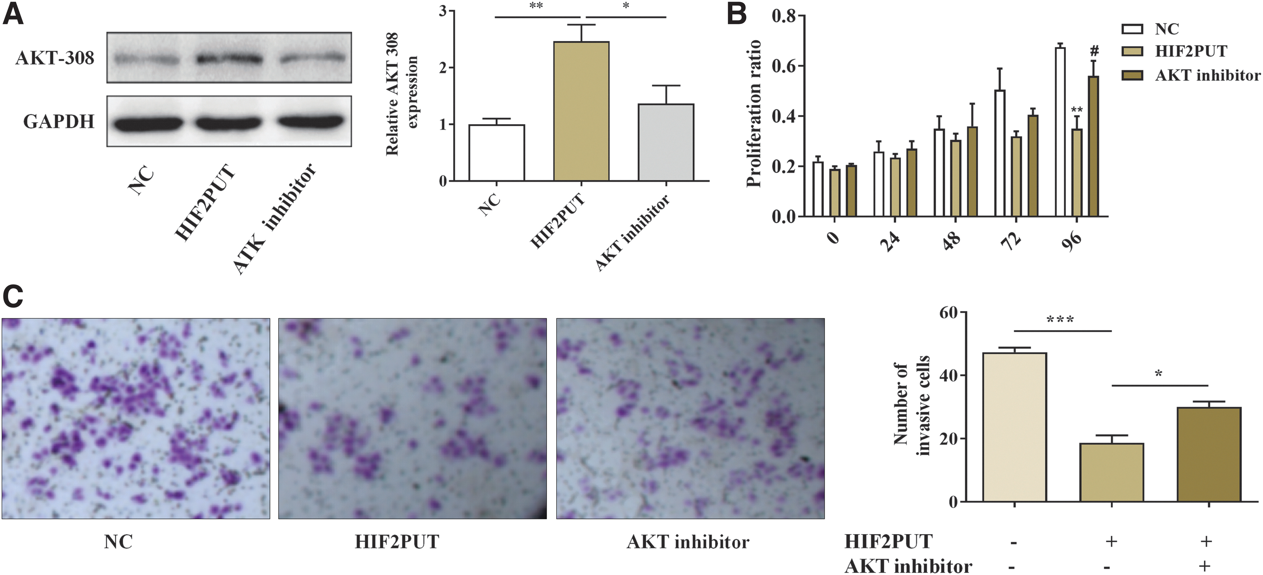
HIF2PUT suppresses NSCLC cell proliferation, invasion through AKT pathway.
Discussion
In recent years, some achievements have been made in the research of noncoding RNA, which also promotes the research of molecular biological mechanism of cancer. 14 lncRNAs are closely related to many diseases, including cancers; many studies have also confirmed that lncRNAs as key regulators affect the occurrence and development of cancers. 15,16 However, the understanding of the role of LN in cancer treatment and prognosis is limited. Therefore, the authors' findings also provide an important basis for the study of the relationship between HIF2PUT and NSCLC.
lncRNA, as a kind of transcript with extensive functions, contains abundant information, and the molecular mechanisms involved in expression regulation are diverse. 17 With the deepening of research, it has been shown that lncRNA, originally considered a by-product of transcription and not functioning biologically, plays an equally important role in the occurrence and development of tumors. 18 Its mechanism of regulating tumors is similar to that of microRNAs (miRNAs), and may be more complex compared with miRNAs. 19 However, only a few functional characteristics of lncRNAs have been confirmed in tumors. Several studies on IncRNAs have confirmed that the abnormal expression of lncRNAs is associated with the occurrence of various types of cancers. 6,20 However, the specific biological roles and mechanisms they play in tumorigenesis, development, invasion, and migration are still unclear.
In this study, the authors examined NSCLC tissue specimens and found that the expression of lncRNA HIF2PUT in NSCLC tissue was significantly reduced. They further explored the function and mechanism of HIF2PUT in NSCLC cell lines, hoping to understand the role of HIF2PUT in the prevention of NSCLC. The authors also revealed that low expression of HIF2PUT was significantly related to TNM stage and histological type of NSCLC. These data suggest that HIF2PUT may be closely related to the predicted prognosis of NSCLC. HIF-2, also known as EPAS1, has been proved to be a critical gene associated with the evolution of a variety of cancers. 21 Previous studies revealed that HIF2PUT was significantly related to HIF-2a expression in some other cancer tissues 13,22 Consistent with their findings, they have also demonstrated that HIF2PUT and HIF-2a are positively correlated in lung cancer, and HIF2PUT has a negative regulatory role in HIF-2a expression. In addition, the authors also found that HIF2PUT inhibits the proliferation and invasion of lung cancer cells through AKT pathway. These data suggest that HIF2PUT overexpression can inhibit many kinds of tumors by regulating HIF-2a/AKT and may have broad-spectrum antitumor effects.
In conclusion, the role of lncRNAs in the field of biology has been paid more and more attention in recent years, but the research on lncRNAs in the field of cancer treatment and prevention is still very limited. The authors' findings will help us better understand the role of lncRNA HIF2PUT in NSCLC and provide a new perspective for the treatment of NSCLC.
Footnotes
Disclosure Statement
No competing financial interests exist.
Funding Information
This work was supported by the Natural Science Foundation of Shanxi Province, China (Grant No. 2018JM7044), the Health and Family Planning Research Foundation of Shanxi Province, China (Grant No. 2018D019), the Health and Wellness Committee Research Foundation of Shanxi Province, China (Grant No. 2016D036), and the Administration of Traditional Chinese Medicine Foundation of Shanxi Province, China (Grant No. JCPT039).