
Abstract
Ovarian cancer (OC) is known to be the most malignant gynecologic cancers. Wnt2B, a member of the Wnt family, plays a critical role in tumor development. However, the effect of Wnt2B on the occurrence and development of OC remains largely uncharacterized. In this study, immunohistochemistry assay indicated that Wnt2B was increased in our study cohort (OC). In addition, the expression of Wnt2B was positively correlated with TNM stages and metastasis of OC patients. Wnt2B markedly mediated the regulation of OC proliferation, invasion, and angiogenesis. Moreover, Wnt2B knockdown inactivated the Wnt/β-catenin signaling pathway. More importantly, the Wnt/β-catenin signaling pathway activator LiCl reversed the effect of Wnt2B knockdown on OC cell proliferation, angiogenesis, and invasion. Our data indicated that Wnt2B silencing could inhibit the proliferation, invasion, and angiogenesis of OC cells through downregulating the activity of Wnt/β-catenin pathway.
Introduction
Ovarian cancer (OC) is the most common lethal gynecologic malignancies and ranked as the fifth leading cause of cancer death worldwide in women. 1 Despite the improvement of surgery and chemotherapy for OC, the 5-year overall survival rate for patients with OC remains unsatisfactory. 2 The occurrence and development of OC involved in a wide variety of cellular processes, such as apoptosis, proliferation, angiogenesis, migration, metastasis, and invasion. 3 Therefore, further study on the underlying molecular mechanisms for OC is essential to improve its prognosis.
The Wnt/β-catenin signaling pathway is implicated in diverse physiological processes, including embryonic development, proliferation, and adult tissue homeostasis. 4 Previous studies have reported that aberrant Wnt/β-catenin signaling is involved in the occurrence and advancement of several cancers, including OC. 5 Chen et al. found that STAT3 signaling maintained cancer stemness by targeting miR-92a/DKK1 and activating Wnt/β-catenin signaling subsequently in OC. 6 c-Kit knockdown could decrease the chemoresistance and tumor-initiating activity of OC cells through activating Wnt/β-catenin signaling. 7 Cyclin G2 protein suppressed cell proliferation, invasion, migration, and spheroid formation, as well as tumor formation and invasion by disrupting Wnt/β-catenin signaling in epithelial OC. 8
Wnt2B, an important Wnt family member, was first discovered in 1996, which played key roles in tumor development. 9 Jiang et al. reported that the high expression of Wnt2B might be correlated with poor prognosis of pancreatic cancer. 10 miR-577 inhibited cell growth and epithelial–mesenchymal transition (EMT) by regulating Wnt2B in non-small cell lung cancer (NSCLC). 11 The high expression of miR-338-3p promoted cell sensitivity to cisplatin through regulation of Wnt2B expression in OC cells. 12 Silencing Wnt2B by siRNA interference inhibited cell counting and invasiveness in OC. 13 Although the important role of Wnt2B has been shown in drug resistance and metastasis, the effect of Wnt2B on the occurrence and development of OC remains largely invisible.
In this study, the expression levels of Wnt2B were significantly higher in OC tissue samples compared with normal controls. The increased expression of Wnt2B was positively associated with metastasis and TNM stages in OC patients. In addition, Wnt2B knockdown markedly inhibited OC cell proliferation, invasion, and angiogenesis dependent on Wnt/β-catenin signaling.
Materials and Methods
Tissue samples and immunohistochemistry
Sections from paraffin-embedded OC tissues were collected from 34 patients undergoing ophthalmectomy, according to the human subject protocol approved by the Ethical Committee of the Affiliated Huaian No. 1 People's Hospital of Nanjing Medical University. The methods of immunohistochemistry (IHC) for Wnt2B were performed as a previous study. 14 In brief, the slices were dehydrated with gradient alcohol, subjected to antigen retrieval, and incubated with Wnt2B antibody (Abcam) at 4°C overnight. The scoring method of IHC was previously described. 14
Cell culture
The OC cell lines A2780 and SKOV3 were purchased from ATCC and maintained in RPMI-1640 media supplemented with 10% (Gibco) and McCoy's 5A media supplemented with 10% (Gibco), respectively.
RNA isolation and real-time quantitative polymerase chain reaction
TRIzol reagent, purchased from Thermo Fisher Scientific, was used to isolate total RNA from OC cells. M-MLv Reverse Transcriptase kit was used (Takara) to obtain cDNA according to the instructions. To detect Wnt2B mRNA expression, real-time quantitative polymerase chain reaction (PCR) was conducted using the Applied Biosystems (ABI) 7500 Fast Real-Time PCR system with SYBR-Green PCR kit (Applied Biosystems). β-actin served as an internal control. Primers to amplify genes were the following as Wnt2B Forward, 5′-GTTACCCAGACATCATGCGTT-3′, Wnt2B Reverse, 5′-GGGTGGTACAGTTCCAGCG-3′; β-actin Forward, 5′-CATGTACGTTGCTATCCAGGC-3′, β-actin Reverse, and 5′-CTCCTTAATGTCACGCACGAT-3′.
Western blot
Cells were lysed in a lysis buffer with protease and phosphatase inhibitors (Beyotime). Total protein was separated by 10% sodium dodecyl sulfate–polyacrylamide gel electrophoresis and electrophoretically transferred to a polyvinylidene fluoride membrane. After that, the samples were incubated with the primary antibodies of the Wnt2B antibody and GAPDH (Abcam). Finally, the Western blot bands were visualized using chemiluminescence.
Cell counting kit-8 assay
Two × 103 cells were seeded onto 96-well plates, with five replicate wells for incubation. Then, 10 μL cell counting kit-8 (CCK-8) solution was added into each well and plates were incubated at 37°C for another 2 h. The absorbance was measured with a microplate reader (Applied Biosystems) at a wavelength of 450 nm.
Cell counting
Cells were seeded in 24-well microplates at 0.5 × 10⁴ cells/well. After treatment by Wnt2B knockdown and overexpression for 24, 48, and 72 h, cell number was determined with a hemocytometer using 0.45% trypan blue (Sigma-Aldrich, St Louis, MO).
Transwell assay
The invasion assay was performed as previously described. 15 In brief, for migration assays, 2 × 10⁴ cells were seeded in media without serum into the upper chamber precoated with Matrigel (BD Biosciences). Cell media supplemented with 10% fetal bovine serum were added to lower chamber. The penetrated cells were fixed and stained with crystal violet, and counted under microscopy.
Tube formation assay
The tube formation assay was performed as described previously. 15 In brief, 2 × 10⁴ human umbilical vein endothelial cells (HUVECs) were seeded into 96-well plates precoated with Matrigel. After 8 h, tubes were photographed with a microscope. Invaded cells were photographed and counted from five randomly selected fields.
Statistical analysis
Statistical analyses and data visualization were performed using GraphPad Prism 5 (GraphPad, Inc.). All data were expressed as mean ± standard deviation. Student's t-test or one-way analysis of variance was used analyze the difference between two groups. p < 0.05 was considered to be statistically significant.
Results
Elevated protein expressions of Wnt2B in OC tissues
As shown in Figure 1A, the expression of Wnt2B protein was much higher in OC tissue samples than that in the normal tissue. In addition, the Wnt2B protein levels were much higher in later-stage OC patients as compared with early-stage patients with OC (Fig. 1B). Moreover, the Wnt2B protein levels were positively associated with metastasis of OC (Fig. 1C). These results indicated the aberrant elevation of Wnt2B expression in OC patients.
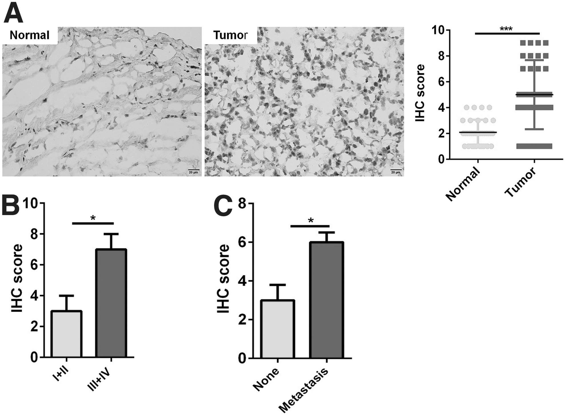
Wnt2B was upregulated in tissue samples of patients with OC.
Wnt2B mediated the proliferation of OC cells
To investigate the role of Wnt2B in the proliferation of OC cells, Wnt2B siRNA-1, Wnt2B siRNA-2, and pc-Wnt2B were used to knockdown and overexpression Wnt2B, respectively, in A2780 and SKOV3 cells. Both Wnt2B siRNA-1 and Wnt2B siRNA-2 markedly reduced the mRNA and protein levels of Wnt2B in A278 and SKOV3 cells; in contrast, pc-Wnt2B significantly increased the mRNA and protein expression of Wnt2B in A278 and SKOV3 cells (Fig. 2A, B). CCK-8 assay and cell counting showed that Wnt2B knockdown inhibited the growth of OC cells, and Wnt2B overexpression promoted the proliferation of OC cells (Fig. 2C, D). These results showed that Wnt2B played an important role in the proliferation of OC cells.
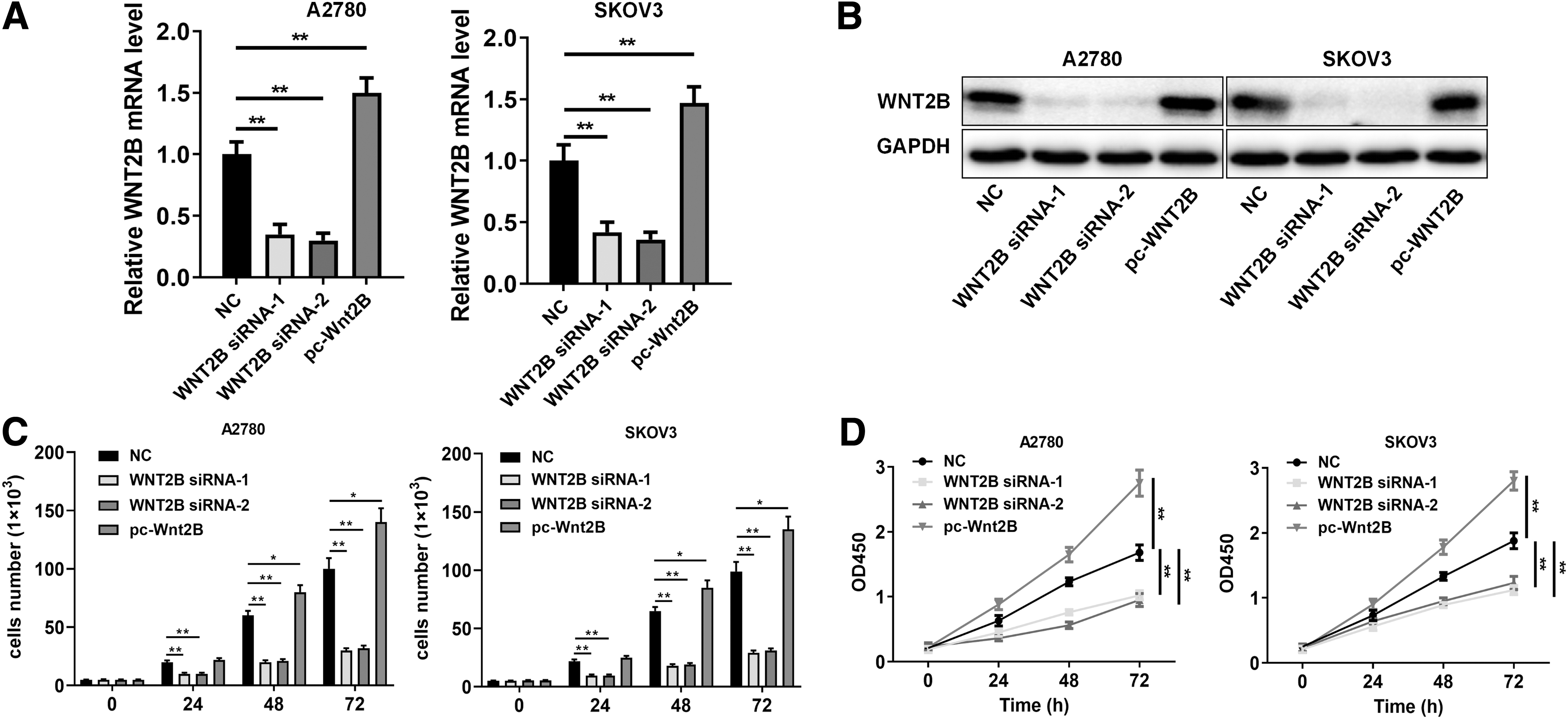
Wnt2B mediated OC cell growth.
Wnt2B mediated the invasion of OC cells
Then, we used the Transwell assay to examine the effect of Wnt2B on OC cell invasion. The number of invasive cells in the Wnt2B siRNA-1 or Wnt2B siRNA-2 group were significantly decreased, and the number of invasive cells in the pc-Wnt2B group were significantly increased compared with the control group, respectively (Fig. 3A, B). The results implied that Wnt2B was crucial to the invasive property of OC cells.

Wnt2B mediated OC cell invasive activity.
Wnt2B mediated the angiogenesis of OC cells
To examine the effect of Wnt2B on angiogenesis in HUVECs, we carried out the tube formation assay in vitro. Conditioned media was collected from SKOV3 cells negative control (NC), Wnt2B siRNA-1, Wnt2B siRNA-2, and pc-Wnt2B, respectively. In Wnt2B siRNA-1 and Wnt2B siRNA-2 group, the number of angiogenetic cells significantly decreased, whereas in pc-Wnt2B group, the number of angiogenetic cells significantly increased (Fig. 4). The importance of Wnt2B in regulating the angiogenesis of OC cells was verified by these results.
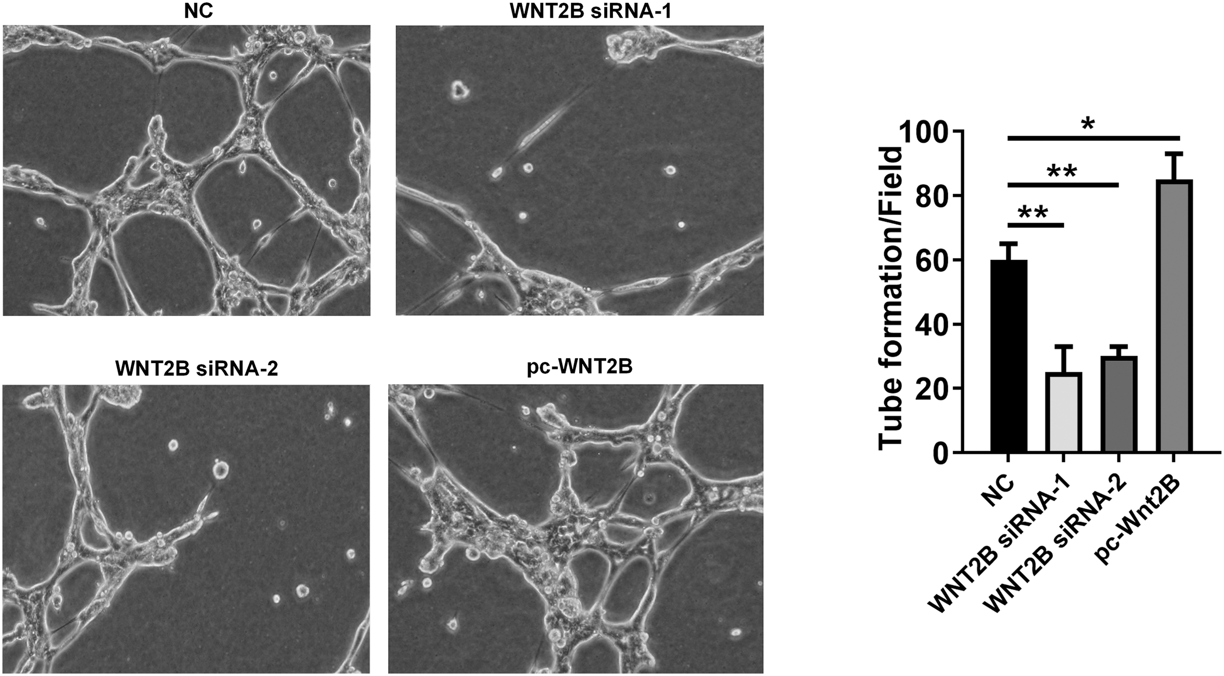
Wnt2B mediated the angiogenesis of HUVECs. Angiogenesis of HUVECs cultured in conditioned media from SKOV3 cells transfected with Wnt2B siRNAs and pc-Wnt2B. The number of angiogenetic cells were calculated. **p < 0.01. HUVECs, human umbilical vein endothelial cells.
Silenced Wnt2B inactivated the Wnt/β-catenin pathway
The protein levels of β-catenin were markedly decreased in A2780 and SKOV3 cells transfected with Wnt2B siRNA-1 or Wnt2B siRNA-2 as compared with OC cells transfected with NC. Moreover, Wnt/β-catenin pathway-regulated target genes, including MMP-9, Cyclin D1, and VEGF, were also decreased in A2780 and SKOV3 cells transfected with Wnt2B siRNA-1 or Wnt2B siRNA-2 (Fig. 5A, B). The results implied that Wnt2B regulates cell proliferation, invasion, and angiogenesis by Wnt/β-catenin pathway in OC cells.
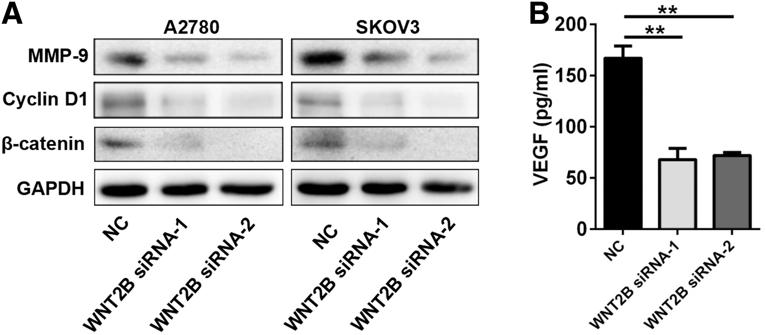
Wnt2B knockdown inhibited Wnt/β-catenin signaling pathway.
Wnt/β-catenin activation reversed the effect of Wnt2B knockdown on cell proliferation, invasion, and angiogenesis
The biological function of OC cell was significantly affected through Wnt/β-catenin signaling.16–18 To further determine whether the biological effect of Wnt2B on cell proliferation, invasion, and angiogenesis in OC depend on Wnt/β-catenin pathway, we co-treated A2780 and SKOV3 cells with Wnt2B siRNA-1 and a canonical Wnt/β-catenin signaling activator LiCl.19–21 As shown in Figure 6A, Wnt2B siRNA-1 obviously inhibited the growth of A2780 and SKOV3 cells, whereas LiCl treatment reversed the inhibition of Wnt2B siRNA-1 on cell proliferation. In addition, compared with the Wnt2B siRNA-1 group, the invasive ability of cells was increased significantly in the Wnt2B siRNA-1+LiCl group (Fig. 6B). Moreover, conditioned media from SKOV3 cells transfected with Wnt2B siRNA-1 decreased the angiogenetic cells numbers, whereas LiCl treatment reversed the effect (Fig. 6C).
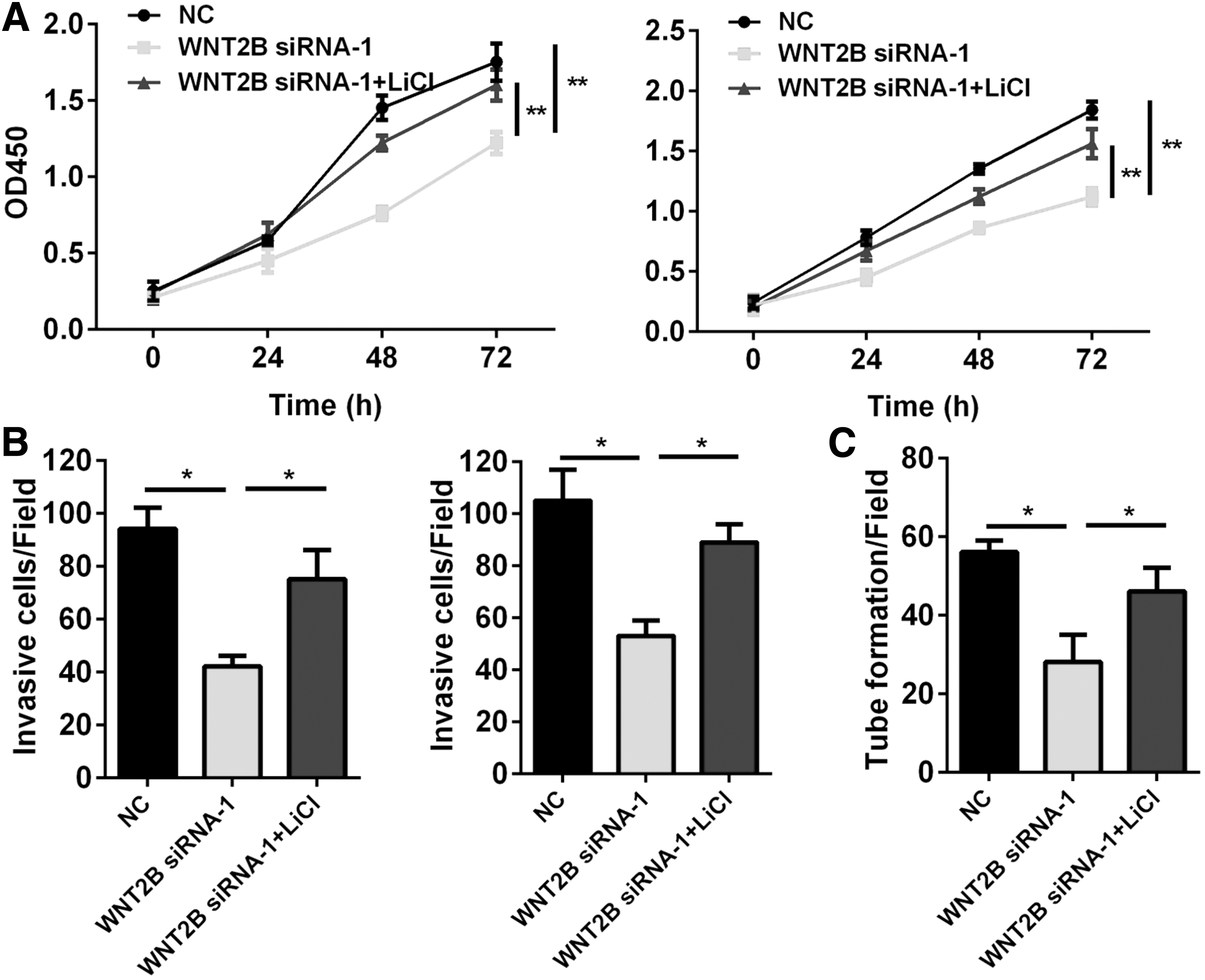
Activation of Wnt/β-catenin signaling abolished the effect of Wnt2B knockdown on cell proliferation, invasion, and angiogenesis.
Discussion
The expression level of Wnt2B protein was significantly increased in OC tissue samples. Previous study showed that the expression of Wnt2B was significantly higher in human pancreatic cancer samples than normal pancreatic samples, indicating a significant correlation with perineural invasion. 10 In nasopharyngeal carcinoma, the mRNA expression of Wnt2B in 15 nasopharyngeal carcinoma tissue specimens compared with noncarcinoma epithelial tissue samples. 22 In gastric cancer, the expression level of Wnt2B was also elevated in gastric cancer tissues compared with normal control. 23 Moreover, Wnt2B protein levels were positively associated with TNM stages and metastasis of OC. Therefore, Wnt2B might be an important oncogene in various cancers, including OC.
Previous studies indicated that Wnt2B played key roles in regulating various biological functions of tumors, including proliferation, EMT, and drug resistance. It has been reported that overexpression of miR-338-3p increased the sensitivity to cisplatin of OC by regulating Wnt2B expression. 12 Previous study showed that decreased expression of Wnt2B suppressed proliferation and invasiveness in OC. 13 miR-577 inhibited proliferation and EMT of NSCLC cell by inactivating the Wnt/β-catenin signaling in a Wnt2B-dependent manner. 11 In osteosarcoma cell, Wnt2B overexpression reversed the effects of sevoflurane treatment on caspase-3 activity cell growth, cell viability, and invasion of U2OS cells. 24 In this study, we showed that Wnt2B knockdown significantly repressed OC cell proliferation, invasion, and angiogenesis.
The Wnt/β-catenin signaling has been identified to play an important role in the regulation of tumor development.25,26 The inhibition of phospholipase D1 (PP1D) suppressed cell growth hyperactivated by the PI3K and Wnt/β-catenin signaling pathways in colorectal carcinoma. 27 RIF1 knockdown inhibited cancer stem cell-like traits and tumor growth in non-small cell lung carcinoma by PP1D-mediated activation of Wnt/β-catenin signaling. 28 Glabridin suppression attenuated angiogenesis by repression of miR-148a in human breast cancer by Wnt/β-catenin signaling pathway. 29 Overexpression of Cyclin G2 suppressed cell growth and metastasis in gastric cancer through inhibiting the Wnt/β-catenin signaling. 30
In OC, SFRP1 knockdown could promote migration and proliferation abilities of epithelial OC cells by activating Wnt/β-catenin signaling pathway. 31 Glycogen phosphorylase B knockdown markedly repressed OC cell proliferation and invasion through the Wnt-β-catenin axis. 32 In this study, we uncovered that Wnt2B knockdown reduced the expression levels of Wnt/β-catenin pathway-regulated genes, such as Cyclin D1, MMP-9, and VEGFs. More importantly, Wnt/β-catenin activation by LiCl reversed the effect of Wnt2B knockdown on cell proliferation, invasion, and angiogenesis. These findings indicated that Wnt2B played key roles in OC progression through Wnt/β-catenin signaling pathway.
In sum, this study revealed that Wnt2B knockdown exerted an inhibitory effect on OC cell proliferation, invasion, and angiogenesis by inactivating the Wnt/β-catenin signaling pathway. Therefore, Wnt2B appears to act as a potential therapeutic target of the treatment of OC potentially.
Footnotes
Funding Information
No funding was received for this article.
Authors' Contributions
H.W. conceived and designed the experiments; X.P. gave an experimental guidance in the laboratory; H.Z. and Q.G. performed the experiments; S.Y. analyzed the data and wrote the article.
Disclosure Statement
There are no existing financial conflicts.