
Abstract
Background:
Lung malignancy is a main source of disease passing all throughout the planet, whereas the transthyretin (TTR) is a specific biomarker for clinical diagnosis. However, its role in lung malignancy stays to be obscure.
Materials and Methods:
In the current study, the authors made an endeavor to research impact of abnormal expression of TTR on nonsmall cell lung carcinoma (NSCLC) by overexpression or knockdown of TTR. To further explore the instruments' fundamental mechanism part of TTR in NSCLC, several signal pathways were searched and verified. To confirm the effect of TTR overexpression on tumors, in vivo experiments were conducted.
Results:
It was found that upregulated TTR clearly stifled cell proliferation, migration, invasion, and expanded apoptosis. Significant suppression of phosphor-extracellular signal-regulated kinase (ERK) was observed in TTR-treated NSCLC cells, implying that TTR was important for cellular progress by regulating mitogen-activated protein kinase/ERK signaling pathway. In in vivo experiment, overexpression of TTR promoted cell apoptosis and inhibited tumor growth.
Conclusion:
Overall, the results suggest that TTR has a potential antitumor effect in human NSCLC progression, which provides theoretical basis for the diagnosis and treatment of NSCLC. Above all, further understanding of TTR was useful for clinical care. Clinical Trial Registration Number: 2016-08.
Introduction
Lung cancer is the most well-known harmful tumor on the planet and is an essential explanation of disease-related demise, in this manner representing an incredible danger to human health. 1 In China, the dreariness and mortality due to lung cancer rank first in harmful tumors. 2 Of note, nonsmall cell lung carcinoma (NSCLC) is the main type of lung cancer. 3 Although great progresses have been made in early detection and treatment strategies in the course of recent many years, the endurance rate actually stays low level. 4 Therefore, further investigation on the molecular mechanisms on NSCLC will be essential for diagnosis and therapy.
Transthyretin (TTR) is a 55-kDa homotetrameric protein that helps transport thyroid hormones through the bloodstream5,6 and plays a role in retinol digestion. It is halfway created by the liver and extrahepatic tissues, such as choroids plexus, retinal shade, epithelium cells, and islet A and B cells.7–9 TTR is a profoundly plentiful protein that ties to coursing retinol binding protein (RBP) to forestall low subatomic mass RBP.10–14 The degrees of TTR could be diminished in extreme liver illness, hunger, and inflammation.15–17 TTR levels were also lower in the serum of patients with ovarian cancer, cervical cancer, and endometrial cancer. 18 As a result, TTR has proven to be a useful biomarker for identifying lung cancer. Notwithstanding, the impact and mechanism of TTR on the lung cancer cells actually stay subtle.
In this study, the authors researched the mechanism underlying abnormal expression of TTR on NSCLC cells, and try to search for efficient signaling pathway in TTR-mediated NSCLC cells. By further investigation into effect of TTR on NSCLC, the authors may provide proof-of-principle evidence for clinical application of TTR.
Materials and Methods
Clinical tissue examples
The tumor tissues and combined contiguous nondisease tissues were gathered from an aggregate of 40 patients determined to have NSCLC at Tianjin Hospital, Tianjin Medical University. The convention of this examination was affirmed by the ethics committee of Tianjin Hospital, Tianjin Medical University (2016-08), and the patients who were enrolled gave their written informed consent.
Transfection and cell culture
The Chinese Academy of Science Cell Bank provided A549 and H1975 NSCLC cell lines, as well as 16HBE standard lung cell lines (Shanghai, China). Cells were refined in RPMI-1640 medium (Hyclone, South Logan, UT) +10% FBS (fetal bovine serum; Biowest, Barcelona, Catalonia, Spain) at 37°C and 5% CO2. TTR overexpression and inhibitor were achieved from GenePharma (Shanghai, China). Sh-TTR were constructed to knockdown TTR, and sh-NC (negative control) were used as control. For TTR overexpression, the full length of TTR coding domain sequence was embedded into pcDNA3.1 vectors (Invitrogen) and the unfilled plasmids filled in as NC. Lipofectamine 2000 (Invitrogen) was used to transfect cells according to the item manuals.
RNA isolation and quantitative reverse transcription-polymerase chain reaction
All RNA was extricated utilizing TRIzol reagent (Invitrogen). Equivalent measures of RNA were interpreted into cDNA utilizing the cDNA first strand synthesis kit. Absolute cDNA was utilized as a beginning material for ongoing polymerase chain reaction (PCR) utilizing the Step One real-time PCR system (Life Technologies Corp), and each example was estimated in threefold. Complete response framework is as follows (20 μL): cDNA 1 μL, SYBR Premix EX Taq 10 μL, each of 1 μL (10 μM) the primers 1 μL, and ddH2O 7 μL. The procedure of PCR was as follows: 94°C for 3 min followed by 35 cycles of 94°C for 10 s, and 63°C for 30 s. Segment overlay changes were determined utilizing the 2-ΔΔCT similar strategy utilizing glyceraldehyde-3-phosphate dehydrogenase (GAPDH) for standardization. The primers used were as follow: TTR: F-TGGGAGCCATTTGCCTCTG, R-AGCCGTGGTGGAATAGGAGTA; Wnt: F-CGATGGTGGGGTATTGTGAAC, R-CCGGATTTTGGCGTATCAGAC; transforming growth factor-β (TGF-β): F-GGCCAGATCCTGTCCAAGC, R-GTGGGTTTCCACCATTAGCAC; extracellular signal-regulated kinase (ERK): F-TACACCAACCTCTCGTACATCG, R-CATGTCTGAAGCGCAGTAAGATT; nuclear factor kappa B (NF-κB): F-AACAGAGAGGATTTCGTTTCCG, R-TTTGACCTGAGGGTAAGACTTCT; GAPDH: F-CTGGGCTACACTGAGCACC, R-AAGTGGTCGTTGAGGGCAATG.
Western blotting
In total, 50 μg all out protein per path was isolated by 10% sodium dodecyl sulfate-polyacrylamide gel electrophoresis gel and smeared onto polyvinylidene fluoride membranes. The membrane was impeded in phosphate buffered saline with Tween 20 and examined with primary antibodies for Bcl-2 (1: 1000; ab32124), Bax (1: 1000; ab32503), caspase-3 (1: 1000; ab13847), caspase-9 (1: 1000; ab13847), matrix metal proteinases-2 (MMP-2) (1: 1000; ab37150), and MMP-9 (1: 1000; ab73734), these antibodies were bought for Abcam (Cambridge, MA). Place Phosphor-ERK (1:2000, no. 9106; Danvers, MA), ERK (1:1000, no. 9102; Danvers, MA), and GAPDH (1: 5000, G8795; Sigma-Aldrich, St. Louis, MOA) at 4°C overnight. Subsequently, appropriate secondary antibodies were used to incubate the membranes (1: 5000, ab6858: Abcam), protein bindings were visualized by ECL kit (Beyotime Institute of Biotechnology, Beijing, China). The groups were checked utilizing a densitometer, and the dark worth of the groups was determined naturally by the ImageJ software version k 1.45 (NIH, Bethesda, MD).
Cell proliferation tests
5-Ethynyl-2′-deoxyuridine (EdU) measure was applied to recognize cell proliferation as indicated by the producers' convention. For colony formation test, cells were cultivated in a six-well plate at a thickness of 1000 cells/well for ∼14 d, after which clones were fixed with methanol and stained with crystal violet (0.1%).
Cell apoptosis tests
FITC-Annexin V/PI apoptotic identification kit (BD ingen, San Diego, CA) was utilized to recognize apoptotic cells by flow cytometry. The cells (1 × 10⁵ cells) were stained with propidium iodide (4.5 mL) and Annexin V-FITC (4.5 mL), trailed by fluorescence assurance by flow cytometer (Beckman, Miami, FL). Terminal deoxynucleotidyl transferase (TdT) dUTP nick-end labeling (TUNEL) assay was also applied to detect the apoptosis in cells with the In Situ Cell Death Detection Kit (Roche). The cells were washed with 1 × phosphate-buffered saline (PBS) and fixed in paraformaldehyde solution (500 μL, 4%) for 15 min at 37°C. All examinations were performed in threefold.
Scratch assay
The cells were grown in six-well plates. Inexact 80% of the cells were placed into the divider and washed in 1 × PBS twice to eliminate gliding cells. Wounds were gotten from sterile pipette tips. Subsequently, cells were washed with 1 × PBS twice, and RPMI-1640 culture medium (2 mL, 10% FBS) was added into the culture plate. Photos were taken 24 and 48 h after the wound was formed.
Transwell invasion measurement
Transwell invasion measure was estimated according to the maker's guidelines. To sum things up, a sum of 3.5 × 10⁴ cells/well in Dulbecco's modified Eagle's medium (DMEM) (100 μL, 0.5% FBS) were put in the upper Transwell chamber (Corning Incorporated, NY), which was precovered with matrigel (Growth factor reduced; BD Biosciences, MD). The bottom chambers were filled with DMEM containing 20% FBS. Once passed through the membranes, the cells were fixed and stained after 24 h. Cells in nine haphazardly chosen fields were checked under a modified magnifying lens (200 × magnification; Carl ZEISS, Jena, German) and a normal worth was utilized as the quantity of invaded cells. For the cell transmembrane relocation examination, except for the method covered by Matrigel (BD Biosciences, Franklin lakes, NJ), each method is completed relative to the method in the invasion test.
Tumor xenograft model in naked mice
The Beijing Vital River Laboratory Animal Technology sold BALB/c nude mice (male, 5 weeks old; 18–20 g) (Beijing, China). Human A549 and H1975 cells were used to create a xenograft model. A549 and H1975 cells transfected with sh-NEAT1 or sh-NC were infused subcutaneously into the back fossa of naked mice (six per group). After 5 d, when normal tumor volume was ∼50 mm3, mice were intraperitoneally infused with paclitaxel (3.5 mg/kg body weight) or PBS every 3 d. The tumor volumes were estimated each week by utilizing calipers: volume (cm3) = width 2 (cm2) × length (cm)/2. Mice were sacrificed at 32nd day after infusion and tumors were eliminated, gauged, and analyzed by quantitative RT-PCR. All animal experiments were performed with the approval of the institutional Ethics Committee.
Statistical analysis
All outcomes were performed in any event three independent replicates and revealed as means ± standard deviation. All measurable examinations were performed utilizing GraphPad software 5.0. Student's t-test or one-way analysis of variance (ANOVA) followed by Tukey's multiple comparison test was used to assess the differences between the two groups or three gatherings, respectively. Statistical significance was defined as a p < 0.05.
Results
The level of TTR expression in NSCLC tissues and cells is reduced
By examining at 40 pairs of human NSCLC and normal tissues, a prominent decrease in TTR expression in NSCLC was found than in the normal tissues (Fig. 1A). At the same time, the authors observed that compared with human bronchial epithelial cells, the TTR expression level in two NSCLC cell lines was significantly reduced (Fig. 1B). The overall survival time of patients with high-TTR expression was significantly higher than those with low-TTR expression (Fig. 1C). These results indicate that TTR may be negatively correlated with NSCLC progression.
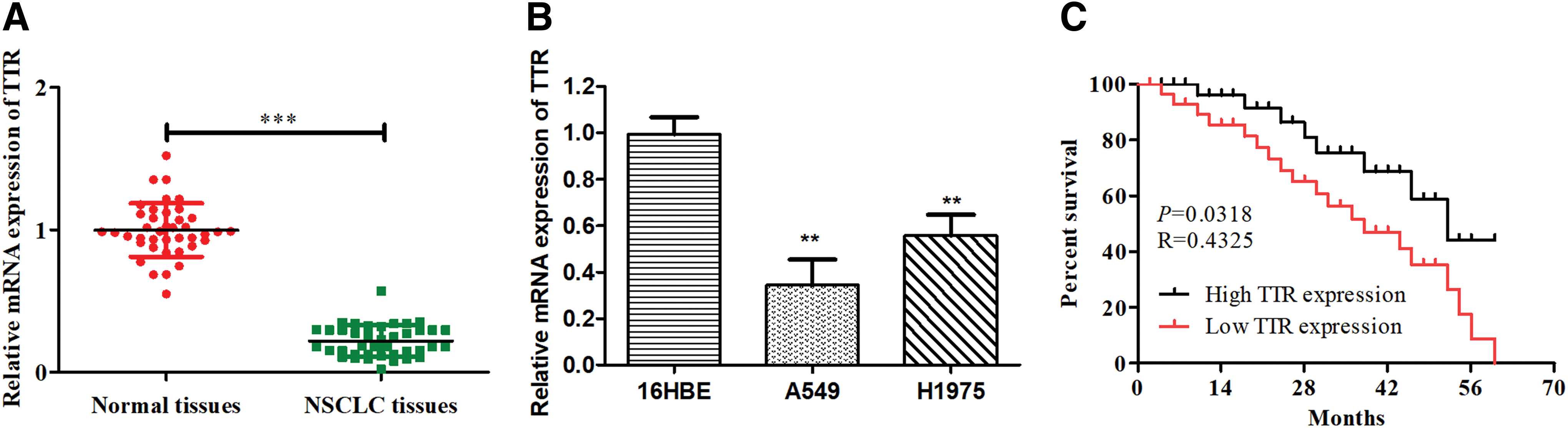
TTR expression in tissues and cell lines of NSCLC.
TTR curbs in vitro NSCLC cell expansion and advances its apoptosis
To analyze the part of TTR in cell proliferation, the authors overexpressed TTR (TTR overexpression) or transfected with TTR inhibitors in A549 cells and H1975 cells, respectively (Fig. 2A). EdU tests indicated that both A549 cells and H1975 cells proliferation capacity were reduced in TTR-overexpressed group compared with NC group, while were increased in the TTR inhibitor-treated group compared with NC group (Fig. 2B).
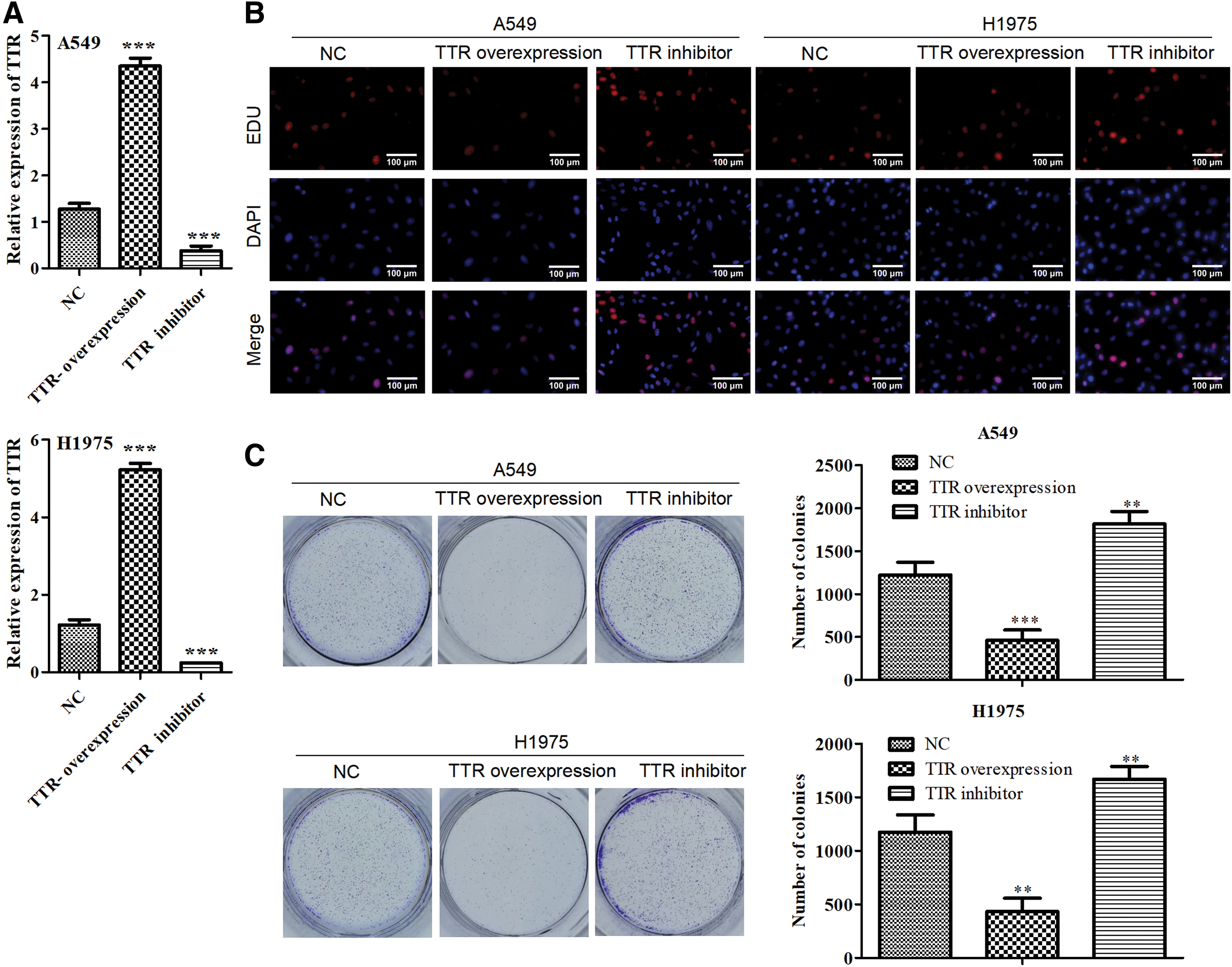
In vitro impact of TTR on cell proliferation of NSCLC in vitro.
Reliably, these outcomes were additionally affirmed by colony formation assay (Fig. 2C).
Furthermore, cell apoptosis by flow cytometry and TUNEL assays was estimated, suggesting that proliferation capacity of both A549 and H1975 cells was decreased in the TTR-overexpressed group (Fig. 3A, B). At the molecular level, we found that proapoptotic protein Bcl2 was downregulated and Bax and caspase-3/9 (antiapoptosis proteins) was up-regulated in TTR overexpression group were determined using Western blot analysis compared with NC group (Fig. 3C). The results indicated that the TTR overexpression group advanced A549 cells and H1975 cells apoptosis.
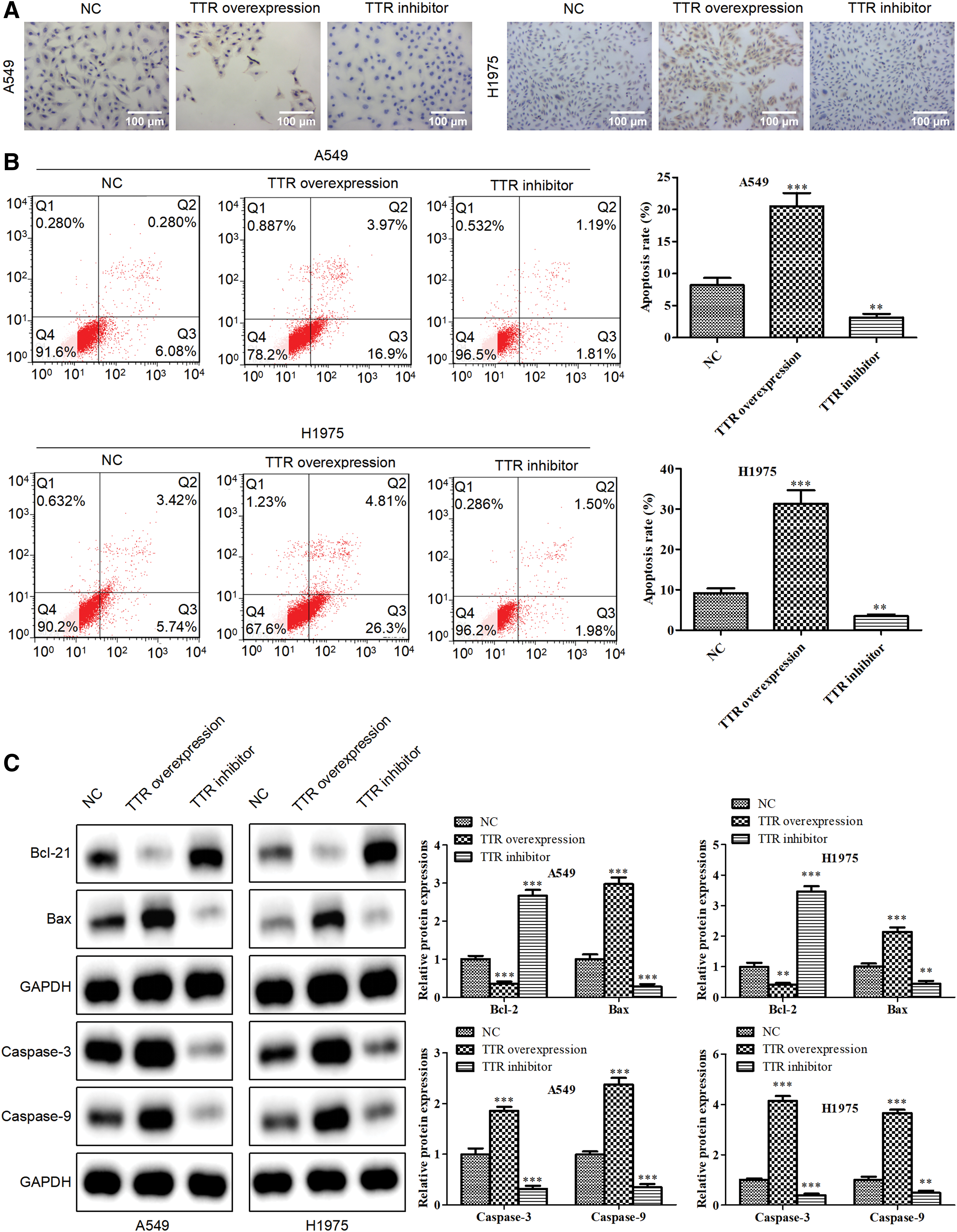
In vitro impact of TTR on cell apoptosis of NSCLC. Cell apoptosis capacity of H1975 and A549 cells was detected by
TTR restrains in vitro NSCLC cell migration and invasion
The impacts of TTR on cell migration and invasion were studied by Transwell chamber and scratch methods. Transwell chamber assay that less cells were moved and attacked to the lower medium in TTR overexpression group compared with TTR inhibitor group (Fig. 4A, B). As shown in Figure 4C, a scratch test revealed that the migrated distance between the control and TTR inhibitor groups differed significantly. Then, Western blotting was performed to detect Matrix metal proteinases (MMP2/9). The MMP-2 and MMP-9 expression levels were downregulated in the TTR overexpression group (Fig. 4D). According to these results, upregulated TTR suppressed NSCLC cell migration and invasion in vitro.
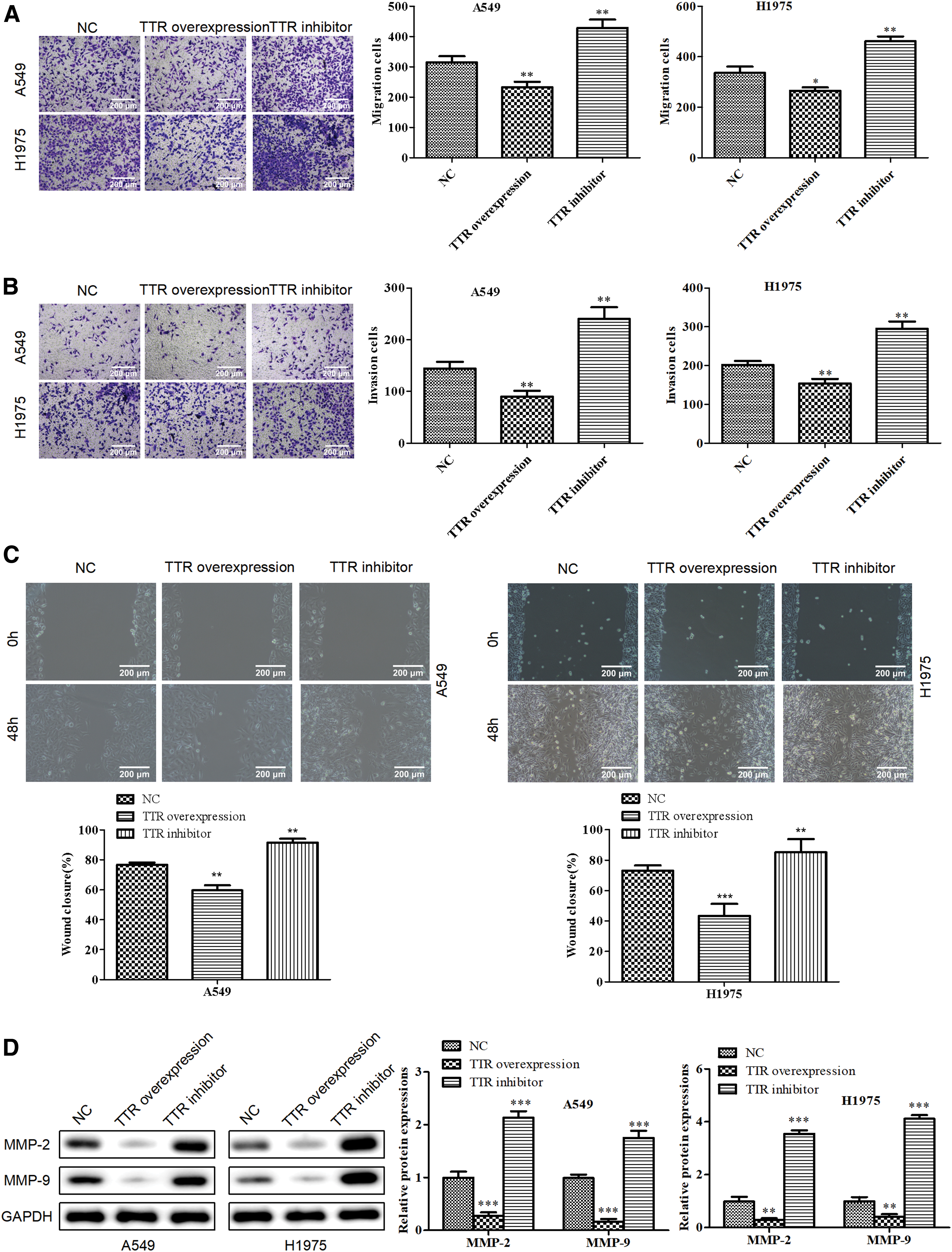
In vitro TTR suppressed NSCLC cell migration and invasion. Transwell migration
TTR represses the in vitro mitogen-activated protein kinase/ERK pathway activation
To further gain insights into the downstream signaling pathways regulation by TTR in NSCLC cells, the functional effects of TTR in several important cancer pathways were analyzed including Wnt, TGF-β, NF-κB, and ERK by RT-PCR. TTR overexpression altogether hindered the declaration of ERK in A549 cells and H1975 cells (Fig. 5A). Western blot results showed phosphor-ERK was observed to be significantly suppressed in the TTR overexpression group (Fig. 5B), demonstrating that the mitogen-activated protein kinase (MAPK)/ERK flagging pathway was fundamental for tumor growth in NSCLC cells. TTR overexpression inhibited ERK phosphorylation, thereby stimulating cell proliferation and cell survival.

In vitro TTR suppressed tumor progression through ERK pathway.
TTR ameliorates in vivo NSCLC progression
In light of the capacity of TTR in vitro, the authors further examined whether overexpression of TTR could suppress NSCLC progression in vivo. The expression of TTR was distinguished by qRT-PCR (Fig. 6A). Accordingly, tumors from TTR overexpression developed clearly more slow than the NC group. The mice were killed 5 weeks after the injection and the tumors were taken out. The tumors were obviously smaller than those in other groups on size and weight (Fig. 6B). These results implied that TTR could inhibit tumor progression. TUNEL assay revealed that the TTR overexpression group had more apoptosis than the NC group. Inhibiting TTR expression, on the other hand, can significantly reduce cell apoptosis (Fig6C).

In vivo upregulated TTR suppressed NSCLC progression.
Discussion
Lung cancer is the most frequent cause of cancer-related death all over the world, and the 5-year survival rate is less than 15%.19,20 Although there have been numerous studies on the resistance, discovery, and treatment of lung cancer, the expectations for lung cancer patients remain pessimistic. In this research, it was demonstrated that TTR suppressed tumor progression in NSCLC by inactivating the MAPK/ERK pathway.
Abnormal expression levels of TTR have been accounted for to assume a significant part in tumor formation and development.21,22 For example, Shimura et al. found a significant negative correlation between TTR levels and tumor size and the number of involved lymph nodes. 23 However, there is still limited understanding of the effect of TTR abnormal expression levels on tumor progression. It was discovered that human NSCLC tissues had lower TTR expression than normal tissue, and that TTR expression was associated with clinic pathological features in this study. Similar results have reported that the expression of TTR in patients with cervical cancer was significantly lower than that in healthy controls. 24 Segment TTR expression in NSCLC cells cultured in vitro can boost cell proliferation, migration, and invasion while decreasing cell apoptosis. Overexpression of TTR, in contrast, is sufficient to inhibit cell proliferation and invasive potential in NSCLC cells, as well as increase in apoptosis. To further investigate mechanisms of TTR on tumor progression in NSCLC, several signaling pathways were searched, and MAPK/ERK signals were found to respond to the TTR manipulation in NSCLC cells. In in vivo experiment, overexpression of TTR obviously inhibited tumor growth and promoted cell apoptosis, implying that TTR was important for regular cellular progression.
The tumor-related pathways, such as Wnt, PI3K/AKT, or NF-kB, may assume a significant part in the advancement of tumorigenesis.24–30 Furthermore, there is expanding proof that common MPAK/ERK flagging pathway is a crucial flagging pathway associated with the improvement of a few tumors.31–36 To additionally investigate the likely atomic system of TTR in A549 cells and H1975 cells, the authors zeroed in on the MAPK/ERK flagging pathway. Past investigations have detailed that a few genes influenced the MAPK/ERK signal pathway. 37 Segment actuation of the MAPK/ERK pathway promoted cell proliferation, migration, and differentiation, and induced cell cycle arrest.38–41 The results suggested that the expression of phosphor-ERK was markedly suppressed in the TTR overexpression group, indicating that the MAPK/ERK signaling pathway was fundamental for tumor development in NSCLC cells. It shows similar results as the previous study, which found that phosphor-ERK expression was significantly reduced in the TTR overexpression group. Therefore, further investigation on the molecular mechanisms of TTR on NSCLC will be essential for diagnosis and therapy.
Taken together, our study exhibited that TTR assumed an imperative part in NSCLC tumorigenesis. TTR stifled cell proliferation, migration, and invasion, and advanced apoptosis at any rate somewhat through hindering MPAK/ERK flagging pathway, which was demonstrated to be an effective diagnostic and predictive biomarker for diagnosis and treatment in NSCLC.
Footnotes
Funding Information
This work was supported by Tianjin Health Industry Key Project (no:16KG138).
Availability of Data and Material
The data that support the findings of this study are available from the corresponding author upon reasonable request.
Disclosure Statement
There are no existing financial conflicts.