
Abstract
Background:
Integrin αvβ6 has become an extremely promising theranostic target for precise delineation of fast-growing malignant cells in the recent years. The aim of the study was to validate the in-house kit-like synthesis of 68Ga-Trivehexin (integrin αvβ6) and to evaluate its uptake in patients with integrin αvβ6 expressing head and neck and pancreatic cancer.
Materials and Methods:
68Ga-Trivehexin was synthesized by adding the variable amount of integrin αvβ6 (30–50 μg) to full volume (4–5 mL) Ga-68 in 0.05 M HCl and heating the reaction mixture at 90°C for 12 min at pH 3.5–4 to obtain the radiotracer with high radiochemical purity (RCP) and high yield. Quality control procedures were done to assess the RCP, stability, pyrogenicity and sterility of the radiotracer. 68Ga-Trivehexin was then administered in patients who met the eligibility criteria. Whole body PET/CT scans were done at variable time points post intravenous (i.v.) injection of 84–185 MBq of 68Ga-Trivehexin to assess its biodistribution and maximum uptake time.
Results:
0.2 mCi of 68Ga/μg of Trivehexin at 90°C for 12 min was the optimal parameter to obtain 85%–88% of noncorrected yield and 99% of RCP. The 68Ga-Trivehexin showed in vitro stability upto 6 h and was also found to be sterile and pyrogen free. Intense radiotracer uptake was noticed in the tumor and no uptake was noticed in healthy tissues. PET/CT imaging at 60 min post injection was found to be the optimal time for imaging the tumors with 68Ga-Trivehexin.
Conclusion:
The protocol for in-house kit-like labeling of 68Ga-Trivehexin was safe, reproducible, and cost-effective. 68Ga-Trivehexin is an extremely promising agent for noninvasive molecular imaging of integrin αvβ6 expressing tumors.
Introduction
Fluoro-2-deoxy-
Over the recent years, the development of probes that can investigate the biological processes other than the glucose metabolism has, therefore, gained momentum. The agents targeted specifically at angiogenesis, hypoxia, and other processes. One such target is αvβ6 integrin. Integrins are a family of heterodimeric cell surface receptors that are expressed in cells, where they mediate cell-cell and cell-extracellular matrix interactions. 1 Because of its well-established role as a promoter of (tumoral) angiogenesis, awareness is mostly limited to the subtype αvβ3 2 –5 ; many other integrins are involved in every step of cancer development. 6
αvβ6 integrin is a driver of invasion and metastasis in epithelial cancers. 7 It acts by activation of the transforming growth factor β (TGFβ). αvβ6 integrin is expressed by the epithelial cells rather than the endothelial cells. It is found in many carcinomas, such as squamous cell, basal cell, lung adeno, and colon. 8 Pancreatic ductal adenocarcinoma (88% of primaries) and head and neck squamous cell carcinomas (95% of primaries 9 ) have been shown to be most closely associated with overexpression of αvβ6 integrin. 10
Expression of αvβ6 integrin is almost always tumor specific with surrounding normal stromal tissue not expressing αvβ6. It represents an extremely valuable theranostic target as αvβ6 is absent in most normal adult human cells. It also potentially enables a precise delineation of carcinoma margins and/or assessment of their invasiveness by molecular (nuclear) imaging as well as therapeutic intervention with targeted radioligands at the most critical locations. Therefore, in recent years, a variety of αvβ6 integrin-targeted PET radiopharmaceuticals have been developed. Most of these compounds showed a good tumor-to-background contrast in rodent models with very low background, but their tumor accumulation was ultimately too low for a successful clinical transfer. 11
Trivehexin is an improved trimerized αvβ6 integrin-selective nonapeptide and is anticipated that it will prove to be clinically useful for specific PET imaging of cancers with high αvβ6 integrin expression, such as ovarian, gastric, pancreatic, and lung carcinoma, as well as invasive head and neck carcinomas. The aim of this study was to validate the robust in-house kit-like synthesis of 68Ga-Trivehexin and to evaluate its uptake in patients with integrin αvβ6 expressing head and neck and pancreatic cancer.
Materials and Methods
68Ga was obtained from 1110 MBq 68Ge-68Ga generator (Isotope Technologies Graching) for all the clinical studies. The trimerized αvβ6 integrin-selective nonapeptide-Trivehexin was synthesized by TRIMT, GmbH (Radeberg, Germany). HPLC-grade ethanol, HPLC-grade water, Suprapure HCl, and acetonitrile were procured from Merck (Germany). Sodium acetate was purchased from Sigma-Aldrich. C-8 Sep-pack cartridges were purchased from Waters (Ireland).
Quality control of 68Ga-Trivehexin was done according to manufacturer's procedures. Radioactivity assay of Ga-68 was done using a properly calibrated dose calibrator (CRC 25 PET; Capintec). Instant thin-layer chromatography-Silica gel (ITLC-SG) paper was purchased from Merck. Sep-Pak C18 light cartridges were purchased from Waters. Syringe filters (0.22 μm pore size) were procured from Millipore. Tryptic soy broth medium (Himedia) was used for sterility testing. Pyrogenicity was checked by the endotoxin reader (PTS, Charles River). The patient imaging was done using a dedicated PET/CT scanner (Philips TruFlight).
Radiolabeling and quality control of Trivehexin with Ga-68
The radiosynthesis of 68Ga-Trivehexin was done applying an updated previously described protocol. 10 For validation of the synthesis protocol in the clinical setting and to optimize it for high yield and high radiochemical purity (RCP), variable amount of Trivehexin (30–50 μg), in sodium acetate buffer (60 mg in 1.5 mL sterile H2O), was used. The 68Ge-68Ga generator was eluted manually with 5 mL of 0.05 M HCl.
Initially, 1.5 mL of the elute was discarded and 2.5 mL of the elute was added to the sodium acetate buffer with the peptide. pH was maintained at 3.5–4 and the reaction mixture was incubated at 90°C for 12 min. The reaction mixtures were cooled to room temperature. Subsequently, the solution was passed through the light C-8 column preconditioned with 5 mL 70% ethanol solution with a flow rate of 1 mL/min and then rinsed with 10 mL of ultrapure water. After cooling at room temperature, the reaction mixture was passed through a preconditioned light C-8 cartridge to release free Ga-68. The labeled product was finally eluted with 1 mL of 50% ethanol from the C-8 cartridge, followed by 9 mL of normal saline. The final product was collected in a sterile vial after passing through a 0.22 μm filter. Sterility was tested on a “post hoc” basis.
Visual appearance
The product was inspected visually to confirm no visible particle in a colorless solution. pH was determined using the pH paper.
Radiochemical purity
Based on the precursor manufacturer´s information on RCP testing and correlation of HPLC and TLC results, the QC was done in the most efficient way, best applicable for daily routine in radiopharmaceutical production. RCP of 68Ga-Trivehexin was determined in a short time using only radio-TLC with an NaI detector (Lablogic), using silica gel-impregnated aluminum strips (ITLC-SG strips) as stationary phase and 0.1 M aq. sodium citrate as mobile phase. A 3–4 μL drop of sample solution was spotted about 1 cm from one end of the strip and the strip was developed in the sodium citrate buffer (0.1 M) till the solvent migrated to the top.
Stability studies
To evaluate the tracer's stability, 68Ga-Trivehexin in its formulation was stored at room temperature and in human serum at 37°C, and its stability was estimated by performing ITLC at regular time intervals using ITLC strips hourly up to 6 h.
Sterility and pyrogenicity
The labeling procedure was performed in a laminar flow hood to maintain sterility. However, to test the sterility of 68Ga-Trivehexin, the sample solution was incubated in Tryptic soy broth at 37°C. Turbidity in the incubated samples was observed for up to 3 d to check the presence of any microorganism. The level of pyrogenicity in the sample was evaluated by the Pyrogen Plus Limulus amebocyte Lysate Kit (Charles River) in accordance with USP XXX1 (Sensitivity 0.125 EU/mL). Twenty-five microliters of the sample was placed on the PTS™ cartridge reader, followed by incubation at 37°C for 15–20 min. This test was performed on a “post hoc” basis.
Clinical evaluation
Twenty patients, 18 years of age or older, with suspected or newly diagnosed or previously treated malignant tumors of the head and neck or pancreas, were included in the study. Patients with nonmalignant lesions and females with pregnancy were not eligible.
The institutional review board approved this study and all the human participants provided written informed consent, before the investigation.
All the patients underwent a 68Ga-Trivehexin PET/CT scan to confirm the overexpression of integrin αvβ6. Whole-body images (base of skull to mid-thigh) were acquired in 3D mode, 10, 60, and 90 min after intravenous (i.v.) injection of 74–185 MBq of 68Ga-Trivehexin to estimate the maximum uptake time and high tumor/bkg ratio. Data acquisition was performed on a hybrid PET/CT scanner (Philips TruFlight select time of flight PET scanner) with CT parameters being 120 kV, 350 mA, slice thickness of 3.75 mm for diagnostic CT, rotation time of 0.5 s, 512 × 512 pixel matrix, and pixel size of about 1 mm. CT-based attenuation correction of emission images was employed. Iterative reconstruction of PET images was done using ordered set expectation maximization algorithm with an attenuation correction. The patients were asked to void before acquiring the imaging. The subsequent CT images were acquired using low current (10 mA).
Image analysis was performed by a Nuclear Medicine physician using an appropriate workstation Philips TruFlight. Volumes of interest were automatically drawn over the organs of moderate to intense physiologic uptake and/or tracer accumulation. Tumor-to-background ratios were also calculated for all the tumors in relation to different tissue types like liver, heart, blood vessel, and muscle. Tracer biodistribution was then quantified by the maximum standardized uptake value (SUVmax). Results were then presented as mean, median, interquartile range (25th quantile–75th quantile), and range (minimum–maximum).
Statistical analysis
Descriptive analyses was used for demographics and tumor characteristics. For description of SUV, arithmetic mean, median and standard deviation, were used. SUVs in tumor and normal tissue and tumor-to-background ratios (TBRs) was compared with the Wilcoxon signed-rank test. A p-value <0.05 was considered statistically significant. All statistical analyses were performed using SPSS Statistics Version.
Results
Optimization of radiolabeling parameters
A series of manual radiolabeling studies was performed to obtain high radiochemical yield and RCP values by changing the peptide amount (20–60 μg) as shown in the Table 1. In all the experimental studies, medium of pH was maintained at 3.5–4 with sodium acetate. Compared to the previously published radiolabeling procedures, 10 the authors employed sodium acetate as a standard buffer to avoid the presence of HEPES buffer in the formulation. HEPES buffer might be a good choice for the automated labeling with various purification steps, but it is recommended to test for its residues, which means another QC procedure and prolonged time before release.
Details of Synthesis of 68Ga-Trivehexin
RCP, radiochemical purity; RCY, radiochemical yield.
The reaction temperature and the reaction time were kept constant at 90°C and 12 min, respectively, which provide the most suitable reaction condition for the complexation of 68Ga with a peptide according to the previously optimized parameters for well-known 68Ga-prostate specific membrane antigen (PSMA) and 68Ga-DOTA-octreotide (DOTANOC). In this study, trimerized αvβ6-specific,68 Ga-Trivehexin was labeled with full eluate of 68Ga with good RCP and yields. Around 0.2 mCi of 68Ga/μg of Trivehexin at 90°C for 12 min were the optimal parameters to obtain around 90% of noncorrected yield and 99% of RCP. Below this concentration, the radiolabeling yield was somewhat low (50%–70%).
Clarity and pH
The final radiolabeled peptide was obtained as a clear solution with no milky appearance or presence of any particulate matter. The pH of the final product was between 3.5 in all the batches.
RCP analysis
According to ITLC findings, 68Ga-Trivehexin was retained at the point of spotting, whereas free Ga-68 moved to the solvent front as shown in Figure 1. The radiochemical purities of the labeled product prepared under optimized conditions before and after C-8 cartridge purification, respectively, as determined by the ITLC, were 90%–92% and 98%–99%, respectively. All reactions were tried three times for validation of RCP and radiochemical yield.
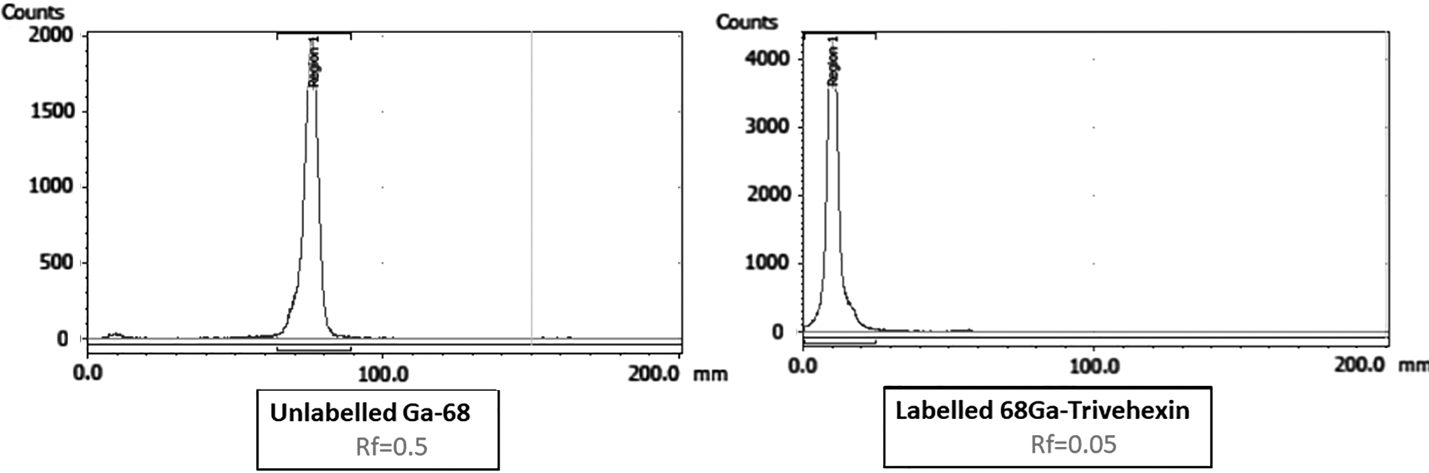
Typical thin-layer chromatography patterns of
Stability studies
Stability studies were conducted by incubating the radiolabeled peptide in human serum at 37°C and laboratory medium at room temperature for up to 6 h. RCP results indicated that the radiolabeled peptide was highly stable at room temperature and human serum up to 6 h confirmed by R-TLC (Fig. 2).
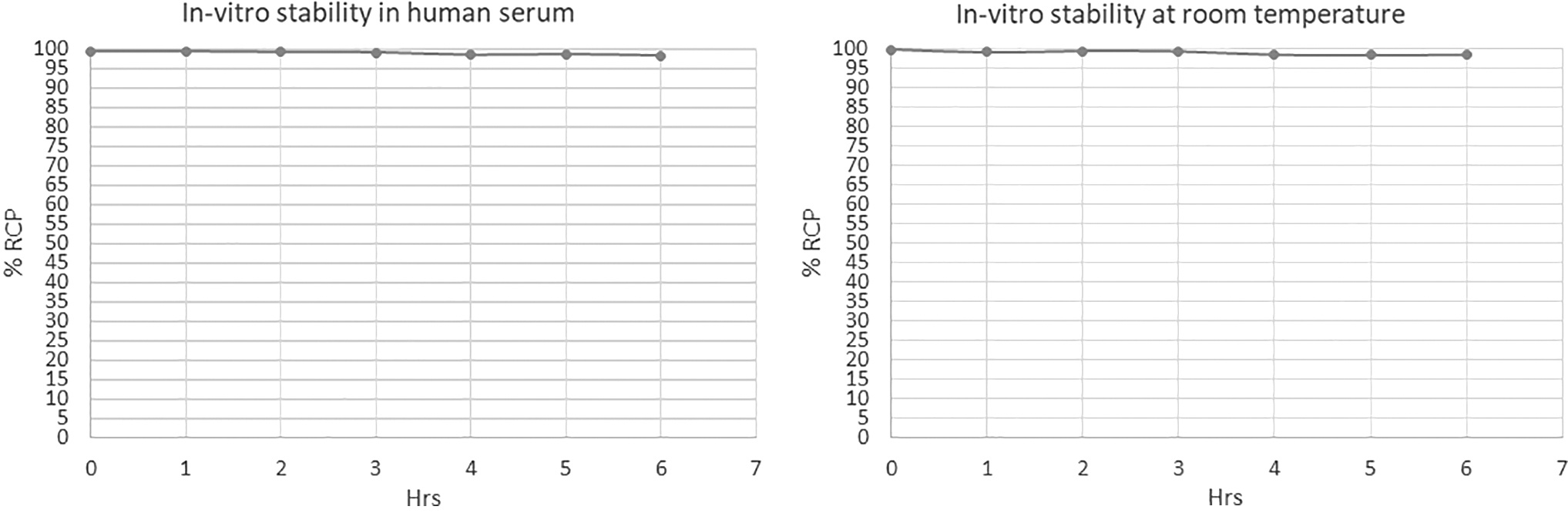
Radiochemical purity of Ga-68 Trivehexin stored in human serum at 37°C and laboratory medium at room temperature up to 6 h. RCP, radiochemical purity.
Sterility and pyrogenicity
Incubation of the sample in Tryptic soy broth for 7 d did not show any turbidity and that confirmed the sterility of the product. Bacterial endotoxin level in all the samples was <0.1 EU/mL. The endotoxins in all the decayed samples tested were found to be within the permissible limits (<17.5 EU), between 0.178 and 0.212 EU/mL.
Patient studies/study population
A total of 20 patients were analyzed. PET/CT imaging was performed in patients with head and neck cancer and head of the pancreas after injecting 110 MBq (range 84–185 MBq) 68Ga-Trivehexin at 10, 60, and 90 min to assess the biodistribution of the peptide 68Ga-Trivehexin and to estimate the maximum uptake time (Fig. 3). Following i.v. administration of 68Ga-Trivehexin, there was no adverse or clinically detectable pharmacologic effect.

The MIP images of 68Ga-Trivehexin PET/CT scan at different time points (10, 60, and 90 min) postadministration. MIP, maximum intensity projection; PET/CT, positron emission tomography/computed tomography.
Biodistribution studies
Maximum physiological radiotracer uptake was demonstrated in the kidneys as the radiotracer was observed to be excreted exclusively through kidneys. Liver, spleen, pancreas, stomach, lungs, heart, and bladder were the other organs showing diffuse physiological uptake of the radiotracer (Fig. 4). High radiotracer uptake with good tumor-to-background ratio was noticed in tumors in all patients at all time points. Quantitatively, the mean SUVmax of the pancreatic lesions and head and neck tumor lesions in 68Ga-Trivehexin PET/CT scans at three time points is shown in Table 2. On the basis of Tumor/Liver, Tumor/Heart, Tumor/Blood pool, and T/Muscle, the best delineation of the Tumor/Backround was observed at 60 min (Fig. 5). So, PET/CT imaging at 60 min postinjection was considered to be the optimal time for imaging the tumors with 68Ga-Trivehexin.

SUVmax in organs at different time points postadministration. SUVmax, maximum standardized uptake value.
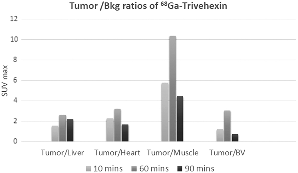
Tumor-to-background ratios at different time points postadministration.
Mean Maximum Standardized Uptake Values of Pancreatic Ductal Adenocarcinoma and Head and Neck Tumors
Discussion
A recent study of trimerized αvβ6 integrin-specific Trivehexin, labeled with Ga-68 in an automated setup showed a remarkable selectivity for αvβ6 over other RGD-binding integrins. 12 Trivehexin was synthesized to exploit the multimer effect to achieve enhanced uptake, a higher target affinity, and longer retention. An automated synthesis of radiopharmaceuticals allows standardization, safety dose, stability, reproducibility, and high yield. 13,14 Moreover, this process allows a Good Manufacturing Practice compliance production in clinical studies and prevents cross-contamination from tubing systems with the usage of disposable cassette systems, which lead to high sterility and purity. 15 However, the centers with high throughput, which require multiple elutions of the 68Ge-68Ga generator to label different peptides in a single day, manual elution allows a better alternative to meet the logistics.
In this study, 68Ga-Trivehexin was synthesized in-house with kit-like manner with good radiochemical yields and RCP using the prefilled low-bioburden portions of precursor .68Ge-68Ga generator selected for this study was optimal as its unique metal-free design and low acidic eluent allowed the fast and convenient onsite production of carrier-free Ga-68 for radiolabeling, which obviated the requirement of prior prepurification or fractionation of the eluate as is the case in previously published labeling procedures for Trivehexin. Low acidity and volume of the eluate allowed for only a small amount of biologically compatible buffer to reach the optimal labeling, as well as injection pH.
Crucial parameters in obtaining good labeling yield are pH of the reaction, peptide amount, and the reaction time. In these experiments, the pH of the medium, reaction time, and temperature were kept constant due to the previously optimized parameters for well-known 68Ga-PSMA and 68Ga-DOTANOC synthesis. In this study, the appropriate amount of the peptide for obtaining good radiolabeling yield was 0.2 mCi/μg (i.e., 45–50 μg for ∼10 mCi 68Ga). Below this concentration, the radiolabeling yield was somewhat lower (50%–60%) (Table 1). However, it was observed that the further increase in peptide amount did not affect the labeling yields. The authors are in line with the results of the previous study, which suggested 10 nmol, that is 43 μg as a minimum peptide for the clinical application. 12
A varied pH range has been reported in literature for labeling of peptides with 68Ga; however, pH range of 3.5–4.0 has been found to be the most consistent range by the authors, 16 –18 and therefore, the authors have also used the same range based on the authors' experience of labeling Ga-68 with other peptides such as PSMA-11 and DOTANOC. In this study, the optimal parameter for the manual synthesis of 68Ga-Trivehexin to obtain 85%–88% of yield and 99% RCP was 0.2 mCi of 68Ga/μg of Trivehexin heated at 90°C for 12 min. The authors' findings suggest that 50 μg is the right amount of the precursor to obtain robust kit radiolabeling setup with good yields and RCP.
The endotoxin content in all the decayed samples tested was within the permissible limits (<17.5 EU), ranging from 0.178 to 0.212 EU/mL. No microbial growth was observed in the Tryptic soy broth after incubation for 3 d, which indicated sterility of the samples. All the physiochemical and biological quality control tests were well presented and all final product specifications were obtained within limits and acceptable criteria, which indicated that the formulation was safe for intravenous administration in human.
In this study, 68Ga-Trivehexin showed good in vivo stability and demonstrated good tumor-to-background ratio. Substantial tumor uptake was noticed at all the three time points, postinjection, with maximum tumor-to-background ratio at 60 min. The deciding factor for the maximum uptake time was tumor-to-background ratios, which were maximum at 60 min. On the basis of this result, PET/CT acquisition was conducted at 60 min postinjection in future clinical studies. This was in line with the results of the earlier study. 12
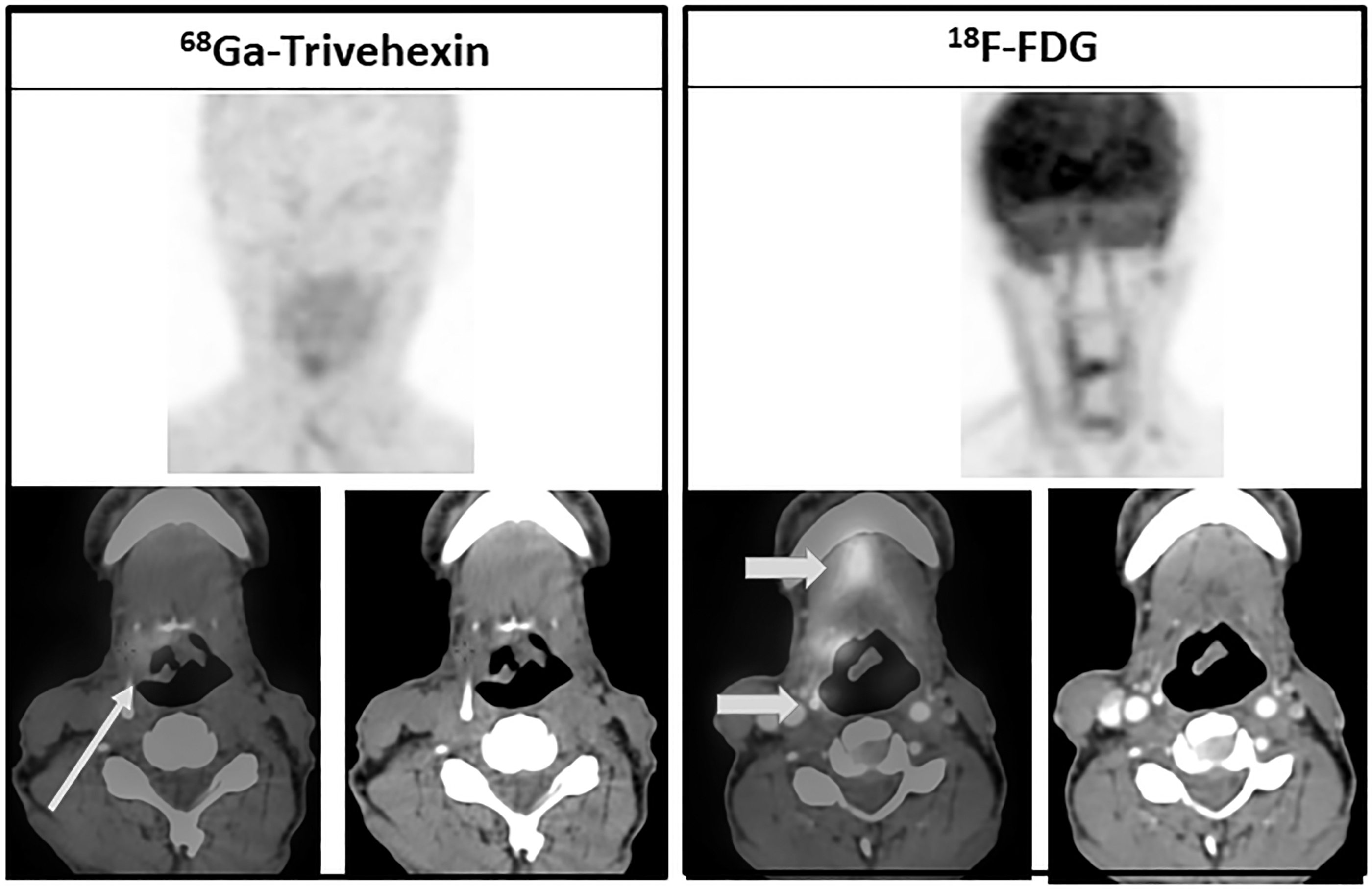
Maximum physiological radiotracer uptake was observed in the kidneys as the radiotracer was observed to be predominantly excreted through the renal route in all patients followed by liver and spleen along with diffuse uptake in stomach, lungs, and so on. The 68Ga-Trivehexin was injected in patients of pancreatic ductal adenocarcinoma (PDAC) and head and neck squamous cell carcinoma (HNSqCC) and was also compared with 18 F-FDG to evaluate its incremental value, as shown in the Figures 6 and 7; however, the clinical results will be shown in a separate study.
68Ga-Trivehexin PET/CT MIP and fused images (left) and
18
F-FDG PET/CT MIP and fused images (right) of patient with SqCC of BOT. 68Ga-Trivehexin PET/CT shows focal uptake in the BOT on the right side, consistent with b6 receptor expression by the lesion and not by the adjacent normal tissues (Thin arrow).
18
F-FDG PET/CT images, in contrast, demonstrate focal FDG uptake by the lesion in the BOT on the right side, with diffuse nonspecific uptake seen in the adjacent lateral pharyngeal wall and the floor of mouth (thick arrow). BOT, Base of tongue; FDG, fluoro-2-deoxy-
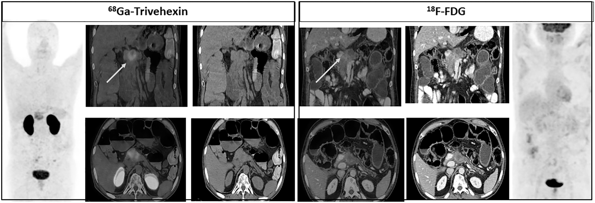
68Ga-Trivehexin PET/CT MIP and fused images (left) and
18
F-FDG PET/CT MIP and fused images (right) of patient with pancreatic ductal adenocarcinoma. 68 Ga- Trivehexin PET/CT shows increased uptake by the pancreas head mass (white), consistent with b6 receptor expression by the lesion.
18
F-FDG PET/CT images, in contrast, are negative for uptake (white arrow). FDG, fluoro-2-deoxy-
As shown in Figure 6, in one patient of HNSqCC, the target lesion was masked by nonspecific uptake in the oral cavity and oropharynx in 18 F-FDG PET/CT scan, while it could be clearly defined in 68Ga-Trivehexin PET/CT scan due to complete absence of any nonspecific uptake of the radio tracer in the surrounding structures. In another patient (Fig. 7) with PDACs, 18 F-FDG PET/CT scan was proved to be suboptimal due to its low uptake owing to low GLUT receptors.
Tumor to background ratio was higher in 68Ga-Trivehexin PET/CT scans compared to 18 F-FDG PET/CT scans for both HNSqCC (68Ga-Trivehexin: 18 F-FDG = 3.7: 2.7) and PDAC (68Ga-Trivehexin: 18 F-FDG = 3.3: 2.6) patients. 68Ga-Trivehexin PET/CT showed a favorable tumor-to-background contrast compared to 18 F-FDG PET/CT with sharper images and practically no uptake in the normal tissue surrounding, and due to high uptake in GLUT receptors. 68Ga-Trivehexin is indeed a suitable agent for noninvasive mapping of αvβ6 integrin expression in a clinical setting with good in vivo stability, less background, and high tumor-to-background ratio compared to 18 F-FDG.
The authors' work will contribute to a safe, reproducible, cost-effective, and easy in-house manual radiosynthesis of a radiotracer to target Integrin αvβ6” to image neoangiogenesis.
Conclusions
Kit-like synthesis of 68Ga-Trivehexin has been presented for the first time. The study validates the protocol for in-house manual synthesis of 68Ga-Trivehexin for providing high yield, reproducibility, stability, and >99% RCP. The study also advocates the potential utility of 68Ga-Trivehexin as a noninvasive molecular imaging agent for tumors expressing avb6 integrins over standard-of-care radiotracer 18 F-FDG in the diagnosis of various cancers. Owing to its specificity for tumors expressing αvβ6 integrins, 68Ga-Trivehexin PET could serve as a surrogate investigation to IHC in the future to determine β6 integrin expression by tumor.
Footnotes
Acknowledgment
We acknowledge Dr. Jakub Simecek of TRIMT, GmbH, Radeberg, Germany, for providing the peptide (Trivehexin) and for helping us with the labeling technique, which made this work possible.
Authors' Contributions
All authors have contributed significantly to the content of the article. The article has been seen and approved by all authors.
Disclosure Statement
No competing financial interests exist.
Funding Information
No funding was received for this article.