
Abstract
Background
: Early detection of skeletal metastasis is of great interest to determine the prognosis of cancer. Positron emission tomography-computed tomography (PET-CT) imaging provides a better temporal and spectral resolution than single photon emission computed tomography-computed tomography (SPECT-CT) imaging, and hence is more suitable to detect small metastatic lesions. Although [18F]NaF has been approved by U.S. FDA for a similar purpose, requirement of a medical cyclotron for its regular formulation restricts its extensive utilization. Efforts have been made to find suitable alternative molecules that can be labeled with 68Ga and used in PET-CT imaging.
Objective:
The main objective of this study is to synthesize and evaluate a new [68Ga]Ga-labeled NOTA-conjugated geminal bisphosphonate for its potential use in early detection of skeletal metastases using PET-CT.
Methods:
The authors performed a multistep synthesis of a new NOTA-conjugated bisphosphonic acid using thiourea linker and radiolabeled the molecule with 68Ga. The radiolabeled formulation was evaluated for its in vitro stability, affinity for hydroxyapatite (HA) particles, preclinical biodistribution in animal models, and PET-CT imaging in patients.
Results:
The bifunctional chelator (NOTA)-conjugated bisphosphonate was synthesized with 97.8% purity and radiolabeled with 68Ga in high yield (>98%). The radiolabeled formulation was found to retain its stability in vitro to the extent of >95% up to 4 h in physiological saline and human serum. The formulation also showed high affinity for HA particles in vitro with Kd = 907 ± 14 mL/g. Preclinical biodistribution studies in normal Wistar rats demonstrated rapid and almost exclusive skeletal accumulation of the complex. PET-CT imaging in a patient confirmed its ability to detect small metastatic skeletal lesions.
Conclusions:
The newly synthesized [68Ga]Ga-labeled NOTA-conjugated bisphosphonate is a promising radiotracer for PET-CT imaging for skeletal metastases.
Introduction
Tumors, particularly of breast and prostate origin, metastasize into bone, leading to poor prognosis of cancer. 1,2 Bone metastasis patients experience severe loss in quality of life, mainly originating from severe pain, loss of mobility, and hypercalcemia. 3 Early diagnosis of bone metastasis is important to determine the therapeutic modalities. 4 Currently, bone-seeking radiopharmaceuticals having a bisphosphonate moiety are the agents of choice for diagnosis of bone metastasis.
In principle, bone metastasis may be visualized by either single photon emission computed tomography (SPECT) or positron emission tomography (PET). Among these, PET offers a better spectral and temporal resolution and, therefore, leads to the detection of very small metastases, which otherwise could not be imaged by SPECT. 5 Although [99mTc]Tc-labeled bisphosphonate namely [99mTc]Tc-MDP (MDP = methylene diphosphonate) is extensively used in clinics for SPECT imaging, use of PET imaging agents for detecting bone metastases are limited.
Among the PET tracers, [ 18 F]NaF is the only formulation approved by U.S. FDA and considered a gold standard in detection of skeletal metastases due to its excellent human pharmacokinetics. 6 Although [ 18 F]FDG PET is useful in detection of bone malignancies, it is often found ineffective in detection of prostate cancer-derived bone metastasis due to low glycolytic rates in metastatic lesions. 7 In addition, despite the advantages of [ 18 F]NaF, the requirement of a medical cyclotron for its regular formulation restricts extensive utilization. Consequently, development of an alternative PET radiotracer with suitable properties for early diagnosis of skeletal metastasis and that can be easily made available at the PET clinic is of great importance.
Introduction and availability of the 68Ge/68Ga generator system has opened up many alternative paths for designing new PET tracers for efficient detection of small metastatic skeletal lesions, which otherwise was not possible using conventional [99mTc]Tc-MDP scintigraphy. Although ethylenediamine tetramethylene phosphonic acid (ETDMP) has emerged as a bone-targeting radiopharmaceutical ligand, particularly with particulate emitting radiolanthanides such as 153Sm and 177Lu, 8 suboptimal accumulation of [68Ga]Ga-EDTMP in skeleton has limited its diagnostic uses. 9
In the past decade, geminal bisphosphonates have emerged as the most preferred choice in the design of bone-seeking metalloradiopharmaceuticals owing to their strong affinity for Ca(II) ions prevalent in hydroxyapatite (HA), the main inorganic component of bone surface. 10 The bisphosphonates preferentially accumulate at the metastatic bone lesion sites, where the bone resorption and desorption occur much aggressively as compared with normal bones. However, a bifunctional chelator where the metal chelation sites have been separated from the HA binding sites is generally advantageous.
Recently, the authors reported a bifunctional 1,4,7,10-tetraazacyclododecane-1′,4′,7′,10′-tetraacetic acid (DOTA)-bisphosphonate moiety, which could be radiolabeled with either 68Ga or 153Sm, for diagnosis and therapy of bone metastasis. 11 However, it has been well established that NOTA, a hexadentate ligand capable of chelating 68Ga with high thermodynamic stability [log K = 30.98] by virtue of its matching cavity size with the hydrodynamic radius of Ga+3, has an advantage in in vitro stability over similar DOTA derivatives. 12 Several NOTA-based bisphosphonates namely NOTA-BP, NOTAMBP, NO2APBP, and NO1A2P were prepared and evaluated earlier. 13,14
Among these, geminal bisphosphonates namely NOTAMBP and NO2APBP were found to be more effective in preclinical studies. 14 Although [68Ga]Ga-NOTAMBP was not suitable for clinical applications due to its hydrolytic instability, 14 [68Ga]Ga-NO2APBP was successfully tested in humans. 14,15 However, the synthetic difficulty of NO2APBP with a concomitant poor yield (∼17% overall yield) 15 motivated us to find a suitable alternative to the ligand. In general, in most of these radiotracers, the chelating moiety is linked to the bisphosphonate through a two-third carbon length linker.
Moreover, synthesis of a tracer with a bisphosphonate having an α-hydroxy group needs a rigorous multistep synthesis, which would eventually increase the cost of the radiotracer. The authors envisaged that a bisphosphonate with a minimal length linker will keep the overall size of the molecule as small as possible, which may lead to favorable pharmacokinetics, namely faster clearance from blood resulting in more favorable bone–blood ratio.
Herein, a potent bisphosphonate bifunctional ligand 2,2′,2’″-(2-(4-(3-(diphosphonomethyl)thioureido)benzyl)-1,4,7-triazonane-1,4,7-triyl)triacetic acid (
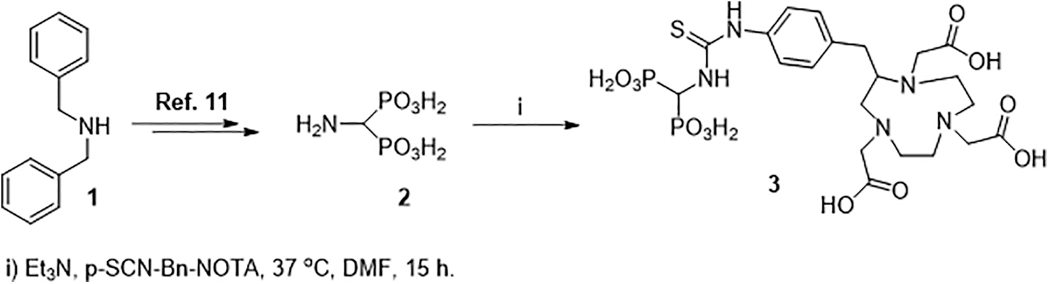
Scheme for synthesis of ligand
Materials and Methods
2-(4-Isothiocyanatobenzyl)-N,N′,N″-triazacyclononane-1,4,7,-triacetic acid (p-SCN-Bn-NOTA) was purchased from Macrocyclics Inc., USA, and used as such. Dibenzylamine (
High performance liquid chromatography (HPLC)-grade methanol and water were procured from Merck, Germany, for HPLC studies and were filtered and degassed before use. For all the radiolabeling studies, MilliQ water (resistivity higher than 18.2MΩ.cm) was used. HA microparticles used for adsorption studies of the radiotracers were synthesized and characterized following the procedure reported by the authors elsewhere. 16
Gallium-68 in the form of [68Ga]GaCl3 in 0.1 M HCl solution was sourced from a 740 MBq (20 mCi) 68Ge/68Ga generator prepared in-house using CeO2-PAN composite sorbent as the column matrix (GALGEN-1) 17 for use in all radiochemical and preclinical studies. Doses for human clinical uses were formulated at the hospital radiopharmacy using [68Ga]GaCl3 eluted from a commercial 1110 MBq (30 mCi) 68Ge/68Ga generator (Isotope Technology Garching GmBH). 18
Thin-layer chromatography (TLC) was performed on TLC aluminum sheets with silica gel 60 F254 (Merck KGaA, Germany) to monitor the progress of reactions during synthesis. Purification of reaction intermediates (Fig. 1) was carried out by column chromatography using silica gel (100–200 mesh; Sisco Research Laboratories Pvt. Ltd., India) column. For the purification of ligand
A gradient elution technique using 0.1% trifluoroacetic acid (TFA) in water (A) and 0.1% TFA in 60% aqueous methanol (v/v) (B) as the solvents was used with the following gradient: 5% B to 95% B in 30 min. The flow rate was maintained at 2.0 mL/min. The absorbance was monitored at a wavelength of 254 nm. The analysis of the purified
All the organic intermediates including ligand
Assay of 68Ga activity was carried out using Curiementor 3 (PTW Freiburg, Germany) isotope dose calibrator. For radionuclidic purity assay, γ ray spectra of 68Ga sample aliquots were recorded using an HPGe detector (Canberra Eurisys, France) coupled to a 4K multichannel analyzer (MCA) system. An 152Eu reference source, obtained from Amersham, Inc. (USA), was used for energy and efficiency calibration of the HPGe detector. All other radioactivity measurements were carried out by using a well-type NaI(Tl) scintillation counter (Mucha, Raytest, Germany).
Instant thin layer chromatography (ITLC) was performed using ITLC-SG paper strips obtained from Agilent Technologies Pvt. Ltd. (India) for analyses of the radiochemical purities of the 68Ga-labeled radiotracer synthesized.
Millex-GP syringe filter (0.22 μm) units used for sterile filtration of 68Ga-labeled radiotracer were procured from Merck-Millipore, Germany. The integrity of the filters before and after use was ascertained at 3.45 × 10⁵ N/m2 pressure adopting bubble point method. 18 Sterility of the solutions was tested in tryptic soya broth media and fluid thioglycollate media using Himedia Labs sterility test kits. Pyrogenicity of the solutions was checked by PTS (point-of-use test system; Charles River, USA). Clinical PET images were recorded by using a dedicated Siemens Biograph 6 LSO PET/CT scanner.
Synthesis, purification, and characterization of ligand 2,2′,2″-(2-(4-(3-(diphosphonomethyl)thioureido)benzyl)-1,4,7-triazonane-1,4,7-triyl)triacetic acid (3 )
The scheme for synthesis of ligand
Thereafter, the solvent was evaporated under vacuum, and the solid residue was dissolved in 1 mL of 10% aqueous solution of K2CO3 (w/v). The product (K-salt of compound
1
H NMR (D2O, 800 MHz): δ 2.49–2.58 (m, 1H), 2.60–2.70 (m, 2H), 2.87–3.17 (m, 9H), 3.17–3.30 (m, 4H), 3.31–3.40 (m, 1H), 3.42–3.53 (m, 3H), 3.69 (t, J = 15.0 Hz, 1H), 7.09–7.28 (m, 4H) (Supplementary Fig. S2). 31P NMR (D2O, 600 MHz): δ 14.38 (s) (Supplementary Fig. S3
Synthesis of [68Ga]Ga–3
Under the optimized reaction protocol, [68Ga]Ga-2,2′,2″-(2-(4-(3-(diphosphonomethyl)thioureido)benzyl)-1,4,7-triazonane-1,4,7-triyl)triacetic acid complex ([68Ga]Ga–
To achieve maximum yield of 68Ga–
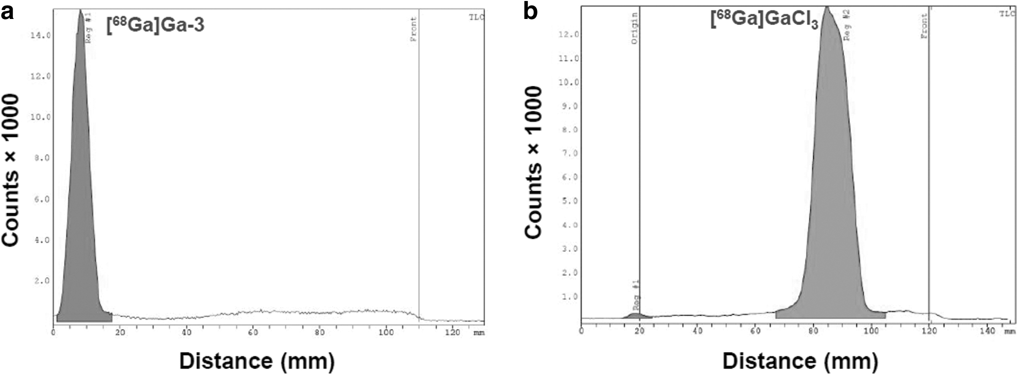
Typical ITLC radiochromatograms of
The optimized reaction protocol for synthesis of [68Ga]Ga–
Determination of partition coefficient (log p) of [68Ga]Ga–3
To ascertain the lipophilicity of [68Ga]Ga–
In vitro stability studies
The in vitro stability of [68Ga]Ga–
Aliquots were withdrawn from the mixtures at different time intervals, ethanol was added to precipitate the serum protein, centrifuged, and the supernatant was analyzed by ITLC. Kinetic rigidity of the [68Ga]Ga–
Adsorption of radiotracers on HA particles
Affinity of [68Ga]Ga–
The same aliquot was withdrawn from “blank” and 68Ga activity was measured. The “blank” consists of a 0.1 mL aliquot of the respective radiotracer diluted with 1.9 mL of MilliQ water, where no HA particle was added. The percentage adsorption of the complex on HA was calculated from Equation (1) using the data.
where “Ai
” is the measured 68Ga activity per milliliter of the blank solution and “A
eq” is the measured 68Ga activity per milliliter of supernatant solution of the test mixture under equilibrium condition. The distribution ratio (Kd
) of [68Ga]Ga–
where “V” is the volume of the solution in milliliters and “m” is the mass of the adsorbent (HA) in grams.
Biodistribution studies
Biodistribution studies of the [68Ga]Ga–
Blood samples were withdrawn from the heart of the animal immediately after sacrifice into a previously weighed syringe. Various other organs and tissues were excised after sacrifice, washed with normal saline, dried using absorbent paper, weighed, and the radioactivity associated with each organ and tissue was determined using a flat-type NaI(Tl) counter (Electronic Enterprises, Mumbai, India). The weight of the organs and samples of various tissues was also determined by using an analytical balance.
The percentage injected activity (dose) in various organs/tissue (%ID/organ) was calculated from the above data. Appropriate decay correction on the activity injected into each animal initially was employed at the time of counting the radioactivity at various time points. This gives the biodistribution pattern of the formulation at various time points postadministration specified above. The total uptake in blood, bone, and muscles was calculated by assuming that 7%, 10%, and 40% of the body weight are constituted by these organs/tissue, respectively. 21,22
Clinical studies
Doses of [68Ga]Ga–
Required ethical clearance for clinical studies with [68Ga]Ga–
Results and Discussions
Synthesis of ligand 3
Commercially available p-SCN-Bn-NOTA and the in-house prepared aminomethylbisphosphonic acid (
To assess whether compound
In Ga–
Synthesis of [68Ga]Ga–3
Variation of radiolabeling yield of [68Ga]Ga–
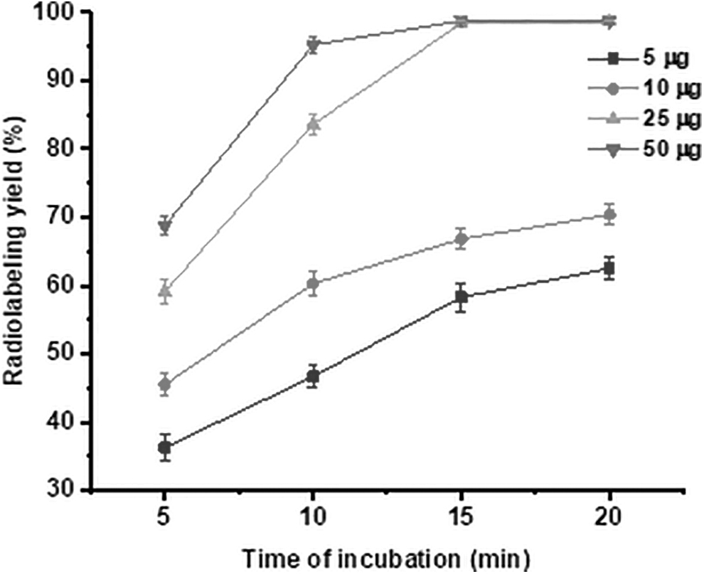
Variation of radiolabeling yield [% radiolabeling yield ± SD (n = 3)] of [68Ga]Ga–
When the reactions were carried out at different pH, it was found that the yield of [68Ga]Ga–
Determination of partition coefficient (log p) of [68Ga]Ga–3
The log p value of [68Ga]Ga–
In vitro stability studies
Figure 4a shows the radiochemical purities of [68Ga]Ga–
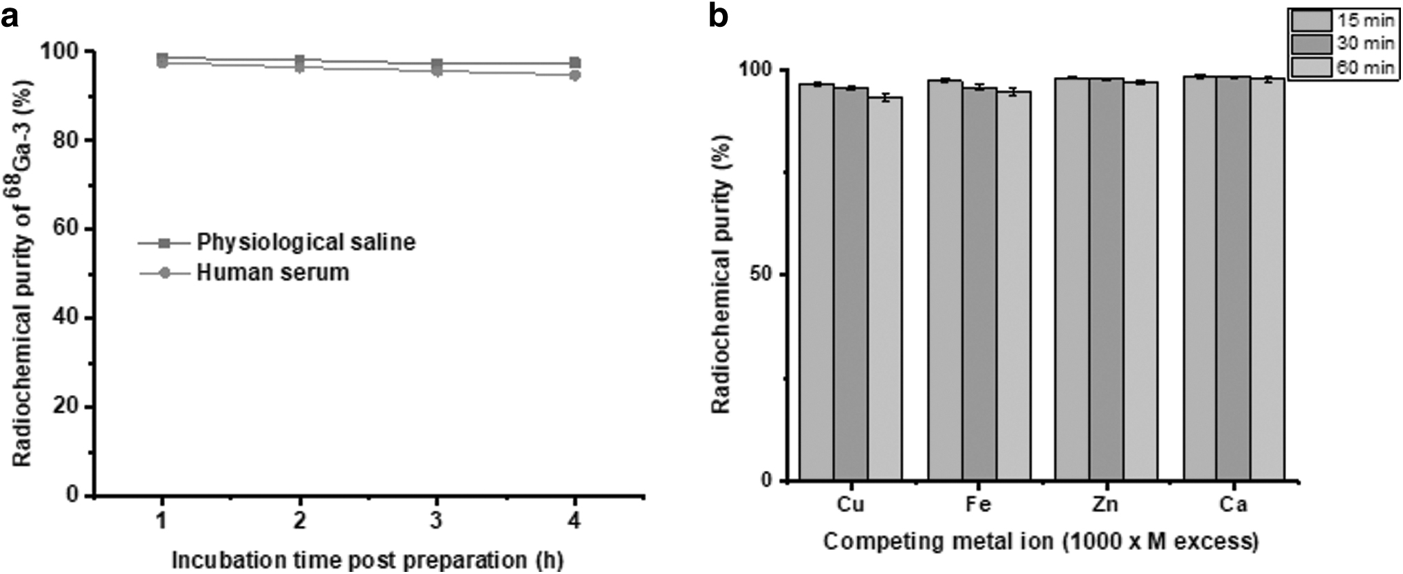
When [68Ga]Ga–
Adsorption of [68Ga]Ga–3 on HA particles
The percentage adsorption of [68Ga]Ga–
Biodistribution studies
The results of biodistribution studies carried out in normal Wistar rats after intravenous administration of [68Ga]Ga–
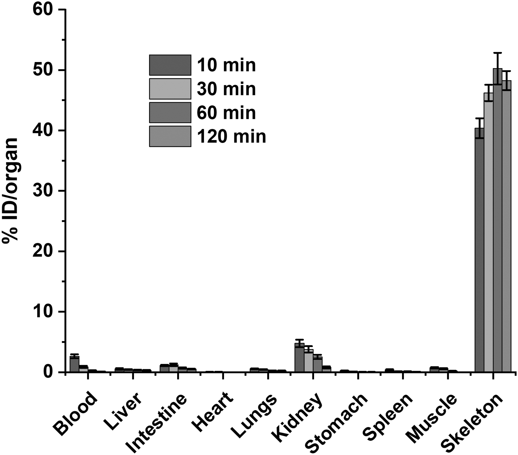
Biodistribution pattern of [68Ga]Ga–
Clearance of the formulation through the renal route was demonstrated by initial uptake of the radiotracer in the kidneys (4.76% ± 0.62% ID at 10 min postadministration, n = 3, p < 0.05) that gradually decreased with time (3.77% ± 0.33% ID at 10 min, 2.53% ± 0.28% ID at 60 min and 0.79% ± 0.12% ID at 120 min post injection, respectively, n = 3, p < 0.05). Comparative uptake (% ID/g) of [68Ga]Ga–
Comparative Uptake (%ID/g) of [68Ga]Ga–
Mean ± standard deviation data are presented. Four animals were used in each time point.
ID, injected dose.
Clinical studies
Doses of [68Ga]Ga–

Whole body PET images of
The PET scan shows that the synthesized radiotracer ([68Ga]Ga–
The radiotracer was prepared at the hospital radiopharmacy setup following the procedure reported earlier.
22
The skeletal lesion sites in the PET scan using [68Ga]Ga–BPAMD were found to exhibit SUV eight to nine times higher than that of normal bones. In comparison with [68Ga]Ga–BPAMD, the developed radiotracer [68Ga]Ga–
Comparison of Radiosynthesis, Biodistribution, In Vivo Pharmacokinetics, and Positron Emission Tomography Imaging Results of Other Promising [68Ga]Ga–Bisphosphonate Radiotracers
PET, positron emission tomography; SUV, standardized uptake value.
Moreover, preclinical investigations indicated that a higher bone-to-blood ratio was obtained for [68Ga]Ga–
Conclusions
In vitro, in vivo, and clinical evaluations revealed that the 68Ga complex of NOTA-conjugated bisphosphonic acid ([68Ga]Ga–
Footnotes
Acknowledgments
The authors from Bhabha Atomic Research Centre are grateful to the Department of Atomic Energy, Government of India for financial support. The valuable service of the staff members of the animal house facility of Bhabha Atomic Research Centre in carrying out animal biodistribution studies is also acknowledged.
Authors' Contributions
S.C. contributed to conceptualization, data curation, formal analysis, writing—original draft, review and editing, and supervision. S.C., H.D.S., and R.N. were involved in data curation and formal analysis. R.C. carried out data curation, formal analysis, and writing—review and editing.
D.G. was in charge of conceptualization, data curation, formal analysis, and writing—review and editing. A.J. carried out formal analysis. M.R.A.P. took charge of formal analysis, writing—review and editing, and supervision.
Disclosure Statement
No competing financial interests exist.
Funding Information
No funding was received for this article.
Supplementary Material
Supplementary Figure S1
Supplementary Figure S2
Supplementary Figure S3
Supplementary Figure S4
Supplementary Figure S5
Supplementary Figure S6
Supplementary Figure S7
Supplementary Figure S8
References
Supplementary Material
Please find the following supplemental material available below.
For Open Access articles published under a Creative Commons License, all supplemental material carries the same license as the article it is associated with.
For non-Open Access articles published, all supplemental material carries a non-exclusive license, and permission requests for re-use of supplemental material or any part of supplemental material shall be sent directly to the copyright owner as specified in the copyright notice associated with the article.