
Abstract
Aim:
ChiTn, a mouse/human chimeric anti-Tn monoclonal antibody, was radiolabeled with iodine-131 (131I) and technetium-99m (99mTc) to assess its biodistribution and internalization in Tn-expressing (Tn+) and wild-type (Tn-) LL/2 lung cancer cells.
Results:
Selective accumulation and gradual internalization of ChiTn were observed in Tn+ cells. Biodistribution in mice with both Tn+ or Tn− lung tumors indicated that the uptake of radiolabeled ChiTn within tumors increased over time. Dual-labeling experiments with 99mTc and 131I showed different biodistribution patterns, with 99mTc exhibiting higher values in the liver, spleen, and kidneys, while 131I showed higher uptake in the thyroid and stomach. However, tumor uptake did not significantly differ between Tn+ and Tn− tumors. To improve tumor targeting, Losartan, an antihypertensive drug known to enhance tumor perfusion and drug delivery, was investigated. Biodistribution studies in Losartan-treated mice revealed significantly higher radiolabeled ChiTn uptake in Tn+ tumors. No significant changes were observed in the uptake of the control molecule IgG-HYNIC™99mTc.
Conclusions:
These findings demonstrate the enhanced tumor targeting of radiolabeled ChiTn in Losartan-treated mice with Tn-expressing lung tumors. They highlight the potential of ChiTn as a theranostic agent for cancer treatment and emphasize the importance of Losartan as an adjunctive treatment to improve tumor perfusion and drug delivery.
Introduction
Monoclonal antibodies (mAbs) have been widely used in clinical practice for the diagnosis and treatment of various human pathologies, including cancer and infectious diseases, as well as for the modulation of the immune response. 1 –3 The development of therapeutic mAbs in cancer has been facilitated by the identification of cell surface structures. These structures, which are either present or overly expressed in abnormal cells compared with their normal counterparts, act as targets for the effective function of mAbs. 4
Aberrant glycosylation is a common change in cancer cells, which causes incomplete elongation of the carbohydrate chains and unmasking of tumor-associated carbohydrate antigens on the cell surface. 5 The Tn-antigen (CD175) is formed by an N-acetyl galactosamine (GalNAc) residue O-linked to the amino acids serine or threonine, present in the amino acid sequence of different glycoproteins, especially those of the mucin-type. 6 This antigen is a cryptic determinant in cancer with high expressions in various epithelial cancers (ovarian, breast, prostate, colorectal, and lung cancers). 7,8 Tn is associated with metastasis and cancer aggressiveness. 9,10
Indeed, in Lewis lung cancer (LL/2), Tn antigen promotes tumor growth, immune evasion, and angiogenesis, resulting in larger and highly vascularized tumors. 11 This may be explained by the ability of LL/2 Tn+ cells and LL/2 Tn+ tumors to produce higher amounts of vascular endothelial growth factor (VEGF) compared with LL/2 wild type (Tn−). 11 VEGF is considered to be the primary angiogenesis factor and has been shown to play a role in inducing extracellular matrix (ECM) synthesis and the angio-fibrotic switch in fibrosis. 12 –14
Fibrosis, characterized by the excessive deposition of collagen, hyaluronan, and other ECM components, plays a critical role in tumor progression. 14 The fibrotic ECM has been identified as a significant contributor to the elevated solid stress observed in tumors. 15 Unlike fluid pressure, this physical force is specifically contained within and transmitted by the solid phase of the tumor, resulting in the compression of blood vessels. This compression, coupled with the structural abnormalities of tumor blood vessels, renders them highly susceptible to collapse under the influence of the increased compressive forces.
Consequently, the overall blood flow throughout the tumor mass is compromised. 15 –18 This reduction in perfusion has two major consequences: (1) compromised efficacy of treatment due to reduced drug delivery to tumors, 19 and (2) increased tumor hypoxia, which promotes aggressive phenotypes, immunosuppression, and resistance to chemotherapy, radiation, and immunotherapy, all of which require oxygen to be effective. 15
Therefore, strategies aimed at reducing solid stress and improving tumor perfusion are being explored to improve the delivery and efficacy of cancer therapeutics. One such strategy is the use of Losartan, an FDA-approved antihypertensive agent that has been shown to improve tumor perfusion and drug delivery in mouse models of breast and pancreatic cancer by reducing the collagen and hyaluronan content within tumors. 17,20,21
Several mAbs targeting the Tn-antigen with different specificities have been developed, some of which have demonstrated increased survival in mice inoculated with Jurkat, 22 mammary, 7 and lung carcinoma cell lines. 23 In addition to their inherent ability to inhibit tumor growth by inducing or stimulating the immune system, 24,25 the antitumor efficacy of mAbs can be significantly enhanced by conjugation with drugs, 26 oligonucleotides, 27 or radionuclides. 28 –31
The chimeric mouse/human IgG1 anti-Tn mAb, ChiTn, developed as an antibody-drug conjugate, effectively targets and internalizes in Tn+ tumor cells, showing selective accumulation in vivo. 8 With the Tn-antigen present in both species and ChiTn's reduced immunogenicity in humans, this antibody is poised for a more efficient transition from preclinical studies to human trials.
Imaging and biodistribution studies utilizing radioactive tracers have emerged as a highly sensitive and versatile approach, making it one of the main approaches for in vitro and in vivo studies of molecular-level processes. It allows analysis of pharmacokinetics, heterogeneity, antigen expression and engagement, and uptake of radiolabeled mAbs in tumors and other organs. 31 –33 Iodine-131 (131I), a widely used radionuclide in nuclear medicine, serves as a dual β and γ emitter (8.02-d half-life).
Its gamma energy of 364.5 keV make it suitable for gamma spectrometry, whereas its beta emission has potential for cancer treatment. 34 In addition, technetium-99m (99mTc), characterized by a 6-h half-life and a 140.5 keV γ-ray emission, 35 enables distinct gamma emission detection in gamma spectrometry, differentiating it from 131I for in vitro and biodistribution studies.
In this study, the authors aimed to explore the potential of a radiolabeled mouse/human IgG1 chimeric anti-Tn mAb (ChiTn) as a theranostic agent for cancer. The authors radiolabeled ChiTn with 131I and 99mTc and evaluated its biodistribution and internalization in Tn+ and Tn− (wild-type) cells. Furthermore, they investigated the potential use of Losartan as an adjunctive treatment to enhance the delivery and targeting of radiolabeled ChiTn to Tn+ tumors in murine models of Tn+ and Tn− LL/2 lung cancer.
Methods
ChiTn and IgG linker formation
The mouse/human IgG1 chimeric anti-Tn mAb (Chi-Tn) 36 was obtained from Laboratorio de Glicobiología e Inmunología tumoral/Institut Pasteur Montevideo. A human IgG (Sigma-Aldrich) was used as a control molecule, which has no declared antigen-specificity. HYNIC conjugation to antibodies followed established protocols, typically yielding 3–5 chelators per molecule. 31
The conjugation reaction was initiated by adding an N-succinimidyl 6-(trifluoroacetyl) hydrazinopyridine-3-carboxylic acid hydrazino-protected form of succinimidyl ester (NHS-HYNIC-Tfa), synthesized in the authors' laboratory, 37 and 100 nmol in 7 μL dimethylsulfoxide (DMSO; J.T. Baker) were added to purified ChiTn or IgG control (10 nmol) in, 0.15 M pH 8 NaC2H3O2 buffer and incubated in darkness for 30 min at room temperature (RT). The ChiTn-HYNIC and IgG-HYNIC were subsequently purified by size-exclusion chromatography using NaCl 0.9% and a Sephadex G-25 (size exclusion chromatography [SEC]) column (PD-10; Amersham Biosciences).
ChiTn and IgG radiolabeling
Radiolabeling of ChiTn-HYNIC or IgG-HYNIC with 99mTc was performed by adding 33.5 mol of tricine, 33 mol of SnCl2.2H2O, and 5 nmol of the antibody conjugate. Immediately, a Na99mTcO4 − solution was added, and the mixture was incubated at RT for 30 min. Radiolabeling of ChiTn or IgG with 131I was achieved using the Pierce precoated iodination tubes (PPCIT; Thermo Fisher Scientific, Waltham, MA), and the “Example Protocol I: The Chizzonite Indirect Method for Iodination” protocol according to Thermo Fisher Scientific manual 0016379.
In brief, the PPCIT was wetted with 1 mL of Tris iodination buffer and decanted. Then, 10 MBq of 131I were added to the PPCIT and activated for 6 min at RT. Subsequently, 50 μg of ChiTn or IgG were added and the reaction mixture was incubated for 10 min at RT.
ChiTn and IgG radiolabeling controls, purification, and stability
Both radiolabeling were evaluated for radiochemical purity by (1) SEC equilibrated and eluted with normal saline, at 1.0 mL/min flow rate, and (2) high-performance liquid chromatography with a Protein-Pak 300 SW 7.5 mm × 30 cm column (size exclusion-high-performance liquid chromatography [SEC-HPLC]; Waters) using isocratic mode with 0.05 M pH 7.0 phosphate buffer, 1 mL/min flow rate, equipped with ultraviolet and NaI(Tl) absorbance and scintillation detectors, respectively.
99mTc radiolabeling was assessed using instant thin-layer chromatography (ITLC) chromatography with various systems: instant thin layer chromatography medium impregnated with a silica gel (ITLC-SG) (Pall Corporation) combined with 0.9% NaCl, ITLC-SG saturated with bovine serum albumin (BSA; Sigma-Aldrich) and EtOH:NH3:H2O (2:1:5), as well as Whatman 1 paper (Whatman International Ltd.) with methyl ethyl ketone (MEK). To ensure the integrity of the radiolabeled antibodies, the same SEC-HPLC system was employed. The radiolabeled antibodies were incubated in serum at 37°C for different time intervals, followed by high-performance liquid chromatography analysis to evaluate their stability.
Cell culture
Wild-type (Tn−) murine lung cancer cell line LL/2 (ATCC), and LL/2 cells expressing the Tn antigen (Tn+; Cells transfected by CRISPR guide targeted to Cosmc [c1galt1c1] and characterized at the Laboratorio de Inmunomodulacion y Desarrollo de Vacunas, Facultad de Medicina UdelaR
11
), were grown in RPMI-1640 medium (Capricorn Scientific) supplemented with 10% fetal bovine serum (FBS; Capricorn Scientific), 2 mM
Membrane-bound and cell internalization
Membrane-bound and cell internalization assay was performed by seeding 0.5 × 10⁶ Tn− or Tn+ cells with culture medium in 24-well plates and allowed to grow overnight. After washing once with culture medium, cells were incubated with ∼4 nM of ChiTn and/or IgG radioconjugated with 99mTc and/or 131I for 1, 2, 3, 4 (quadruplicates), and 24 h (octuplicate) at 37°C.
Then, the reaction medium was aspirated, and cells were washed twice with 500 μL cold phosphate-buffered saline (PBS). Cells were washed with cold acid buffer solution (0.2 M pH 2 glycine buffer) for 5 min to remove membrane-bound radioconjugated antibodies. The remaining internalized radioconjugated antibodies were obtained by lysis with 1 M NaOH for 5 min. Membrane-bound and internalized radioactivities were measured, and the results were analyzed, according to Radioactivity Measurements and Statistical Analysis sections.
Animals and tumor induction
C57BL/6 female mice were purchased from URBE Laboratories. Mouse handling, care, and experiments were in accordance with ethical principles adopted by National Committee on Animal Research (CNEA; protocol approved by the Commission of Ethics in the Use of Animals, Facultad de Ciencias [CEUA] No. 240011-001327-18). Tn− or Tn+ LL/2 cells (2 × 10⁵/100 μL) were inoculated on the flank region of each mouse (8 weeks old, 19.8 g average weight) through subcutaneous (s.c.) or intradermal (i.d.) injection (5 mice/group). The animals were followed daily for at least 1 month, evaluating tumor growth. All procedures were confirmed to comply with ARRIVE guidelines.
Losartan treatment
Losartan pills (50 mg/pill; Losartan potassium) were ground using a mortar and pestle, and dissolved in PBS to obtain a concentration of 2.5 mg/mL. The solutions were then filtered and stored in a sterile container. Animals were treated daily with an intraperitoneal (i.p.) injection of Losartan (20 mg/kg) or saline (control) for 9 d. 17,20,21
Biodistribution studies
When tumors reached 500–1000 cm3 in size (mean and standard deviation [SD] mass (g): 0.35 ± 0.1 and 0.72 ± 0.2, Tn− and Tn+, respectively; 15–20 d), mice from the same cohort received 0.74–3 MBq of ChiTn-HYNIC-99mTc or IgG-HYNIC-99mTc (1–4 μg, in 100 μL PBS), and/or ∼185 kBq of ChiTn-131I or IgG-131I (1–4 μg, in 100 μL PBS) through the tail vein. The amount of radiolabeled antibody used falls within the range typical for biodistribution studies. 69
After 4, 24, and 48 h, the animals were euthanized with anesthetic drugs (xylazine—100 mg/kg and ketamin—300 mg/kg). Blood, urine, and feces were collected, tumor and selected organs were excised, rinsed of residual blood, weighed and their radioactivity was measured according to the Radioactivity measurements section. Results were expressed as percent injected dose (% ID) and per gram of tissue (% ID/g).
Radioactivity measurements
γ-ray measurements were acquired using a high-purity germanium detector (HPGe; Canberra) and appropriate low-background shielding. The HPGe has the following features: 20% counting efficiency (at 1332 keV) and full width at half maximum resolution <0.85 keV (at 122 keV), enabling simultaneous in vitro and/or biodistribution measurements of multiple γ-emitting radionuclide. 37,38 Results are reported for 99mTc (140.5 keV, 89%) lower limit of detection (LLD) 110 Bq, and 131I (364.5 keV, 81.7%) LLD 1.0 kBq for 30-s measurements.
Statistical analysis
To evaluate the statistical significance of differences in medians, the authors used the Mann–Whitney U test with Bonferroni correction. The following annotation was used for statistical significance, ns: p ≤ 1e+00; *: 1e−02 < p ≤ 5e−02; **: 1e−03 < p ≤ 1e−02; ***: 1e−04 < p ≤ 1e−03; ****: p ≤ 1e−04, also, a 95% confidence interval was applied. The authors performed all statistical analyses and data visualizations using multiple packages within Spyder v. 4.1.5, 39 an integrated development environment (IDE) for scientific programming in Python v. 3.8.5 (Python Software Foundation). The packages utilized include Pandas 40 for data manipulation, Matplotlib 41 for data visualization, NumPy 42 for numerical computations, Seaborn 43 for advanced data visualization, and Statannotations 44,45 for statistical annotations.
Results
Radiolabeling and in vitro quality controls
Both 131I and 99mTc radiolabeled ChiTn or IgG antibodies were purified and their chemical identity was determined by SEC, SEC-HPLC, and ITLC. SEC analysis showed a retention volume of the radiolabeled antibodies between 3 and 4 mL, whereas free 99mTc and free 131I showed a retention volume between 8 and 9 mL. SEC-HPLC profile showed retention times of 8.5, 8.9, and 9.0 min for ChiTn-HYNIC-99mTc, ChiTn-131I, and unlabeled ChiTn, respectively (Fig. 1). Radiochemical purity exceeded 92% in all the experiments.

SEC-HPLC profile and the in vitro radiochemical stability of the radiolabeled ChiTn. The upper panel displays the retention profile of ChiTn when radiolabeled with either 99mTc (red) or 131I (blue), as well as unlabeled ChiTn (gray and dashed lines) under UV at a wavelength of 280 nm. The lower panel illustrates the gamma signal of the radiolabeled ChiTn, measured in counts per second. In the bottom right box, the average and SD of the percentage radiochemical stability of the radiolabeled ChiTn over time in serum at 37°C. 131I, iodine-131; 99mTc, technetium-99m; SD, standard deviation; UV, ultraviolet.
The analysis conducted by ITLC demonstrated that within the residual 8%, nearly two-thirds were attributed to 99mTcO2·H2O, whereas the remaining one-third was associated with 99mTc-tricine. The radiochemical stability exhibited a gradual decrease over time, with an average of 89% (SD 2.1) and 90% (SD 2.08) of antibodies retaining their radiolabeling with 99mTc and 131I, respectively, after a 48 h incubation period in serum at 37°C (Fig. 1, lower right). Similar results of radiochemical purity and stability were obtained for IgG-HYNIC-99mTc (data not shown).
Membrane-bound and cell internalization
Results for the membrane-bound internalization and uptake ratio between Tn+ and Tn− LL/2 cells for radioconjugated ChiTn and/or IgG are shown in Figure 2, and data are available at https://zenodo.org/record/8110446A gradual time-dependent increase in both membrane-bound and intracellular activities was observed for ChiTn-HYNIC-99mTc during the 24 h experiment at 37°C (Fig. 2A). This increase was found to be significantly higher in Tn+ cells compared with Tn− cells (p ≤ 1e−04 at 24 h).
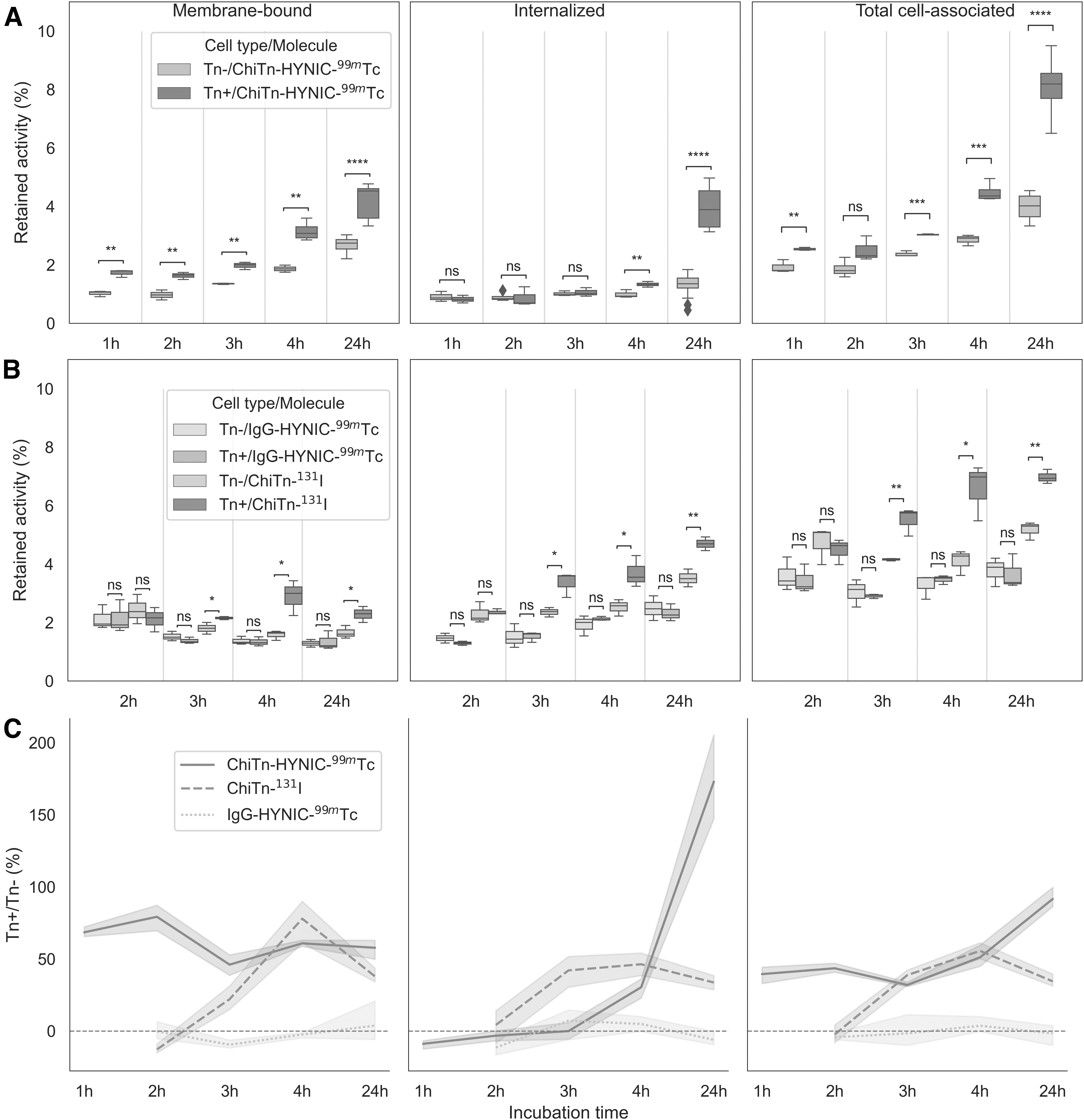
Membrane-bound, internalization, and total cell association of radiolabeled antibodies in LL/2 Tn− or Tn+ cells, measured at different incubation times. The figure includes
Similarly, when IgG-HYNIC-99mTc and ChiTn-131I were co-incubated, a similar trend of gradual increase was observed for ChiTn-131I, with significantly higher activities observed in Tn+ cells (1e−03 < p ≤ 1e−02 at 24 h). However, no specific trend or significant differences were observed between Tn+ and Tn− cells when using IgG-HYNIC-99mTc (Fig. 2B). The total cell-associated uptake of ChiTn-HYNIC-99mTc was ∼100% higher in Tn+ cells compared with Tn− cells, whereas for ChiTn-131I, the difference was ∼50% (Fig. 2C at 24 h). In contrast, no significant difference favoring Tn+ cells was observed for IgG-HYNIC-99mTc (Fig. 2C).
Biodistribution studies
The results of the biodistribution of ChiTn-HYNIC-99mTc in female C57BL/6 inoculated s.c. or i.d. with Tn− or Tn+ LL/2 cells obtained at 4, 24, 48 h postinjection, are shown in Figure 3 and Supplementary Figure S1, and data are available at https://zenodo.org/record/8110446 An increase in Tn+ tumor uptake occurred between 4 h (4.4% ± 1.6 ID/g) and 24 h (10.5% ± 2.3 ID/g) postinjection, remaining stable at 48 h (11.3% ± 2.5 ID/g). No significant differences in the uptake were found between Tn− and Tn+ or between s.c. or i.d. tumors.

Furthermore, there were no significant differences between Tn− and Tn+ or between s.c. or i.d. tumors concerning the ratios of tumor-to-blood (T/B) and tumor-to-muscle (T/M). The remaining organs showed a typical antibody biodistribution profile, with liver uptake and hepatobiliary elimination (Supplementary Fig. S1).
The biodistribution of double-labeled ChiTn with 99mTc and 131I at 48h postinjection (Fig. 4 top panel) resulted in a profile with higher values for 99mTc in the liver, spleen, and kidneys, and a tendency to show higher values in Tn− and Tn+ tumors, although this was not significant. The 131I shows a high preference for the thyroid and is also significantly higher in the stomach. No significant differences were found in the T/B and T/M ratios.
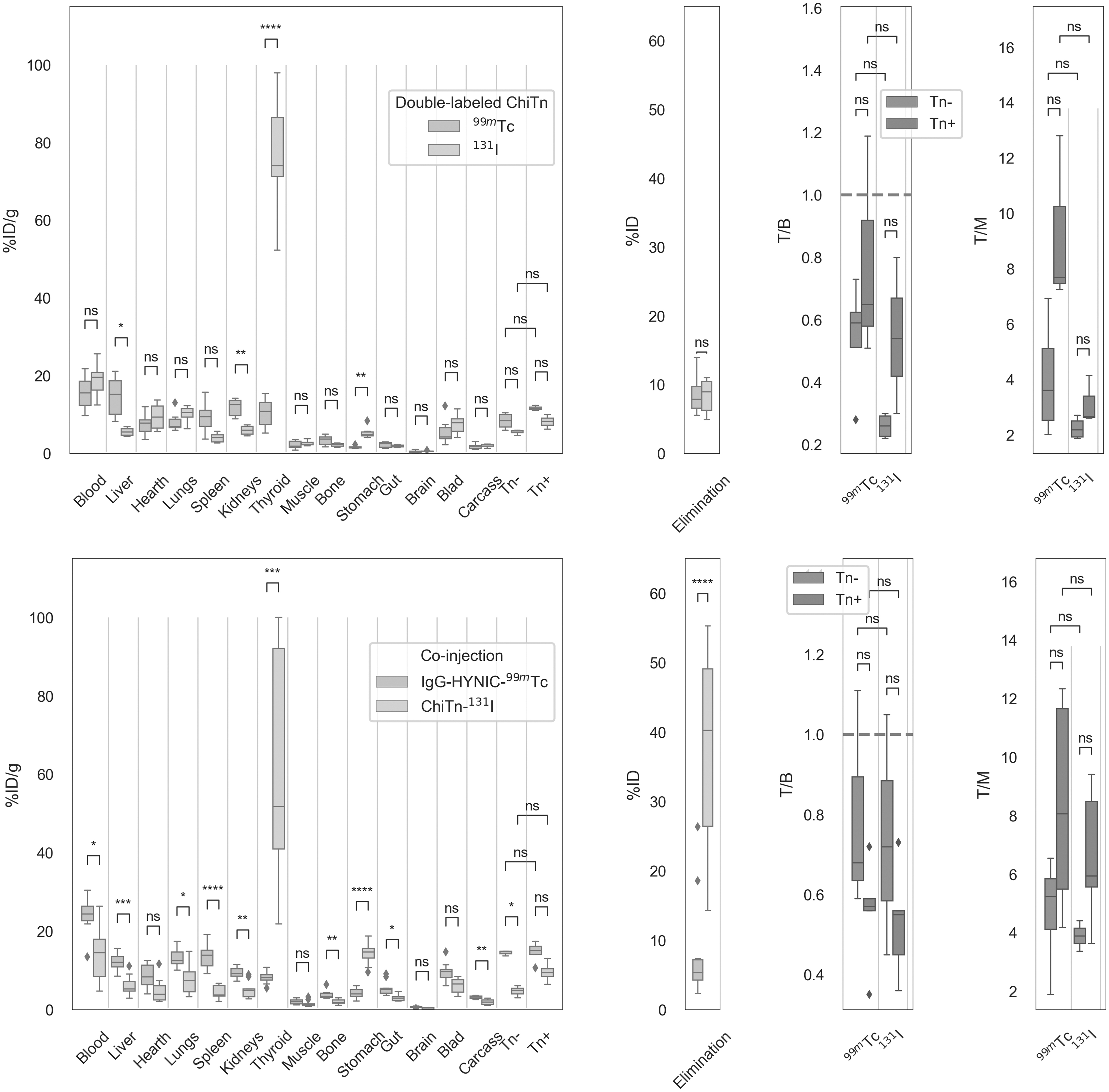
Biodistribution profile of double-labeled ChiTn (99mTc and 131I; top panel) and co-injection of IgG-HYNIC-99mTc and ChiTn-131I (bottom panel) at 48 h, in mice with SC Tn− or Tn+ tumors. The figure displays elimination (urine+feces), T/B, and T/M ratios for each tracer. ns: p ≤ 1e+00; *, 1e−02 < p ≤ 5e−02; **, 1e−03 < p ≤ 1e−02; ***, 1e−04 < p ≤ 1e−03; ****, p ≤ 1e−04.
Also, the biodistribution of the co-injection of IgG-HYNIC-99mTc and ChiTn-131I was performed at 48 h (Fig. 4 bottom panel). Most of the organs showed a higher % ID/g of IgG-HYNIC-99mTc compared with ChiTn-131I, even in the Tn− tumor, but not in the Tn+. Increased uptake was also found in the thyroid by ChiTn-131I. No significant differences were found in the T/B and T/M ratios, for both biodistributions. All values, except for thyroid, are comparable with Figure 3 and Supplementary Figure S1 (Data available at https://zenodo.org/record/8110446).
Finally, the biodistribution of ChiTn-HYNIC-99mTc and IgG-HYNIC-99mTc at 48 h in mice pretreated with Losartan is shown (Fig. 5) (data available at https://zenodo.org/record/8110446). Although the profile was similar to Figures 3 and 4, interestingly a higher uptake was observed in Tn+ than in Tn− tumors for ChiTn-HYNIC-99mTc. Also, higher T/B and T/M ratios were observed between Tn+ and Tn− tumors for ChiTn-HYNIC-99mTc. No significant differences between Tn+ and Tn− tumors were observed for IgG-HYNIC-99mTc. The average results and their SD are summarized in Table 1.

Biodistribution profile of ChiTn-HYNIC-99mTc and IgG-HYNIC-99mTc in mice treated with Losartan, at 48 h, with SC Tn− or Tn+ tumors. The figure displays elimination (urine+feces), T/B, and T/M ratios for both tracers. ns: p ≤ 1e+00; *, 1e−02 < p ≤ 5e−02; **, 1e−03 < p ≤ 1e−02; ***, 1e−04 < p ≤ 1e−03; ****, p ≤ 1e−04.
Total 48 h Biodistribution Results for Tn+ and Tn− Tumors, Including % ID/g Averages, Standard Deviations, Tumor-to-Blood, and Tumor-to-Muscle Ratios
% ID/g, percent injected dose per gram of tissue; 131I, iodine-131; 99mTc, technetium-99m; SD, standard deviation; T/B, tumor-to-blood; T/M, tumor-to-muscle.
Discussion
This study principally focused on the dynamics of the mAb ChiTn, radiolabeled for potential diagnostic and theranostic applications in cancer. Although various barriers such as interstitial resistance, hypoxia, and acidic conditions are known to impede the penetration of anticancer agents, 46 –50 the administration of Losartan can play an adjunctive role in modifying the tumor stroma and enhancing antibody delivery by alleviating solid stress. 17,20,21 The findings provide insights into the distribution and targeting efficiency of radiolabeled ChiTn to Tn+ tumors, contributing to a better understanding of antibody-based cancer diagnostics and therapy.
The experimental findings demonstrate that the radiochemical purity of both 99mTc and 131I radiolabeled antibodies remained consistently >92% even after a 48 h incubation period in serum at 37°C (Fig. 1). These results align with previous studies that reported similar levels of radiochemical purity and stability over time using the same antibody labeling methodology with 99mTc 30,31 and 131I. 51
Different binding and internalization capacities of the ChiTn mAb to tumor cell lines expressing the Tn-antigen have been observed, 7,8,52 In addition, it is rapidly internalized by Tn+ tumor cells and primarily localizes in early endosomes. 7,8 The results show that membrane-bound and internalization of the labeled antibody in Tn+ and Tn− LL/2 cells exhibit slower kinetics than the unlabeled antibody, but are similar to those of other radiolabeled antibodies. 30,53
The 99mTc labeling process involves the reaction of HYNIC with, for example, the ɛ-amino group of antibody lysines. 54,55 Similarly, for the chemical oxidation process used in 131I labeling, sodium iodide is converted into a reactive form that can be incorporated into the tyrosyl groups of the antibody. 56,57 Therefore, modification by radioactive labeling can alter the physical and chemical properties of the antibody, which can affect its interaction with cells. 58
ChiTn-HYNIC-99mTc showed persistent membrane binding after 24 h, favoring Tn+ cells significantly (Fig. 2A). Notable Tn+ internalization was observed after 4 h (Fig. 2A). ChiTn-131I yielded similar results unlike the control IgG-HYNIC-99mTc, which showed minimal binding and internalization with no significant Tn+ and Tn− differences (Fig. 2B). Figure 2C illustrates the disparities in Tn+/Tn− ratios.
Although intermediate affinities of radiolabeled antibodies may hinder binding and internalization, studies by Rudnick et al. have shown that low-affinity mAbs can penetrate solid tumors more efficiently. 59 To investigate the uptake and retention ability of radiolabeled ChiTn and IgG in Tn+ and Tn− tumors in vivo, the authors conducted various biodistribution studies.
The biodistribution results of ChiTn-HYNIC-99mTc indicated a typical distribution pattern for 99mTc radiolabeled antibodies through HYNIC, as reported by Camacho et al. 30 The radiotracer exhibited slow clearance in the bloodstream, liver, heart, and lungs, and sustained tumor uptake up to 48 h (11.3 ± 2.5% ID/g), with no significant differences between Tn+ and Tn− tumors or T/B and T/M ratios (Figs. 2 and 3 and Supplementary Fig. S1). Similar results were seen for i.d. and s.c. injections of LL/2 Tn+ and Tn− cells in mice. These results indicate that the uptake of ChiTn-HYNIC-99mTc is not an artifact of the injection route of the tumor models. Tumors injected i.d. and s.c. may differ in their microenvironment due to increased immunogenicity in i.d. tumors because of a higher density of dendritic cells. 68
It should be clarified that no ChiTn excess biodistribution studies were conducted, relying on the specificity established in prior work. 8,11 Future experiments will consider this, contingent on increased ChiTn production.
To assess the impact of the radioactive labeling method on biodistribution, 60 the authors performed dual-labeling of the ChiTn antibody with 99mTc and 131I. This enabled simultaneous measurement of biodistribution using gamma spectrometry, 39 thereby minimizing inter-mouse, inter-measurement, and handling variability. 38 Double-labeled ChiTn exhibited higher values for 99mTc in the liver, spleen, and kidneys, with a slight tendency for higher values in both Tn− and Tn+ tumors (Fig. 4 top panel), but this was not significant. The 131I exhibited a high affinity for the thyroid and was also significantly higher in the stomach. The T/B and T/M ratios did not show any significant differences.
When radioiodinated antibodies are taken up by cells, they are quickly broken down in lysosomes, resulting in the release of monoiodotyrosine into the extracellular space. This metabolite is further broken down by deiodination enzymes, which release free radioiodide into the bloodstream. 61 The radioiodide is then taken up by any tissues expressing the sodium-iodide (Na+/I−) symporter, which is present in the thyroid gland and stomach. The lysosomal degradation of radioiodinated antibodies leads to the rapid clearance of radioiodine from all tissues, except those that metabolize or process iodine. 62 This occurrence leads to diminished activity concentrations within the tumor tissue in comparison with residualizing radiolabels that employ the radiometal 99mTc. 63
The accumulation of the antibody within tumors ideally would solely rely on the specific target antigen. However, challenges arise when nonspecific factors contribute to overall tumor uptake. One example is the enhanced permeability and retention (EPR) effect, which occurs due to rapid and irregular angiogenesis, resulting in antibodies passively extravasating into the tumor tissue through the leaky vasculature. 64,65 These nonspecific contributions to tumor uptake can vary significantly between tumor models, within a single tumor (intratumoral heterogeneity), or because of different responses to treatment (intertumoral heterogeneity). Consequently, the sensitivity of these techniques may be reduced, increasing the likelihood of false discoveries. 66
The authors conducted an additional experiment to assess nonspecific uptake by comparing the biodistribution of Tn-specific ChiTn-131I with that of nonspecific IgG-HYNIC-99mTc. Most of the organs showed a higher % ID/g of IgG-HYNIC-99mTc compared with ChiTn-131I (Fig. 4 bottom panel). This is expected since the radioactive label with 131I tends to be more unstable in vivo, leading to increased uptake in the thyroid and stomach, as well as greater clearance from organs, as previously discussed.
Nevertheless, the Tn+ tumor demonstrated a comparable uptake of ChiTn-131I to that of IgG-HYNIC-99mTc, whereas a reduced uptake was observed in Tn− tumors. Once again, no distinct advantage in uptake was noted for the Tn-specific ChiTn as compared with the nonspecific IgG. This observation is further corroborated by the similarities in the T/B and T/M ratios (Fig. 4 below).
The presence of solid stress may be a contributing factor hindering the efficient delivery of antibodies to tumors, particularly in highly vascularized tumors such as LL/2 Tn+. These tumors produce higher levels of VEGF compared with LL/2 wild type (Tn−), as demonstrated by da Costa et al. 11 VEGF is recognized as a key factor in angiogenesis, capable of inducing ECM synthesis and promoting the angio-fibrotic switch in fibrosis, as documented by Larsson-Callerfelt et al., 12 Kuiper et al., 13 and Zhang and Chu. 34 The fibrotic ECM significantly contributes to elevated solid stress within tumors. 15
To assess this phenomenon, biodistribution studies were conducted after pretreatment with Losartan, a compound known to alleviate solid stress in fibrotic tumors. 17,20 Previous research has indicated that Losartan treatment does not alter VEGF levels or microvessel density. However, it has been shown to significantly enhance the percentage of perfused blood vessels. 21
The % ID/g values indicate that the biodistribution profiles of normal tissues in the Losartan pretreated mice (Fig. 5) were comparable with those of the untreated counterparts at 48 h postinjection for both IgG-HYNIC-99mTc and ChiTn-HYNIC-99mTc, which aligns with the findings of Chauhan et al., 17 where Losartan treatment does not affect accumulation in normal tissues. However, a slightly higher uptake in the kidneys and elimination through urine and feces were observed for ChiTn-HYNIC-99mTc (Fig. 5).
In addition, ChiTn-HYNIC-99mTc demonstrated a higher blood clearance compared with IgG-HYNIC-99mTc, as evident in Figure 5, consistent with the outcomes of the previous biodistribution studies where ChiTn-HYNIC-99mTc values ranged from 15% to 20% ID/g and IgG-HYNIC-99mTc values ranged from 22% to 30% ID/g at 48 h in bloodstream (Figs. 3–5).
In mice pretreated with Losartan, a significantly higher uptake of ChiTn-HYNIC-99mTc was observed in Tn+ tumors (% ID/g 14.9 ± 2.1) compared with Tn− tumors (% ID/g 7.9 ± 1.2; p ≤ 0.05), as indicated in Table 1 and Figure 5. However, there was no significant change in the uptake of IgG-HYNIC-99mTc in either tumor type.
These findings are further supported by the T/B and T/M ratios (Table 1 and Fig. 5). ChiTn-HYNIC-99mTc demonstrated significantly higher ratios in favor of Tn+ over Tn− tumors (p ≤ 0.01 and p ≤ 0.05, respectively), whereas IgG-HYNIC-99mTc did not show the same pattern. Specifically, the T/B ratio in Tn+ tumors increased from 0.8 ± 0.5 in untreated mice to 1.2 ± 0.2 in mice pretreated with Losartan for ChiTn-HYNIC-99mTc. The T/M ratio also exhibited a moderate increase from 7.7 ± 3.3 to 10.4 ± 3.3.
These values indicate a substantial increase in Tn+ uptake compared with Tn− in mice pretreated with Losartan, with an average of 88% higher uptake in Tn+ tumors. In addition, there was a significant enhancement in the T/B ratio, with a 50% increase. Similarly, the T/M ratio exhibited a 35% increase in Tn+ tumors. All of these values closely align with the findings reported by Chauhan et al., 17 where Losartan treatment resulted in a 74% increase in the accumulation of the small-molecule chemotherapeutic 5-FU in AK4.4 pancreatic tumors.
The findings indicate that LL/2 Tn+ tumors exhibit elevated solid stress compared with their wild-type (Tn−) counterparts. As a result, Losartan is expected to be more effective in alleviating this stress specifically in Tn+ tumors. In addition, the radiolabeled ChiTn mAb demonstrates enhanced penetration into this tumor type, allowing for increased interaction with the Tn antigen. In contrast, nonspecific IgG lacks the capability to undergo specific binding and subsequent internalization into tumor cells, leading to a lack of this effect. Its uptake primarily occurs through nonspecific binding or due to its higher concentration in the bloodstream.
Although Losartan may increase the blood flow to the tumor by alleviating solid stress, and this could lead to an increase in tumor size, the use of Losartan should be understood as an adjuvant treatment in conjunction with a therapeutic drug. Even so, Kumar et al. 67 found that the administration of 5FU, Doxil, or Losartan alone did not affect the growth of pancreatic tumors, but tumors were significantly smaller in mice treated with Losartan combined with either Doxil or 5FU.
The authors' data suggest a differential modulation by Losartan when used as an adjuvant therapy on Tn− versus Tn+ tumors, with an observed augmentation in ChiTn antibody uptake specifically in Tn+ tumors. This phenomenon implies that Losartan may selectively enhance the delivery and therapeutic efficacy of ChiTn in Tn+ tumor environments. It is essential, therefore, that the authors' observations serve as a catalyst for subsequent targeted investigations dedicated to elucidating the distinct mechanisms by which Losartan interacts with and potentially alters the tumor microenvironment in both Tn− and Tn+ malignancies. Such focused studies are critical to fully understand the role of Losartan in modifying ChiTn uptake and to determine the translational potential of these findings in the context of therapeutic interventions.
In conclusion, this study provides insights into the potential use of radiolabeled ChiTn in combination with Losartan for improved delivery and targeting of Tn+ tumors, which may have clinical implications for the treatment of epithelial cancers expressing the Tn-antigen. Further studies are warranted to explore the theranostic efficacy of this approach and optimize the radiolabeling and targeting strategies.
Data Availability Statement
Comprehensive data sets for in vitro and biodistribution assays can be accessed through this link: https://zenodo.org/record/8110446
Footnotes
Acknowledgments
This study is part of the Master's and Doctoral Program in Medical Sciences (Proinbio) from the Facultad de Medicina at the Universidad de la Republica, Uruguay, where M.T., MSc, is pursuing his doctorate in medical science. The authors would like to extend special thanks to Roger Chammas, MD, PhD, professor of oncology and deputy dean at the faculty of medicine of the University of São Paulo and the Cancer Institute of the State of São Paulo, for his insightful advice on the use of Losartan for tumor delivery.
Authors' Contributions
M.T., X.C., C.P., M.C., M.F.G., M. F., J.P.G., and P.C. participated in the antibody radiolabeling and in vitro/in vivo studies in both models. T.F., V.C., and E.O. developed cell lines, tumor models, and the ChiTn antibody. M.T. drafted the article. All authors participated in the editing and review of the article.
Disclosure Statement
The authors declare no competing interests.
Funding Information
This work was funded by “Comisión Sectorial de Investigación Científica CSIC I+D 2018 - Imagenología Molecular en Oncología”, Uruguay.
Supplementary Material
Supplementary Figure S1
References
Supplementary Material
Please find the following supplemental material available below.
For Open Access articles published under a Creative Commons License, all supplemental material carries the same license as the article it is associated with.
For non-Open Access articles published, all supplemental material carries a non-exclusive license, and permission requests for re-use of supplemental material or any part of supplemental material shall be sent directly to the copyright owner as specified in the copyright notice associated with the article.