
Abstract
Introduction:
The expression of alpha-five beta-three (αVβ3) integrins is upregulated in various malignancies undergoing angiogenesis. The development of integrin antagonists as diagnostic probes makes the αVβ3 integrin a suitable candidate for targeting tumor angiogenesis. The goal of this study was to optimize the radiolabeling and evaluate the potential of conjugated integrin antagonist carbamate (IAC), a peptidomimetic, as a theranostic radiopharmaceutical for targeting tumor angiogenesis.
Methodology:
Radiolabeling of DOTAGA [2,2′,2”–{10-(2,6-dioxotetrahydro-2H-pyran-3-yl)−1,4,7,10-tetraazacyclododecane-1,4,7-triyl} triacetic-acid]-IAC with [68Ga]Ga, [177Lu]Lu, and [225Ac]Ac was optimized. The binding affinity (Kd) of DOTAGA-IAC for the αVβ3 receptor and cancer cell lines was quantified. The biodistribution studies were conducted in healthy Wistar rats. Dosimetry analysis was performed on [177Lu]Lu-DOTAGA-IAC distribution data. A pilot study of [68Ga]Ga-DOTAGA-IAC and [18F]FDG Positron Emission Tomography (PET/CT) imaging was performed in five patients with histopathologically confirmed breast cancer. PET/CT findings were compared between [68Ga]Ga-DOTAGA-IAC and [18F]FDG in these patients.
Results:
Radiopharmaceuticals were prepared with high radiochemical purity (>99.9%). Kd and Bmax measurements were 15.02 nM and 417 fmol for αVβ3 receptor protein: 115.7 nM and 295.3 fmol for C6 glioma cells. Biodistribution studies in rats suggested the excretion via kidneys and partially through the hepatobiliary route. The effective dose of [177Lu]Lu-DOTAGA-IAC was found to be 0.17 mSv/MBq. The dynamic study in patients revealed the optimal imaging time to be 30–35 mins postadministration. Out of the cohort, [68Ga]Ga-DOTAGA-IAC detected the primary lesions in all five patients with a mean standard uptake value (SUVmax) of 3.94 ± 0.58 compared with [18F]FDG (SUVmax 13.8 ± 6.53).
Conclusion:
The study demonstrates that DOTAGA-IAC exhibits strong binding to αVβ3 integrin, positioning it as a promising PET agent for assessing primary and metastatic cancers. The outcomes from the pilot study suggest the potential of [68Ga]Ga-DOTAGA-IAC PET/CT in breast carcinoma diagnosis. While recognizing the theranostic potential of DOTAGA-IAC for αVβ3 integrin-expressing tumors, further clinical investigations are warranted to comprehensively assess therapeutic efficacy.
Introduction
Angiogenesis plays a pivotal role not only in physiological processes but also in tumor growth, invasion, and metastasis. Tumor expansion beyond 2 mm3 results in heightened interstitial pressure, leading to reduced nutrient and metabolite supply that results in halting tumor growth. 1 This hypoxic state triggers the angiogenic switch by upregulating proangiogenic proteins like vascular endothelial growth factor (VEGF), platelet-derived growth factor (PDGF), and tumor necrotic factor-α (TNF-α). Subsequent steps involve the removal of pericytes and degradation of the extracellular matrix leading to angiogenesis. Mesenchymal stem cells differentiate into pericytes, conferring stability to the newly formed blood vessels and promote tumor growth. 2,3
Various classes of cell adhesion molecules (CAMs) such as integrins, cadherins, selectins, immunoglobulins, vitronectin, fibronectin, and tenascin play a key role in tumor angiogenesis. 4 Among CAMs, αVβ3 integrins are key promoters of tumor angiogenesis, binding to various extracellular matrix proteins, immunoglobulins, and growth factor receptors (insulin receptor, PDGF, and VEGF) via the recognition of the short peptide sequence Arg-Gly-Asp (RGD). 5,6 Overexpression of αVβ3 integrin has been noted in breast cancer, glioblastoma, pancreatic tumor, prostate carcinoma, and cervical cancer. 7 –12 The nonexpression in normal or nonproliferating cells makes αVβ3 integrin an attractive therapeutic target. 13
Various RGD-based ligands such as [18F]FGalacto-RGD, [18F]F-AH111585, [68Ga]Ga-NOTA-RGD, cyclic pentapeptide (RGDfV), and [68Ga]Ga-DOTA-RGD2 have been explored for their utility in targeting angiogenesis. 14 –18 These RGD ligands show promise in diagnosing αVβ3 integrin-expressing tumors. However, their peptide nature may limit their therapeutic potential due to susceptibility to chemical and proteolytic degradation, leading to short physiological half-lives.
To address this limitation, peptidomimetics have gained attention due to their higher receptor affinity, enhanced chemical and proteolytic degradation resistance, and longer physiological half-lives. 6,19 Integrin Antagonist Carbamate Derivative (IAC) is a promising peptidomimetic agent with high affinity for αVβ3 integrin, indicating its potential in detecting αVβ3 integrin-expressing tumors. 20 –23 However, the therapeutic potential of previously developed 1,4,7-triazacyclononane-N-glutamic acid-N’,N”-diacetic acid (NODAGA)-IAC is limited due to the absence of a suitable bifunctional chelator for Lutetium-177 (177Lu) and Actinium-225 (225Ac). 24
This study explored the theranostic potential of IAC conjugated with DOTAGA [2,2′,2”–{10-(2,6-dioxotetrahydro-2H-pyran-3-yl)−1,4,7,10-tetraazacyclododecane-1,4,7-triyl} triacetic-acid].
The DOTAGA-IAC radiolabeling was optimized and characterized with 68Ga, 177Lu, and 225Ac followed by their quality control assessment. The biodistribution and dosimetry study was performed for [177Lu]Lu–DOTAGA-IAC. Whereas, with [68Ga]Ga-DOTAGA-IAC, along with in vitro receptor binding assays and preclinical biodistribution study, a pilot study was conducted in patients with breast cancer to explore its diagnostic potential.
Materials and Methods
Radiolabeling with [68Ga]GaCl3
The radiolabeling was optimized for the ligand concentration, reaction pH, temperature, and incubation time. Reactions 1–13 (Supplementary Table S1) were performed using [68Ga]GaCl3 (370 MBq) eluted from a commercially available 68Ge/68Ga generator (ITM Germany) using 0.05 M HCl and incubated with DOTAGA-IAC (2–20 µg) at 95°C (or room temperature) for 10–25 min. The reaction pH was maintained between 3.5 and 7.5 using 0.25 M sodium acetate buffer. The radiochemical yield (RCY) and radiochemical purity (RCP) were determined through instant thin-layer chromatography (radio-ITLC). Purification was performed using a preconditioned C-18 cartridge, as detailed in section purification by solid-phase extraction. To verify the absence of [68Ga]Ga-DOTAGA in the final product, DOTAGA was also radiolabeled with [68Ga]GaCl3. To differentiate between [68Ga]GaCl3, [68Ga]Ga-DOTAGA, and [68Ga]Ga-DOTAGA-IAC, various stationary and mobile phases were assessed for radio-ITLC. The corresponding retention factors (R f ) were recorded (Supplementary Table S2). For, radio-ITLC assessment, a 100 mm TLC pate was used and the origin front and solvent front were marked at 10 and 90 mm, respectively.
To enhance readability and avoid repetition in the following sections, DOTAGA-IAC will be referred to as simply IAC.
Radiolabeling with high specific activity (SA) [177Lu]LuCl3
The high SA [177Lu]LuCl3 (SA: 3388 MBq/µg) was sourced from ITM Germany. IAC (5 µg) in a 0.5 M sodium ascorbate buffer was incubated with 370 MBq of [177Lu]LuCl3 at 100°C for 30 min. As a radio stabilizer, ascorbic acid was added to the buffer solution. The final reaction pH was 4.5. Radiochemical yield and purity were determined by radio-ITLC with sodium citrate (0.5 M, pH 3.0) as the mobile phase and Instant thin layer chromatography with silica gel (ITLC-SG) as the stationary phase. In addition, acetonitrile: water (1:1, v/v) and Whatman No. 3 paper were used as another combination of mobile and stationary phases.
Radiolabeling with low SA [177Lu]LuCl3
The low SA [177Lu]LuCl3 (SA: 740 MBq/µg) was procured from BRIT, India. DOTAGA-IAC (10 µg) in 0.5 M sodium ascorbate buffer was incubated with 370 MBq of [177Lu]LuCl3 at 100°C for 30 min. Gentisic acid (0.25 M) was added to the buffer solution as a radio stabilizer. The final reaction pH was 4.5. The radiochemical yield and purity were assessed using the same method, as described in the previous section.
Radiolabeling with [225Ac] AcCl3
The [225Ac]AcCl3 was procured from ITM Germany. IAC (20 µg) in 1 M sodium ascorbate buffer (pH 5.0) was incubated with [225Ac]AcCl3 (481–999 KBq) at 90°C for 30 min, with final reaction pH toward 5.0. The radiochemical yield and purity were assessed by radio-ITLC. The combination of mobile and stationary phases included sodium citrate and ITLC-SG, as well as acetonitrile: water (1:1, v/v) and Whatman paper 3.
Purification by solid-phase extraction
Purification was performed using sep-pak C-18 cartridges. To condition the cartridge, 70% ethanol (5 mL) was passed through at 1–2 mL/min for activation. Followed by a 10 mL water wash at the same rate for equilibration. The radiolabeled mixture was then passed through the cartridge, trapping radiolabeled IAC via hydrophobic interaction. The product eluted with 50% ethanol (1 mL) at a flow rate of 1 mL/min.
HPLC characterization
68Ga and 177Lu labeled IAC were identified via HPLC analysis with nonradioactive surrogates, 69Ga and 175Lu. The analysis was done on a Shimadzu LC-2030 system equipped with a Photodiode Array (PDA) detector and a Purosphere STAR RP18 end-capped (5.0 µm) RT 125-4 column (Darmstadt, Germany), operating at a flow rate of 1 mL/min. PDA detection ranged from 190 to 800 nm. Before HPLC analysis, nat[69Ga]Ga-IAC and nat[175Lu]Lu-IAC samples were purified using a C-18 cartridge to remove free metals and buffer solutions, followed by elution with 50% ethanol (Supplementary Table S3).
Quality control
Radionuclide purity was evaluated using a thallium-doped sodium iodide [NaI (Tl)]-based γ-ray spectrometer. Characteristic gamma photo peaks were observed for each radiometal: 68Ga (511 and 1022 KeV), 177Lu (113 and 208 KeV), and 225Ac (78, 213, and 470 KeV). The Endpoint Chromogenic Limulus Amebocyte Lysate test was conducted to determine endotoxin levels in accordance with the U.S. Pharmacopeia (USP), Chapter 85 guidelines. Sterility testing was performed following the procedures specified in USP chapter 71, using two growth media: fluid thioglycollate medium (FTM) and tryptic soy broth (TSB). The shelf life and stabilities of [68Ga]Ga-IAC, [177Lu]Lu-IAC, and [225Ac]Ac-IAC were assessed over different time points in normal saline at room temperature and in human serum at 37°C.
Determination of partition coefficient (log P)
The log p-values of [68Ga]Ga-IAC and [177Lu]Lu-IAC were determined using the n-octanol/saline model. Equal volumes of the radioactive drug solution and n-octanol solution were mixed vigorously for 10 min, followed by centrifugation at 1509 Relative Centrifugal Force (RCF) for 10 min to separate the phases. Radioactivity in each fraction was measured, and log p-values were calculated using the following formula:
Plasma protein binding in human blood
To assess plasma protein binding, we used the trichloroacetic acid (TCA) precipitation method. Briefly, 0.1 mL of each radiocomplex was mixed with 0.9 mL of plasma, incubated at 37°C for an hour, treated with 10% TCA for 2–3 min, and then centrifuged at 671 RCF for 5 min. The radioactivity in the pellet and supernatant was measured to calculate % Plasma Protein Binding (PPB).
In vitro radioligand binding study
The binding experiment was performed in MCF-7 (human breast cancer cells), Hep-G2 (human liver cancer cells), and C6 rat glioma cells. The cell lines were procured from the National Center for Cell Sciences, Pune, India. The cells were cultured in RPMI-1640 and Dulbecco's Modified Eagle Medium (DMEM) media supplemented with 10% FBS, penicillin (100 U/mL), and streptomycin (100 µg/mL) antibiotic solutions in a 5% CO2 environment at 37°C. Subsequently, 24-well plates were seeded, and the saturation binding experiment was performed to determine the binding affinity (Kd) and the number of binding sites (Bmax) of [68Ga]Ga-IAC for αVβ3 integrin receptor. See Supplementary Section 1.0. for a detailed experiment design. The saturation binding experiment was also conducted with recombinant human αVβ3 integrin receptor protein purchased from RD (CAT: 3050-AV). A 96-well ELISA plate was coated with the receptor (2.5 µg/mL) in 50 mM carbonate/bicarbonate buffer (pH 9.5) overnight at 4°C. The subsequent steps were the same as followed for the cell lines. The data were processed using Prism software to determine Kd and Bmax.
Biodistribution in healthy Wistar rats
Animal biodistribution studies were conducted with prior ethical clearance from the Institutional Animal Ethics Committee (IAEC-724/IAEC/107/105) of PGIMER, Chandigarh, India. Eighteen healthy female Wistar rats, 16 weeks old, weighing 286 ± 21 g, received intravenous injections of [68Ga]Ga-IAC (7.55 ± 1.3 MBq) and [177Lu]Lu-IAC (4.07 ± 0.1 MBq) via the lateral tail vein. All rats were anesthetized with a mixture of ketamine (40 mg/kg) and xylazine (4 mg/kg) and euthanized through cervical dislocation. For [68Ga]Ga-IAC, groups of rats (n = 3 at each time point) were sacrificed at 0.5, 1, and 2 h postinjection. As for [177Lu]Lu-IAC, rats were sacrificed at 1, 24, and 48 h postinjection. The counts in each organ were recorded using a NaI (Tl)-based gamma-ray spectrometer (CAPTUS 4000). The percentage of injected dose per gram of tissue (%ID/g) was determined using the following formula:
Dosimetry for [177Lu]Lu-IAC Preclinical Biodistribution Data
The radiation dose estimates of [177Lu]Lu-IAC were calculated for human organs from the animal distribution data. As the animal data were presented in concentration (% injected dose/g of tissue), extrapolation to humans was performed using a mass-based extrapolation. In the mass extrapolation method, the animal concentration was normalized by the ratio of the total body weight of animals and humans. 25 Extrapolated data were fit using the SAAM II software. 26 Time integrals of activity were entered into the OLINDA/EXM software to compute organ effective dose, using the adult male model. 27,28 As no excretion data were available, the decay in the remainder of the body was assumed to be solely by radioactive decay.
Patient imaging
[68Ga]Ga-IAC PET/CT imaging was conducted with PGIMER Institutional Ethics Committee clearance (IEC-03/2020-1545). The inclusion criteria included age >18 years, patient consent, histopathologically confirmed breast cancer, and prior [18F]FDG PET/CT, while exclusions included refusal, pregnancy, lactation, or significant illness. The patients were closely monitored for vital parameters and instructed to self-report any adverse events at home.
[68Ga]Ga-IAC dynamic and whole-body PET/CT imaging
A dynamic PET/CT study was conducted with a cohort of three patients to determine the optimal time of maximum uptake of [68Ga]Ga-IAC in the lesion of interest. The lesion was positioned within the scanner’s field of view, and [68Ga]Ga-IAC (111.37 ± 10.81 MBq) was administered. The injected mean mass of IAC per patient was 2.5 ± 0.34 µg. PET data were acquired for up to 60 min using a hybrid 3D PET/CT scanner (Siemens Healthineers Biograph mCT S-64, Germany). A series of 10 PET images were retro-reconstructed at different time intervals, ranging from 5 to 60 min postadministration. A region of interest was drawn around the lesion, and the maximum standard uptake value (SUVmax) was calculated. The SUVmax of the lesion and the ratio of SUVmax of the lesion to SUVmax of the aorta (representing blood uptake) were determined for all reconstructed images. Both parameters were plotted over time to assess dynamic behavior.
In addition to dynamic imaging, three patients also underwent whole-body (WB) PET/CT imaging at 60 min. For the remaining two patients, whole-body PET/CT imaging was acquired at 30 min, as determined by the dynamic study. The PET/CT parameters are referenced in the Supplementary Section 3.0. The [68Ga]Ga-IAC PET/CT findings were then compared with the results obtained from [18F]FDG PET/CT scans (263.44 ± 37.31 MBq) for comprehensive evaluation.
Results
Radiolabeling and quality control of [68Ga]Ga-IAC
Among the different combinations tested, sodium citrate (0.5M, pH 3.0) with ITLC-SG, trifluoracetic acid (TFA): acetonitrile (ACN) (1:1, v/v) with ITLC-SG and ACN: Water (1:1, v/v) with W#3 proved effective in distinguishing the three radiochemical species (Supplementary Fig. S1). The sodium citrate with ITLC-SG combination showed radio peaks at the origin for [68Ga]Ga-DOTAGA (R f 0.0) and [68Ga]Ga-IAC (R f 0.0) and a radio peak at the near solvent front for 68Ga (R f 0.8). This combination was able to identify the presence of free 68Ga in the final product. TFA-ACN with ITLC-SG showed a radio peak at the origin for [68Ga]Ga-IAC (R f 0.0) and a radio peak at the near solvent front for [68Ga]Ga-DOTAGA (R f 0.8), enabling the detection of [68Ga]Ga-DOTAGA in the final product. ACN: Water with W#3 showed a radio peak at R f 0.6 for [68Ga]Ga-IAC, confirming the absence of hydrolyzed 68Ga in the final product, as hydrolyzed species would remain at the origin in any combination of mobile and stationary phases.
The effect of temperature, pH, and IAC concentration was also assessed. Only a 7.5% RCY was achieved when the reactions (1–3) were performed at room temperature for up to 25 min (Supplementary Table S1). The yield was increased to 95% on heating at 95°C within 10 min of reaction time. The RCY was reduced at pH 3.5 and 5.5% to 5% and 18%, respectively, indicating pH and temperature as the major factors in radiolabeling of IAC with 68Ga (Supplementary Fig. S12). Furthermore, the highest labeling yield (> 95%) was noted at ratios of IAC to 68Ga (µg: MBq) of 0.5:37 and 1:37 (Supplementary Fig. S12). Radio-ITLC analysis of reactions displayed >95% radiochemical yield. After purification with a sep-pak C18 cartridge, >99% radiochemical purity was achieved (Fig. 1A). The specific activity for [68Ga]Ga-IAC was achieved up to 74 MBq/µg.

Radio-ITLC chromatograms of [68Ga]Ga-DOTAGA–IAC
Gamma-ray spectra of 68Ga showed energy peaks at 511 KeV and 1022 KeV, indicating >99% radionuclide purity (Supplementary Fig. S2A). No photopeak was observed after 10 half-lives of 68Ga ruling out the presence of 68Ga breakthrough (Supplementary Fig. S2B). A single radio peak at R f 0.0 was consistently observed at each time point for up to 4 h, indicating the high shelf-life of [68Ga]Ga-IAC at room temperature. Similarly, a single radio peak at R f 0.0 for [68Ga]Ga-IAC in human serum at 37°C demonstrated no evidence of decomplexation in serum for up to 4 h (Supplementary Table S4 and Supplementary Figs. S3 and Figs. S4). No observable growth in TSB and FTM medium at 25°C and 30°C, respectively, for up to 14 days indicated the sterility of the in-house synthesized [68Ga]Ga-IAC. Endotoxin levels for [68Ga]Ga-IAC samples were calculated to be 7.2 ± 0.7 EU/V (Supplementary Table S5).
Radiolabeling and quality control of [177Lu]Lu-IAC
The radio-ITLC chromatograms obtained using sodium citrate and ITLC-SG revealed R f values of 0.8 for free 177Lu and R f 0.0 for [177Lu]Lu-IAC. ACN: Water (1:1, v/v) and W#3, R f values were 0.0 for 177Lu and R f 0.6 for [177Lu]Lu-IAC (Supplementary Fig. S6). The absence of a radio peak at the origin in ACN: Water indicated the absence of hydrolyzed 177Lu in the final product. Similar patterns were observed for both low and high specific activities [177Lu]Lu-IAC. Radio-ITLC chromatograms of both low and high specific activities [177Lu]Lu-IAC demonstrated >99% radiochemical purity (Fig. 1B), and the specific activities of both the radio constructs were 37 MBq/µg and 74 MBq/µg, respectively.
The multichannel acquisition spectrum displayed characteristic energy peaks of 177Lu at 113 KeV and 208 KeV, indicating >99% radionuclide purity (Supplementary Fig. S2C). A single radio peak at R f 0.0 for [177Lu]Lu-IAC was observed for up to 14 days when incubated at room temperature in normal saline (0.9%), at 37°C in human serum, demonstrating good stability (Supplementary Table S6 and Supplementary Figs. S7 and Figs. S8). Both culture mediums remained free from visible microbial growth for up to 14 days, confirming the sterile production of [177Lu]Lu-IAC. The endotoxin levels obtained from pyrogenicity experiments were 22.2 ± 2.0 EU/V (Supplementary Table S5).
Radiolabeling and quality control of [225Ac]Ac-IAC
More than 99% RCY was achieved for [225Ac]Ac-IAC (Fig. 1C). Radio-ITLC of free 225Ac showed two peaks at R f 0.7 and 1.0 (Supplementary Fig. S5). The same ITLC-SG strip was reread after 2 h and showed only a single radio peak at R f 0.7 for 225Ac; a radio peak at R f 1.0 diminished. The reaction showed a single peak at R f = 0.0 indicating >99% [225Ac]Ac-IAC. After 15 min of synthesis, a major peak of [225Ac]Ac-IAC at R f = 0.0 (with noise at R f of 0.2–0.5) was observed. However, rescanning the same ITLC-SG strip after 2 h, only a single radio peak at the origin (R f 0.0) was obtained for [225Ac]Ac-IAC. At 1 h of end of synthesis (EOS), multiple peaks were observed (R f 0.0, 0.3, and 0.5), and peaks at R f 0.3 and 0.5 diminished when the same plate was rescanned after 2 h. Radiochromatogram appeared using 2 M HCl: acetone showed a radio peak at R f = 0.5; no peak was observed at the origin. (Supplementary Fig. S9). The [225Ac]Ac-IAC was prepared with a specific activity of up to 50 KBq/µg.
The 225Ac radionuclidic purity was greater than 99.9% (Supplementary Fig. S2D). [225Ac]Ac-IAC incubated in human serum and normal saline demonstrated a single radio peak at R f 0.0 for up to 14 days indicating high stability (Supplementary Table S7). No visible microbial growth for up to 14 days in both culture mediums confirmed the sterility of the prepared drug solution. Endotoxin levels in the final product solution were 0.4 ± 0.0 EU/V (Supplementary Table S5).
HPLC characterization
The HPLC analysis of IAC revealed a purity of 100% with a retention time (tR) of 8.3 min. The labeled nat[69Ga]Ga-IAC and nat[175Lu]Lu-IAC were identified at tR = 8.6 min, along with the unlabeled IAC (tR = 8.3 min) in the reaction mixture (Supplementary Figs. S10 and Figs. S11).
Partition coefficient and plasma protein binding
The log p-values for [68Ga]Ga-IAC and [177Lu]Lu-IAC were found to be −2.23 ± 0.07 and −2.35 ± 0.06, respectively. The plasma protein binding values for [68Ga]Ga-IAC and [177Lu]Lu-IAC were found to be 34.45% and 36.12%, respectively.
In vitro radioligand binding study
The nonlinear regression between bound radioligand (fmol) on the Y-axis and radioligand concentration (nM) on the X-axis estimated the Kd of 15.02 nM and a Bmax of 417 fmol for recombinant human αvβ3 integrin receptor (Fig. 2A, B). For the C6 cell line, the Kd and Bmax values were found to be 115.7 nM and 295.3 fmol, respectively (Fig. 2C, D). However, both Hep-G2 and MCF-7 cells did not exhibit any specific binding for the [68Ga]Ga-IAC radioligand (Fig. 2E).
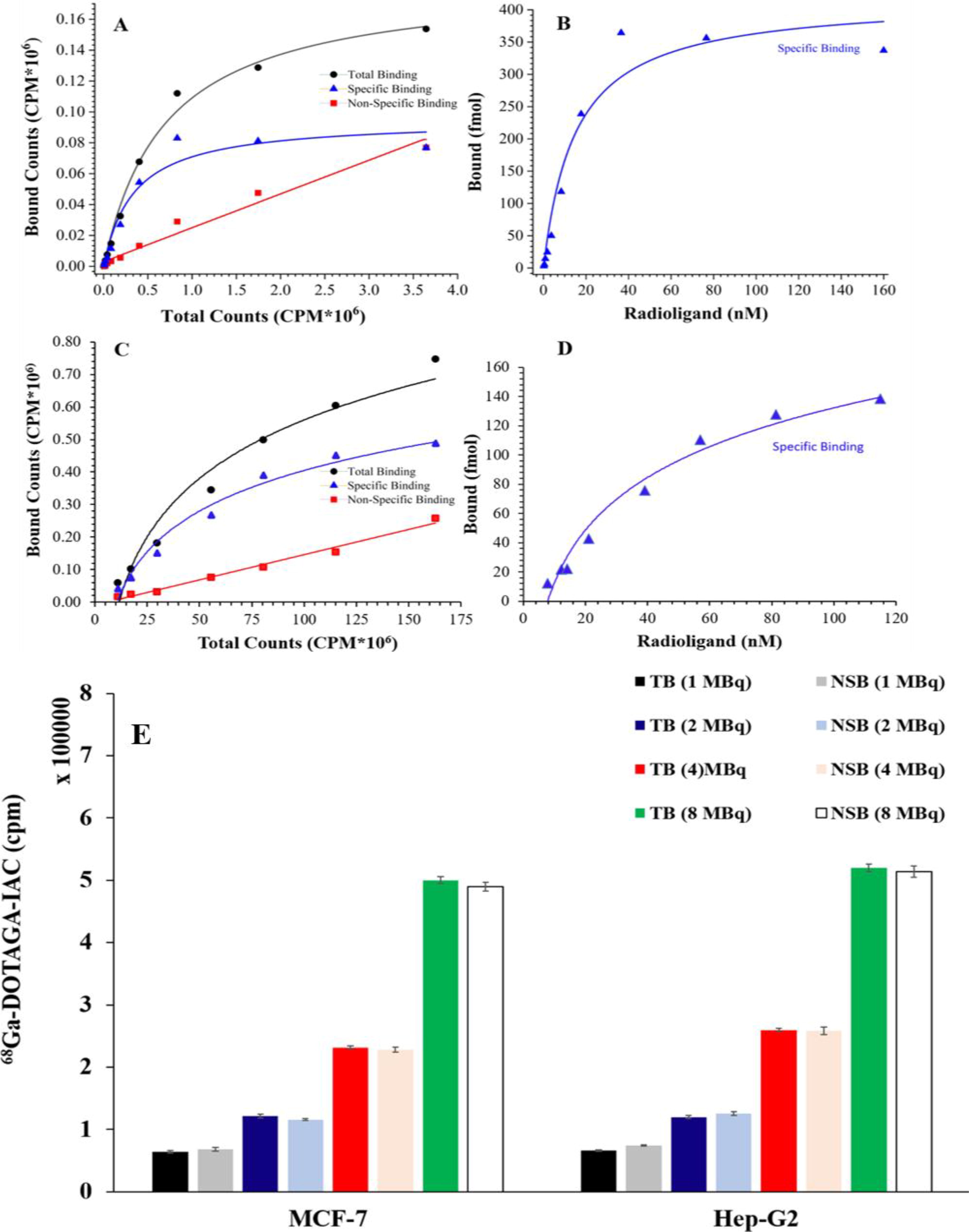
[68Ga]Ga-IAC total, nonspecific, and specific binding with αvβ3 integrin receptor
Biodistribution studies in healthy Wistar rats
The elevated levels of radioactivity detected in the kidneys for both radiotracers indicated that this is the main route of excretion. In addition, moderate levels of radioactivity, observed in the stomach, small intestine, and large intestine, suggested partial excretion through the hepatobiliary route. A significant decrease in blood pool radioactivity was observed at 1 h ([68Ga]Ga
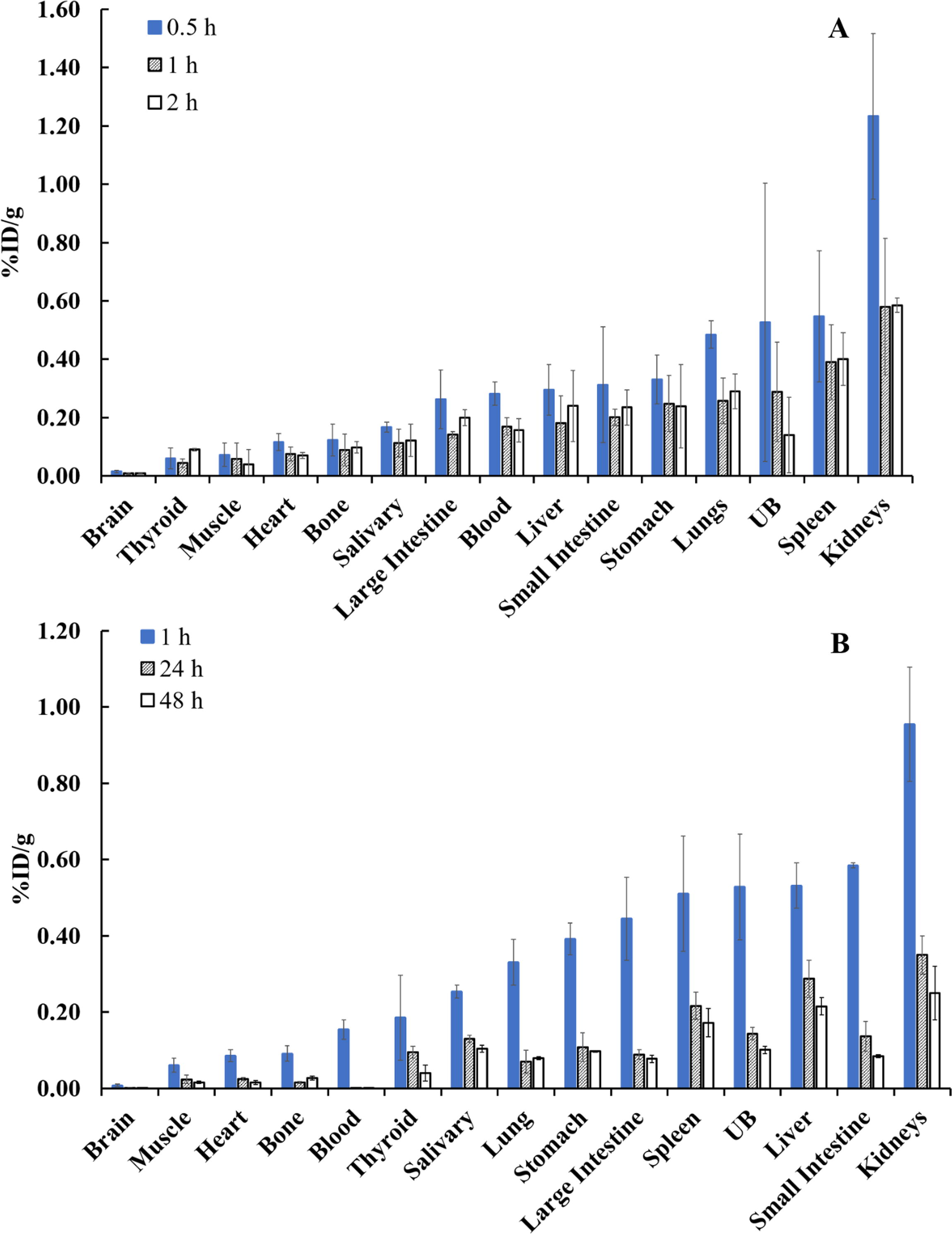
Biodistribution of [68Ga]Ga-IAC in Wistar rats at 0.5 h, 1 h, and 2 h
Dosimetry analysis
The total-body dose was calculated to be 0.28 mSv/MBq, and the effective dose was 0.174 mSv/MBq. The equivalent doses in organs were 0.114 mSv/MBq for kidneys, 0.097 mSv/MBq for liver, 0.082 mSv/MBq for spleen, and 0.041 mSv/MBq for the lungs. The highest radiation dose was found for osteogenic cells (0.313 mSv/MBq) followed by almost similar doses in the intestine, pancreas, prostate, and bladder wall (∼0.284 mSv/MBq). The values of equivalent doses for various organs are summarized in Table 1.
Radiation Dose Estimates (Adult Male Model) for [177Lu]Lu-IAC
PET/CT imaging
Dynamic and whole-body PET/CT imaging
For patient 1, [18F]FDG PET/CT revealed a well-defined, heterogeneously enhancing soft tissue lesion (SUVmax 7.6) in the retroareolar area of the right breast and mild uptake in perilesional fat stranding. No significant hypermetabolic lesion was observed in other parts of the body (Fig. 4M–O). The dynamic [68Ga]Ga-IAC PET/CT of the same breast lesion showed varying SUVmax values, which were 3.77 (at 5 min) and 3.75 (at 60 min), with a peak value of 5.61 at 15 min (Fig. 4A–K). The tumor and blood (aorta) SUVmax ratios ranged from 0.37 (at 5 min) to 1.82 (at 60 min), peaking at 2.08 between 30 and 35 min (Fig. 4L). The normal right breast tissue showed low background activity (SUVmax 0.9), resulting in a good tumor-to-background ratio of 4.2 (Fig. 4P). On whole-body [68Ga]Ga-IAC PET/CT (Fig. 4Q), normal organ distribution demonstrated physiological uptake in the thyroid (SUVmax 2.6), lungs (SUVmax 1.3), liver (SUVmax 3.62), and muscles (SUVmax 1.67). The excretion of [68Ga]Ga-IAC was observed from the kidney (SUVmax 10.2). Compared to [18F]FDG (SUVmax 20.9), [68Ga]Ga-IAC showed minimal to negligible uptake in the brain (SUVmax 0.19).
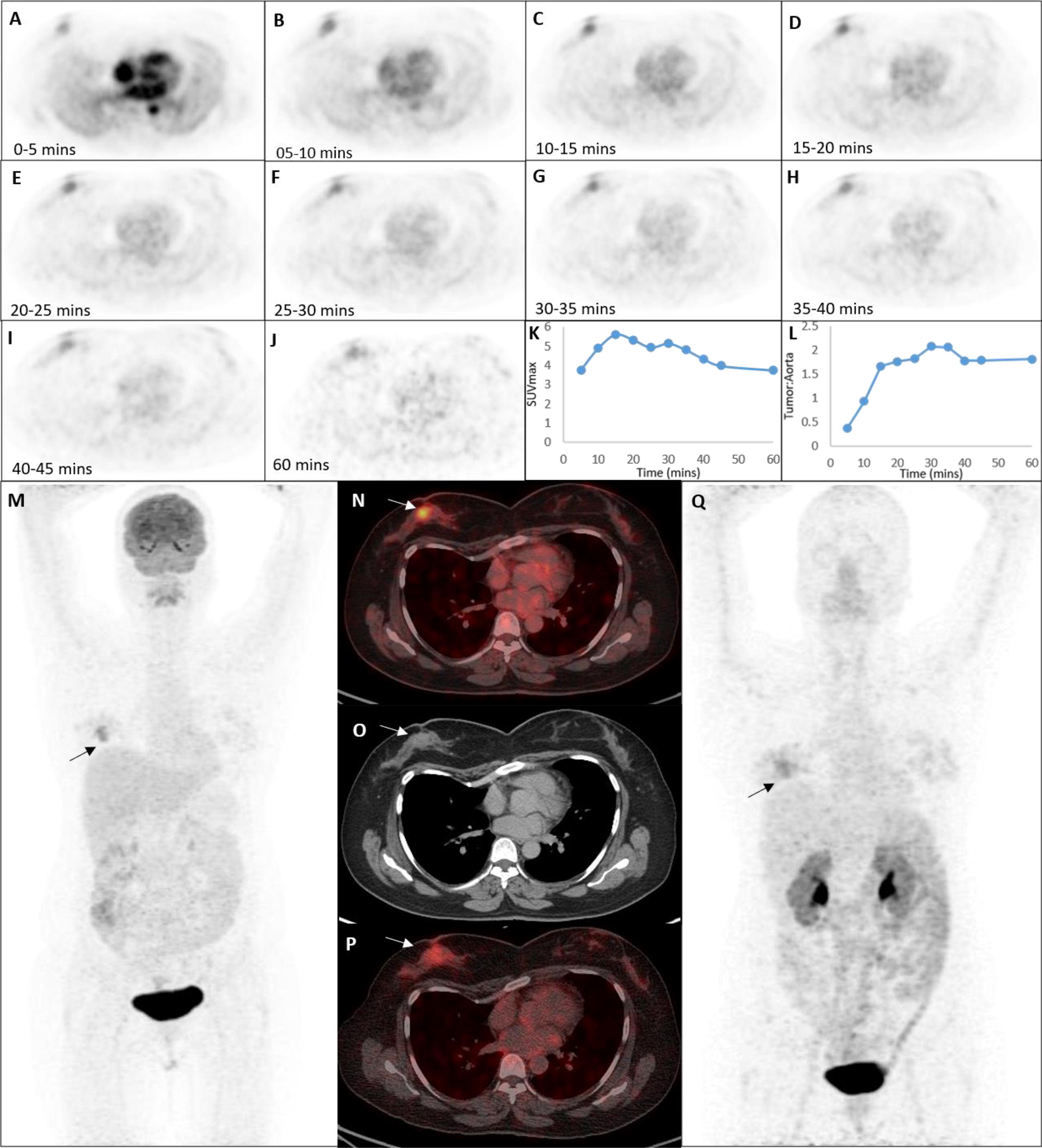
A 41-year-old woman (patient 1) was diagnosed with right breast cancer (T4b N1 M0), biopsy showed invasive ductal carcinoma grade III and immunohistochemistry confirmed the receptor status as Estrogen Receptor Positive (ER+), Progesterone Receptor Positive (PR+), Human Epidermal Growth Factor Receptor 2 (HER2) - with Ki67 value of 20%. Day 1: [18F]FDG (305.25 MBq) WB PET/CT revealed a primary lesion on the right breast
[18F]FDG and [68Ga]Ga-IAC detected primary lesions in all five patients, with mean SUVmax values of 13.8 ± 6.53 and 3.94 ± 0.58, respectively. The detailed patient analysis is summarized in Table 2. Patient 4 presented with extensive metastases, [68Ga]Ga-IAC detected all the primary and metastatic lesions which were revealed on the [18F]FDG (Fig. 5). The [18F]FDG and [68Ga]Ga-IAC demonstrated multiple soft tissue lesions in the right breast as primary (SUVmax FDG vs. IAC: 16.7 vs. 4.7). In addition, multiple right axillary (SUVmax 11.0 vs 3.9), internal mammary (SUVmax 5.0 vs. 2.2), and abdominal (SUVmax 7.7 vs. 4.5) lymph nodes were also noted with liver (SUVmax 15.1 vs. 4.2), marrow-based, and lytic skeletal lesions (SUVmax 13.2 vs. 5.3). Owing to the physiological uptake of [18F]FDG in the brain, skull lesions were not clearly visible. However, the lack of avidity of [68Ga]Ga-IAC in the normal brain enabled the successful identification of multiple tracer-avid skull metastatic lesions (SUVmax 3.7) (Fig. 5G).
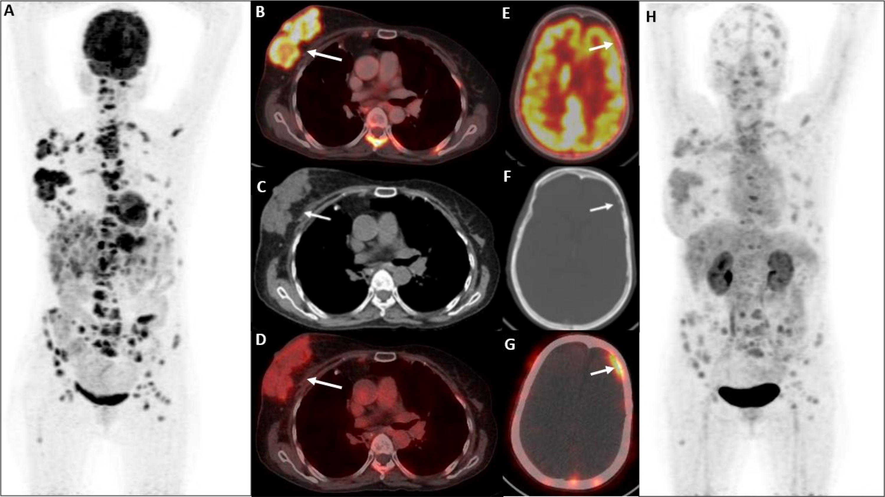
A 50-year-old female (patient 4) presented with disease progression and bone metastasis post 7 cycles of neoadjuvant chemotherapy. [18F]FDG (236.8 MBq) WB PET/CT was performed for disease evaluation post 45 min i.v. administration and [68Ga]Ga-DOTAGA-IAC (96.2 MBq) WB PET/CT was performed post 30 min i.v. administration. The MIP, corresponding transaxial CT
Patient Details
F, female; M, male; SUVmax, Maximum Standardized Uptake Value; FDG, fluorodeoxyglucose; IAC, integrin antagonist carbamate; IDC, Invasive ductal carcinoma; TNM, Tumor, Nodes, and Metastasis; ER+, Estrogen Receptor Positive; PR+, Progesterone Receptor Positive; Her2+, Human Epidermal Growth Factor Receptor 2 Positive; Her2−, Human Epidermal Growth Factor Receptor 2 Negative.
Adverse events
Following the injection of [68Ga]Ga-IAC and during the hospital stay, no adverse events or drug-related side effects were observed in any of the patients enrolled in the clinical study. The vital parameters of the patients remained stable, and no adverse events were observed in patients after their hospital stay.
Discussion
The radio-ITLC analyses revealed more than 95% RCY for [68Ga]Ga-IAC when 740 MBq [68Ga]GaCl3 was incubated with 10–20 µg IAC at 95°C for 10 min and reaction pH 4.5. The pH and temperature were the major factors in the radiolabeling of IAC with 68Ga (Supplementary Fig. S12). The optimization of IAC concentration on the radiochemical yield was important for the judicious utilization of the IAC, particularly in scenarios where the patient pool is limited. In addition, the higher ratio of IAC to 68Ga during radiolabeling can reduce the specific activity and excess of unlabeled IAC and may cause receptor saturation that could lower the PET image resolution. High specific activity and >99% RCP are preferred for high-resolution PET images and to reduce an unnecessary radiation burden to patients. The incorporation of C-18 cartridge purification in the preparation procedure resulted in more than 99.9% RCP. A peak at 8.6 min in HPLC validated the complexation of gallium with IAC.
The [177Lu]Lu-IAC was prepared with a high RCY (>99.9%), allowing the direct administration of the reaction mixture without a purification step. The high SA 177Lu contains only radioactive lutetium atoms in contrast to the low SA 177Lu (carrier added). Therefore, the radiolabeling of high SA 177Lu was achieved at a reasonably low IAC concentration compared with low SA 177Lu. Clinically, the high SA [177Lu]Lu-IAC is advantageous in tumors exhibiting high receptor expression and low receptor expression. However, in the case of low SA 177Lu, the IAC gets labeled with nonradioactive lutetium atoms that may consequently saturate the receptor sites, potentially blocking [177Lu]Lu-IAC binding. The impact of low SA 177Lu is more prominent when the receptor expression is low.
The [225Ac]Ac-IAC was prepared with a high radiochemical yield (>99%). The stability assessment was challenging. When the radio-ITLC was performed immediately after synthesis, a single peak at the origin for [225Ac]Ac-IAC was observed, with a radiochemical purity >99%. However, the delayed ITLC assessment resulted in multiple peaks at R f 0.0–0.5, and the intensity of these peaks was increasing over time (Supplementary Fig. S5).
This phenomenon could be understood as follows: (1) at the end of synthesis, all 225Ac and daughters were bound with IAC, (2) post-synthesis, the recoil effect from α decay caused the daughters to detach from IAC, and (3) the accumulation of recoiled daughters increased over time and appeared as multiple peaks in the radio-chromatogram which eventually decreased the purity to 76%. This finding emphasizes the importance of injecting [225Ac]Ac-IAC immediately after synthesis to avoid undesired localization and toxicity by the recoiled daughter radionuclides. If there is a time gap between synthesis and injection of [225Ac]Ac-IAC, incorporation of a purification step using a C-18 cartridge may effectively remove recoiled daughter radionuclides.
To confirm that multiple peaks were due to free daughter radionuclides, the same strips (developed at 15 min and 60 min post-synthesis) were rescanned after 2 h and showed a single peak at R f 0.0.
The other peaks were diminished due to the short half-lives (in min) of the recoiled daughter radionuclides. To validate that multiple peaks were due to free daughter radionuclides, the same strips (developed at 15 and 60 min post-synthesis) were rescanned after 2 h. The resulting single peak at Rf 0.0 confirms the decay of the short-lived recoiled daughter radionuclides over time. Our findings are in concordance with the available literature. 29
Another interesting question that could arise is the absence of recoiled daughter peaks in the radio-chromatogram obtained immediately at the end of synthesis. This could be attributed to the possibility of a low concentration of recoiled daughters present in the formulation, rendering them undetectable by the radio-ITLC detector. However, as time passes, the accumulation of daughters increases to the detectable range.
The low Kd (15.02 nM) demonstrates a high binding affinity of IAC to αvβ3 integrin receptor. The two known FDA-approved radiopharmaceuticals such as [68Ga]Ga-DOTATATE and [68Ga]Ga-PSMA-11 also have nanomolar binding affinities for their corresponding receptors, 7.36 and 4.3 nM, respectively. 30,31 The nanomolar affinities of IAC for αvβ3 integrin receptor suggest the strong binding between the radioligand and receptor. The binding of [68Ga]Ga-IAC with the C6 glioma cells line indicates its potential application in glioblastoma (Fig. 2). Although C6 rat glioma cells are considered safe, and effective, and simulate human glioblastoma, there still might be an interspecies variation in the binding of IAC with rat and human integrin. Therefore, additional studies are warranted in human glioblastoma cells (U87 cells) to establish the application of IAC in glioblastoma.
The log p-values of [68Ga]Ga-IAC (−2.35 ± 0.06) and [177Lu]Lu-IAC (−2.23 ± 0.07) indicate the hydrophilic nature of both radiocomplexes. The difference in lipophilicity arises from structural dissimilarities in the [68Ga]Ga-DOTAGA and [177Lu]Lu-DOTAGA complexes. Ga(III) and Lu(III) share similar chemistry but differ in ionic radii: Lu3+ (0.861 Å) forms 8-coordination with 4 amine nitrogens, 4 carboxylate oxygens, and Ga3+ (0.62 Å) forms 6-coordination with 4 amine nitrogens, 2 carboxylate oxygens, leaving 2 unbound carboxylates, hence increasing hydrophilicity. Routinely used DOTANOC/DOTATATE exhibits a similar lipophilicity of −2.88 ± 0.12. 32 The relatively lower hydrophilicity of IAC indicates that it will prefer the renal clearance route and may also show a partial hepatobiliary clearance route.
Fast blood pool clearance of [68Ga]Ga-IAC at 60 min and negligible blood pool activity of [177Lu]Lu-IAC at 24 h makes this radioligand a potential theranostic agent. The biodistribution of [111In]In-DOTA-Bz-SCN-IAC, as reported by Jang et al. in 2007 shows significantly higher values in all organs compared to our findings for [68Ga]Ga/[177Lu]Lu-IAC. 20 The difference in %ID/g for [68Ga]Ga-IAC versus [111In]In-DOTA-Bz-SCN-IAC at 60 min postinjection were as follows: Blood: 0.11 ± 0.05 vs. 1.22 ± 0.40; spleen: 0.39 ± 0.13 vs. 3.17 ± 0.31; bone; 0.09 ± 0.05 vs. 2.20 ± 0.14; liver: 0.20 ± 0.03 vs. 3.01 ± 0.27; kidney: 0.58 ± 0.23 vs. 14.43 ± 1.56. 20 This variation in physiological distribution could be due to the different chelator, linker, and radioisotope used in both studies. Furthermore, our preclinical distribution pattern was in concordance with clinical uptake findings. The clinical studies performed using [68Ga]Ga-IAC show minimal physiological distribution in the whole body.
Notably, slow renal elimination was observed for both [68Ga]Ga-IAC and [177Lu]Lu-IAC in animals. However, therapeutic radionuclide-induced radiation nephrotoxicity is avoidable by appropriate hydration and using renal protection agents. Nowadays, pretargeted radioimmunodiagnosis (PRID) and radioimmunotherapy (PRIT) strategies are also gaining attention for their potential to enhance therapeutic efficacy and reduce toxicity. 33
The effective dose for [177Lu]Lu-IAC in humans, as estimated using animal biodistribution data, is of 0.17 mSv/MBq. The value indicates favorable dosimetry as it is significantly lower than other established 177Lu radiopharmaceuticals. 34 The dose-limiting tissue was found to be osteogenic cells with a dose of 0.31 mSv/MBq. However, the dose appears to be safe in view of the reported permissible cumulative dose of 2 Gy for bone. 35,36
The dynamic imaging in patients revealed peak [68Ga]Ga-IAC uptake at 10–15 min. However, a favorably low blood pool with the highest tumor-to-blood ratio was achieved at 30–35 min. At 60 min, the blood pool activity was minimal, but the tumor uptake was also decreased due to the physical decay of 68Ga. Therefore, 30–35 min is the optimum imaging time for whole-body [68Ga]Ga-IAC PET/CT (Fig. 4).
In patients, [68Ga]Ga-IAC effectively detected all the primary and metastatic lesions positively revealed by [18F]FDG PET/CT (Table 2). [68Ga]Ga-IAC may offer a considerable advantage over [18F]FDG for detecting brain lesions. The high physiological uptake of [18F]FDG in the brain makes it challenging to delineate skull lesions in the [18F]FDG PET/CT imaging. However, for [68Ga]Ga-IAC, the absence of physiological brain uptake makes it a better agent for delineating skull lesions. Besides these findings, minimal physiological distribution in healthy organs and a good tumor-to-background ratio makes IAC a potential diagnostic and therapeutic agent for the αVβ3 expressing tumors. The chemotherapy-resistant, refractory, and metastatic patients may benefit from the IAC when chelated with 177Lu and 225Ac.
Conclusions
This study provides radiolabeling, quality controls, and clinical evidence that [68Ga]Ga-IAC is a useful tracer for the visualization of angiogenic breast cancer tumors and was found to be safe and effective. The bifunctional chelator, DOTAGA, is suitable for radiolabeling with 177Lu and 225Ac as well. The probe was evaluated clinically and demonstrated selective binding and rapid accumulation in the tumor neovasculature. The high receptor binding affinity, significant tumor-to-background ratio, minimal physiological uptake in body organs, and favorable dosimetry suggest theranostic agent potential of IAC. Peptide Receptor Radionuclide Therapy and Targeted alpha-therapy using [177Lu]Lu-IAC and [225Ac]Ac-IAC may have the potential for treating a broad range of early and late-stage cancers expressing αVβ3 integrin. Further clinical investigations with [177Lu]Lu/[225Ac]Ac-IAC are warranted to establish their therapeutic efficacy.
Footnotes
Acknowledgment
This research and development were supported in part by Advanced Innovative Partners, Inc. (AIP), Miami, Florida.
Authors’ Contributions
S.P.: radiolabeling optimization, quality control assessment, cell culture, animal studies, patient recruitment, data analysis, and article writing; G.K.: radiopharmaceutical preparation, cell culture and animal studies; N.R.: patient imaging, image reconstruction and determination of optimal PET/CT imaging time; S.C.: radiopharmaceutical preparation and patient imaging; I.R.: animal studies; R.K.: interpretation of patients image; I.L.: patients evaluation and recruitment; V.D.C.: animal studies; S.S.: conceptualization of application of IAC; M.G.S.: Dosimetry Study; B.R.M.: interpretation of patients image; J.S.: study design, data interpretation, article review and proofreading.
Disclosure Statement
Dr. Stanley Satz receives compensation from Advanced Innovative Partners, Inc. No other author has any potential conflict of interest.
Funding Information
Advanced Innovative Partners, Inc. (AIP), Miami, Florida
Supplementary Material
Supplementary Section
Supplementary Figure S1
Supplementary Figure S2
Supplementary Figure S3
Supplementary Figure S4
Supplementary Figure S5
Supplementary Figure S6
Supplementary Figure S7
Supplementary Figure S8
Supplementary Figure S9
Supplementary Figure S10
Supplementary Figure S11
Supplementary Figure S12
Supplementary Table S1
Supplementary Table S2
Supplementary Table S3
Supplementary Table S4
Supplementary Table S5
Supplementary Table S6
Supplementary Table S7
References
Supplementary Material
Please find the following supplemental material available below.
For Open Access articles published under a Creative Commons License, all supplemental material carries the same license as the article it is associated with.
For non-Open Access articles published, all supplemental material carries a non-exclusive license, and permission requests for re-use of supplemental material or any part of supplemental material shall be sent directly to the copyright owner as specified in the copyright notice associated with the article.