
Abstract
Background:
Long noncoding RNAs (lncRNAs) contribute to the initiation and progression of gastric cancer (GC). This study examined the potential role of lncRNA colorectal neoplasia differentially expressed (CRNDE) in modulating the expression of migration and invasion enhancer 1 (MIEN1) through the suppression of miR-136-5p in GC.
Methods:
The biological roles of CRNDE, miR-136-5p, and MIEN1 in GC were assessed both in laboratory settings and through the examination of clinical samples.
Results:
CRNDE was found to be significantly increased in GC tissues, and this upregulation was associated with an unfavorable prognosis of GC patients. In vitro experiments showed that inhibiting cell growth and migration, along with promoting apoptosis in GC cells, could be achieved by either disabling CRNDE or MIEN1, or by increasing the expression of miR-136-5p. MIEN1 is a specific recipient of miR-136-5p, and the anticancer effects of miR-136-5p can be counteracted by the increased expression of MIEN1. Through the examination of clinical specimens, it has been observed that there is a significant positive correlation between the expression of MIEN1 and CRNDE. In contrast, miR-136-5p expression in GC tissues shows a negative correlation.
Conclusion:
A previously unexplored therapeutic target for GC involves the CRNDE/miR-136-5p/MIEN1 signal transduction cascade.
Introduction
According to global cancer statistics, gastric cancer (GC) is the second leading cause of death from malignant tumors and the fourth most prevalent in terms of morbidity. 1,2 Even though many differentially expressed genes have been discovered and annotated, the full comprehension of the molecular mechanisms that cause GC remains unknown.
Initially, long noncoding RNAs (lncRNAs) were recognized as a group of noncoding transcripts that exceed 200 nucleotides in length. 3,4 At the cellular level, lncRNAs have a pivotal function in regulating cell growth, cellular differentiation, the cell cycle, and apoptosis. 5,6 Furthermore, lncRNAs modulate protein translation through transcriptional and post-transcriptional mechanisms. 7 LncRNAs function as competing endogenous RNAs (ceRNAs) to sponge microRNAs (miRs) to promote the translation of target genes. 8,9 At present, numerous lncRNAs are determined by functional genomics studies as oncogenes or tumor suppressors in cancers. 10,11
LncRNA-colorectal neoplasia differentially expressed (CRNDE), known as an oncogene, promotes cell proliferation and chemoresistance, thereby accelerating colorectal cancer (CRC) progression. 12,13 Furthermore, a rise in the expression level of lncRNA-CRNDE is detected in various clinical tumor tissue samples, including hepatocellular carcinoma, cervical cancer, melanoma, and clear cell renal cell carcinoma. 14 –17 Nevertheless, the role and manifestation of lncRNA-CRNDE in GC remain ambiguous. In this study, the authors present fresh evidence indicating that lncRNA-CRNDE is significantly increased in GC samples and is closely associated with advanced disease stage and unfavorable prognosis. Cell experiments conducted in vitro demonstrate that the suppression of lncRNA-CRNDE markedly hampers the growth and movement of GC cell lines, while promoting cellular apoptosis. Furthermore, the definitive molecular mechanism of lncRNA-CRNDE as an miR sponge has been established, offering a precise understanding of the development of GC.
Methods
Specimen collection
A total of 73 sets of GC tissues and adjacent nontumor tissues were collected and approved from Qionglai Medical Center Hospital in Qionglai.
Cell culture and transfection
The GES-1 cell line, which is a normal gastric mucosal epithelial cell line, and the BGC-823, MKN-28, SGC-7901, MGC-803, and AGS cell lines derived from GC were acquired from the American Type Culture Collection (ATCC) (Bethesda, MD, USA). These cell lines were cultured in Dulbecco's Modified Eagle Medium (DMEM) (Gibco). GenePharm (Shanghai, China) synthesized si-Con, si-CRNDE, sh-Con, and sh-migration and invasion enhancer 1 (MIEN1). Sangon Biotech (Shanghai, China) provided miR-Con, miR-136-5p mimics, and inhibitors. Plasmids with elevated levels of MIEN1 and a control empty vector were acquired from GeneCopoeia, Inc. (Rockville, MD, USA). Lipofectamine 3000 (Invitrogen) was used to transfer plasmids into MGC-803 and SGC-7901 cells.
Real-Time quantitative Polymerase Chain Reaction (RT-qPCR)
The miRNeasy Mini Kit from Qiagen, Inc. (Valencia, CA, USA) was employed for the extraction of total RNA. The First-Strand cDNA Synthesis Kit from TaKaRa Shuzo (Kyoto, Japan) was utilized for cDNA synthesis. PCR analysis was conducted using the TaqMan® Universal PCR Master Mix provided by Thermo Fisher Scientific, Inc.
Cell proliferation, migration, invasion, and apoptosis and luciferase reporter assay
Previously described protocols were used to assess cell proliferation with cell counting kit-8 (CCK-8), migration, invasion, apoptosis, and Luciferase reporter assay. 18,19
Immunohistochemical staining and Western blot
Immunohistochemical (IHC) staining, histological evaluation, and Western blot assays were performed as described previously. 20,21 Anti-MIEN1 primary antibody (Invitrogen; dilution: 1:50) was used to incubate the histological section. In addition, anti-MIEN1 primary antibody (Invitrogen; dilution: 1:500) was used to incubate protein membranes.
Statistical analysis
The data were presented in the form of mean plus the standard deviation. The statistical analysis was conducted using IBM SPSS Statistics Version 19.0 (SPSS Inc., Chicago, IL, USA). To analyze differences between two groups, either the student’s t-test or the nonparametric Mann–Whitney test was employed. The evaluation of CRNDE, miR-136-5p, or MIEN1 expression in GC tissues and paracarcinoma tissues was conducted using the Wilcoxon paired t-test.The analysis of intergroup variances was conducted using one-way ANOVA. The correlation was identified using Spearman’s rank analysis. A p-value below 0.05 suggests a statistically significant distinction.
Results
LncRNA-CRNDE is increased and associated with poor prognosis of GC
According to The Cancer Genome Atlas (TCGA) database, the expression of CRNDE in stomach adenocarcinoma (STAD) is considerably higher when compared with nontumor tissues (Fig. 1A and 1B). A significant upregulation of CRNDE is also discovered in the pairs of GC tissues and nontumor tissues (Fig. 1C). The analysis of the Kaplan–Meier curve demonstrated that individuals with high CRNDE expression had a significantly reduced overall survival (OS) time (Fig. 1D), disease specific survival (DSS) time (Fig. 1E), and progress free interval (PFI) (Fig. 1F) compared with GC patients with low CRNDE expression. Consistent with the bioinformatics findings, the results of this study also validate a significant increase in CRNDE expression in GC tissues (Fig. 1G) and cell lines (Fig. 1H) compared with the control group. GC patients with CRNDE high expression is significantly correlated with shorter OS than those patients with CRNDE low expression (p = 0.023; Fig. 1I). CRNDE high expression is strongly correlated with advanced TNM stages, metastasis to lymph nodes, and recurrence, as indicated in Table 1. Both univariate and multivariate regression analyses confirmed that CRNDE serves as an independent prognostic indicator in forecasting unfavorable outcomes in patients with GC (Table 2).
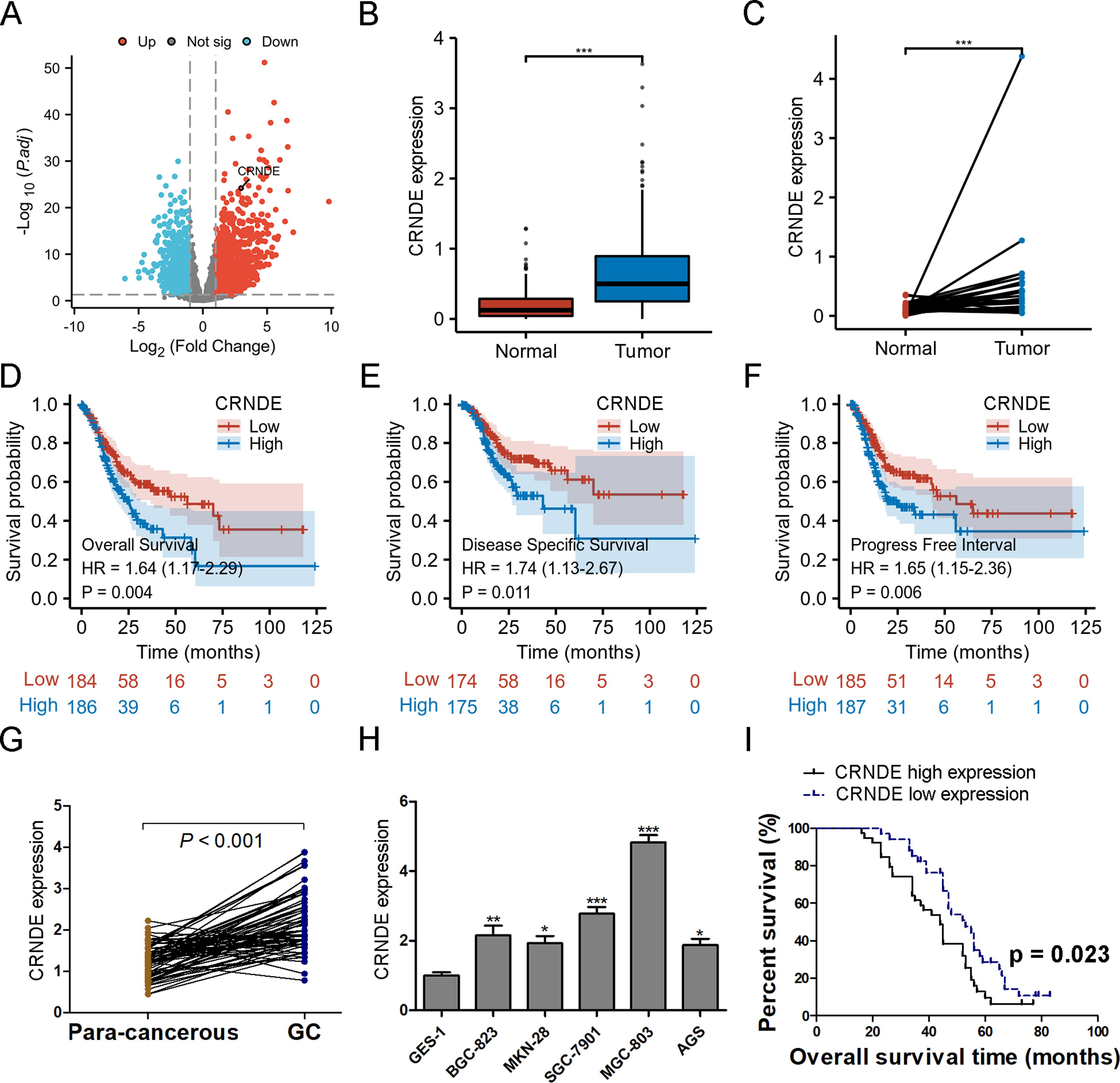
The expression of lncRNA CRNDE is elevated and correlated with an unfavorable prognosis in individuals with GC. CRNDE expression is predicted using TCGA database, with heatmap
Correlation between Clinicopathological Factors and lncRNA CRNDE Expression Levels in GC Patients
GC, gastric cancer; M, male; F, female; Y, years; N, negative; P, positive.
Univariate and Multivariate Regression Analyses of GC Patients for Overall Survival
GC, gastric cancer; N, negative; P, positive; CI, confidence interval.
Knockdown of CRNDE suppresses proliferation, migration, and invasion and induces apoptosis of GC cells
In vitro experiments were conducted to evaluate the antitumor activity of si-CRNDE after the depletion of CRNDE using siRNA transfection. In vitro, si-CRNDE transfection significantly suppressed the growth (Fig. 2A), migration, and invasion (Fig. 2B) of MGC-803 and SGC-7901 cells, as demonstrated by the CCK-8 and Transwell assays. The TUNEL staining results also indicate that transfection with si-CRNDE significantly enhances the amount of TUNEL-positive staining cells in comparison to the si-Con group, implying that the suppression of CRNDE encourages apoptosis in GC cells (Fig. 2C).
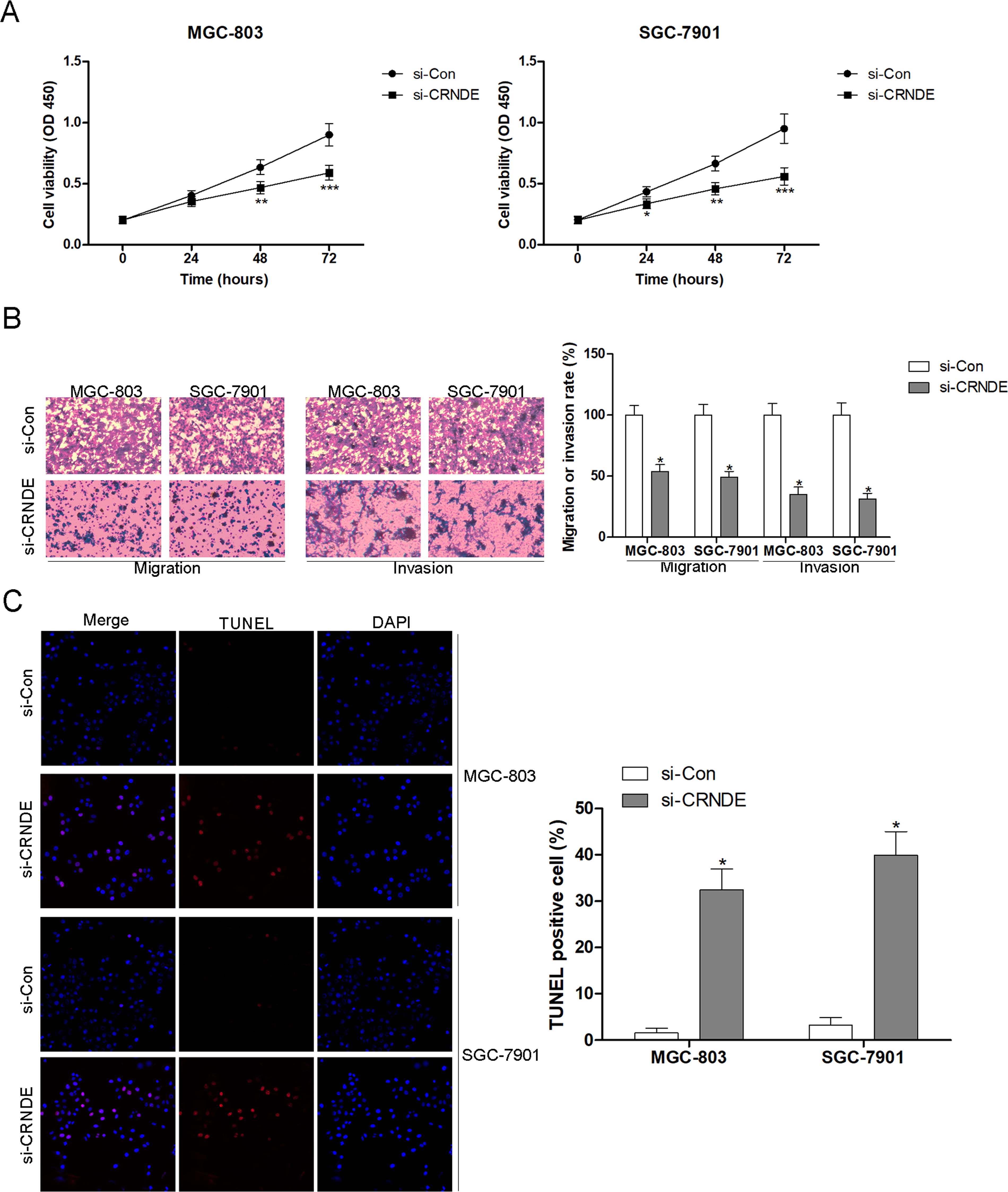
Suppression of CRNDE leads to inhibition of proliferation, migration, and invasion, and triggers apoptosis in GC cells. Following the stable expression of si-Con or si-CRNDE in MGC-803 and SGC-7901 cells, the viability of cells was assessed using CCK-8
CRNDE sponges miR-136-5p and regulates its expression
LncRNAs have been recognized as significant contenders to ceRNAs that control the expression of miRs. 22,23 According to DIANA tools’ LncBase Predicted v.2 (Fig. 3A), the study indicates that CRNDE can potentially bind to two presumed sites of miR-136-5p. Subsequently, luciferase tests were conducted to anticipate the correlation between CRNDE and miR-136-5p in MGC-803 cells that harbored CRNDE plasmids of either WT or Mut, which were cotransfected with miR-136-5p mimics. The findings from the study indicate a notable decrease in luciferase activity in WT MGC-803 cells or when transfected with CRNDE-Mut-1. This suggests that the binding site2 (418–445) of CRNDE has the ability to sequester miR-136-5p, as depicted in Figure 3B. Furthermore, to further confirm miR-136-5p as a CRNDE target, a CRNDE knockout experiment was conducted. According to the data presented in Figure 3C, the RT-qPCR findings indicated a notable reduction in CRNDE expression in MGC-803 and SGC-7901 cells following si-CRNDE transfection. Interestingly, the decrease in CRNDE expression through si-CRNDE leads to a simultaneous increase in miR-136-5p expression in MGC-803 and SGC-7901 cells (Fig. 3D). In comparison to nontumor tissues, the expression of miR-136-5p in GC tissues is notably reduced (as shown in Fig. 3E). The analysis of clinical samples was conducted to confirm CRNDE as a ceRNA that suppresses the expression of miR-136-5p. In 73 GC tissues, the Pearson correlation analysis revealed a significant inverse correlation between CRNDE expression and miR-136-5p (Fig. 3F). The results support the idea that CRNDE acts as a ceRNA to suppress the expression of miR-136-5p in the progression of GC.

CRNDE absorbs miR-136-5p and controls its level of expression. LncBase Predicted v.2 of DIANA tools,
miR-136-5p inhibitors reverse si-CRNDE-mediated proliferation and migration inhibition of GC cells
To examine the regulatory mechanism of the CRNDE/miR-136-5p axis on the proliferation, migration, and apoptosis of GC cells, MGC-803 and SGC-7901 cells were cotransfected with si-CRNDE and miR-136-5p inhibitors. Initially, findings from this study demonstrated that miR-136-5p imitates hinder cellular growth (Fig. 4A) and migration (Fig. 4C) while enhancing cellular apoptosis (Fig. 4E). The inhibition of miR-136-5p counteracts the growth induction (Fig. 4B), migration inhibition (Fig. 4D), and apoptosis (Fig. 4F) caused by si-CRNDE in MGC-803 and SGC-7901 cells, which is of greater significance. The combined findings indicate that CRNDE hinders the action of miR-136-5p, leading to the enhancement of GC cell proliferation and movement while hindering apoptosis.

Inhibition of miR-136-5p reverses the proliferation and migration inhibition of GC cells caused by si-CRNDE. Following the introduction of miR-Con or miR-136-5p mimics into MGC-803 and SGC-7901 cells, they assessed cell viability, migration, and apoptosis using CCK-8
miR-136-5p directly targets MIEN1
The next step was to identify potential targets of miR-136-5p by utilizing DIANA TOOLS and TargetScan, with a direct targeting on MIEN1. According to the information presented in Figure 5A, it was observed that miR-136-5p can potentially bind to a site in the 3'-untranslated region (3′-UTR) of MIEN1. Afterward, the aim was to confirm the location where the binding occurs in GC cells. The luciferase activity is notably suppressed in MGC-803 (Fig. 5B) and SGC-7901 (Fig. 5C) cells when WT or Mut 3′-UTR of MIEN1 is cotransfected with miR-136-5p mimics, indicating that miR-136-5p has the ability to directly target MIEN1. In addition, it was observed that the transfection of miR-136-5p mimics significantly decreases the expression of MIEN1 protein in MGC-803 and SGC-7901 cells (as shown in Fig. 5D and 5E).
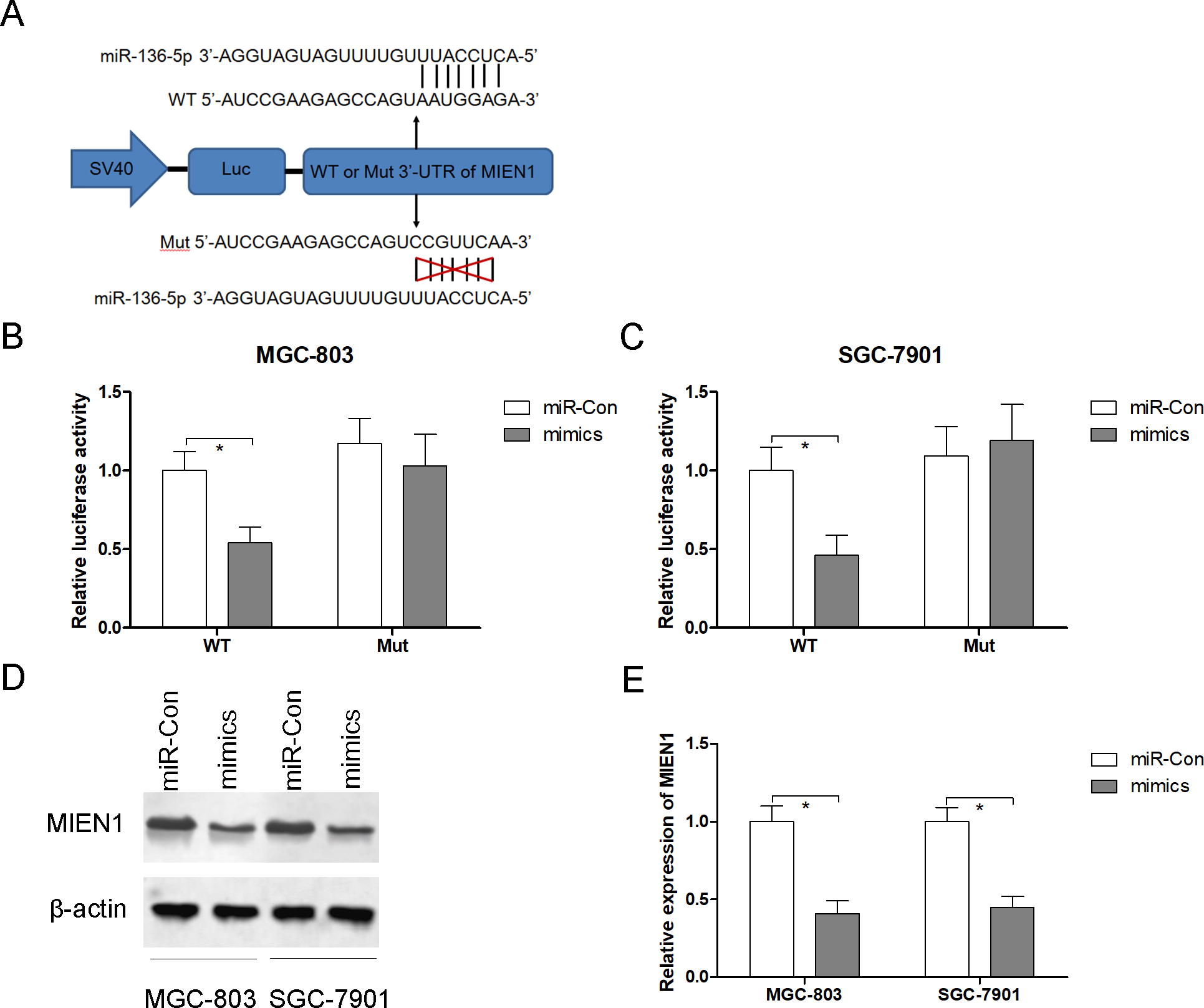
MIEN1 is directly targeted by miR-136-5p. DIANA TOOLS and TargetScan
Overexpression of MIEN1 neutralizes the antineoplastic activities of miR-136-5p in GC cells
To examine the involvement of the miR-136-5p/MIEN1 pathway in the advancement, MGC-803 and SGC-7901 cells were cotransfected with plasmids overexpressing MIEN1 and mimics of miR-136-5p. Initially, findings from this study demonstrated that the suppression of MIEN1 hinders cellular growth (Fig. 6A) and migration (Fig. 6C) while enhancing cellular programmed cell death (Fig. 6E). The overexpression of MIEN1 counteracts the transfection-induced growth (Fig. 6B), migration inhibition (Fig. 6D), and apoptosis (Fig. 6F) in MGC-803 and SGC-7901 cells, which interestingly mimics miR-136-5p. The combined findings indicate that miR-136-5p inhibits the expression of MIEN1 after transcription, leading to the inhibition of GC cell proliferation and migration, and promoting apoptosis in GC cells.
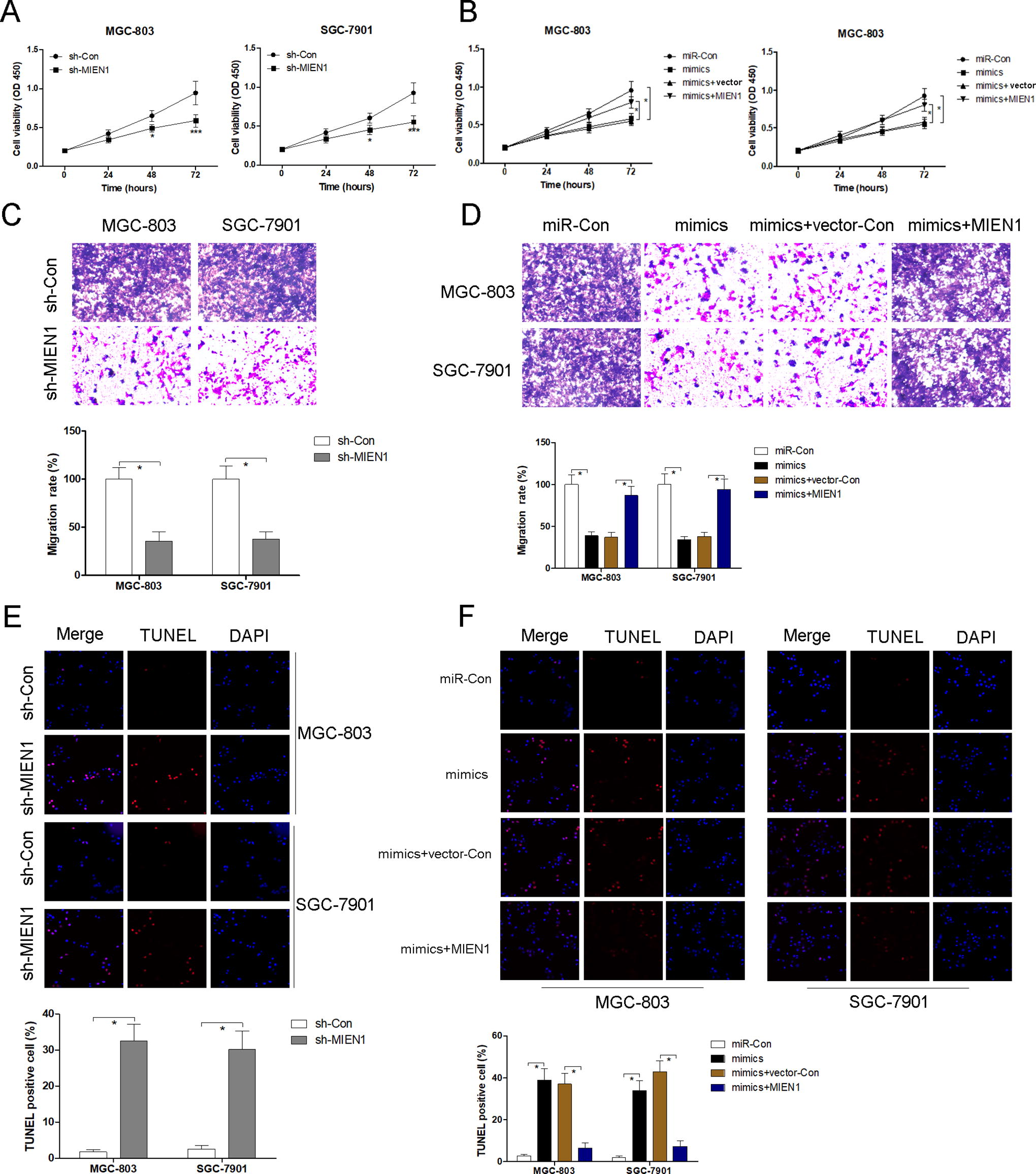
The overexpression of MIEN1 in GC cells antagonizes the anticancer effects of miR-136-5p. Following the introduction of sh-Con or sh-MIEN1 into MGC-803 and SGC-7901 cells, the assessment of cell viability, migration, and apoptosis is conducted using CCK-8
MIEN1 expression is upregulated in GC tissues
Figure 7A confirms that MIEN1 expression is markedly increased in STAD tissues compared with the corresponding control group, as validated by the TCGA and GTEx databases. In Figure 7B, the IHC staining analysis reveals a notable rise in MIEN1 expression in 73 GC tissues when compared with the surrounding nontumor tissues. The examination of Pearson correlation revealed a significant positive correlation between the expression of MIEN1 and CRNDE (r = 0.558; p < 0.001; Fig. 7C). Conversely, there was a negative correlation between the expression of MIEN1 and miR-136-5p in GC tissues (r = −0.594; p < 0.001; Fig. 7D).
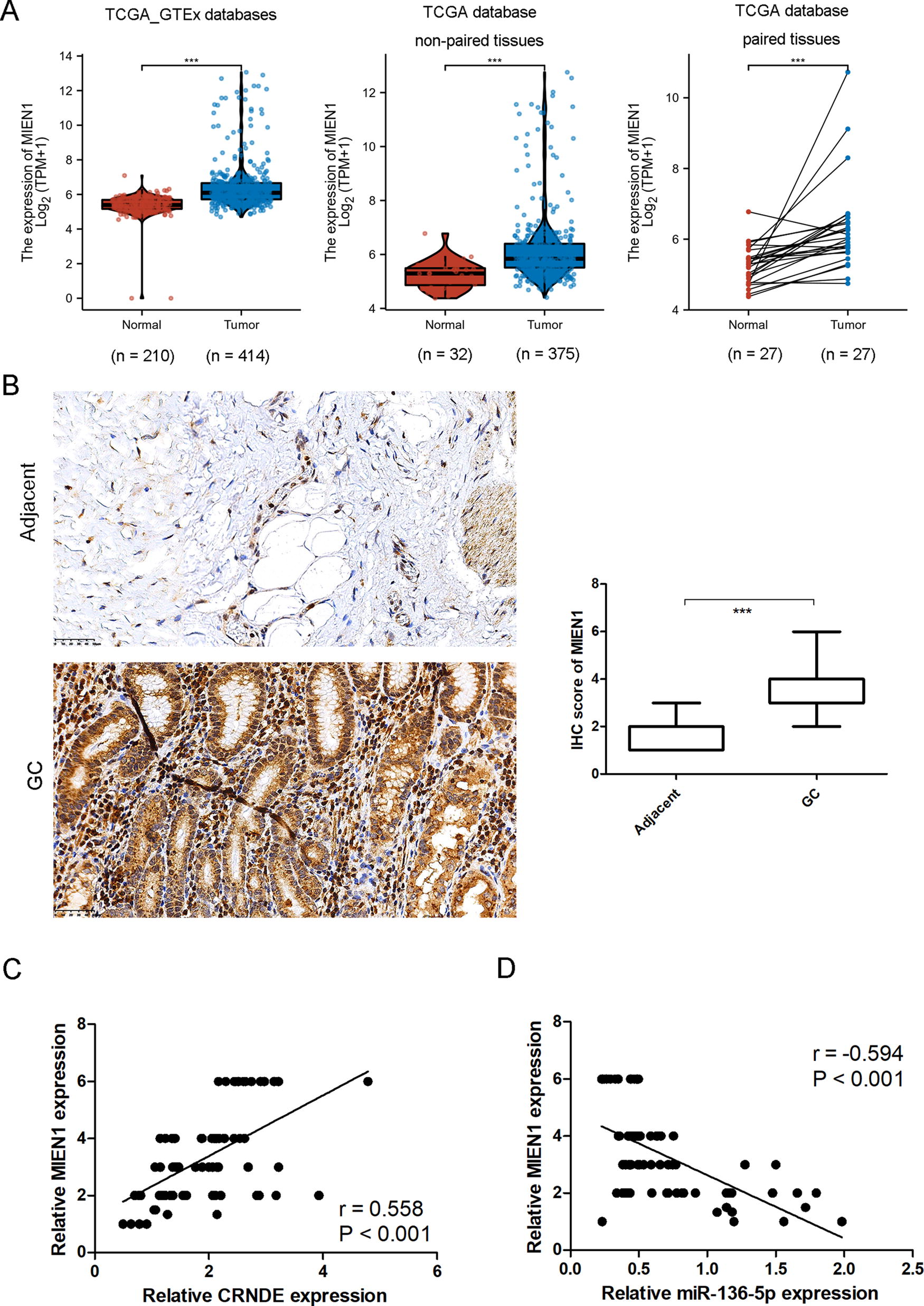
The expression of MIEN1 is increased in gastric cancer tissues. MIEN1 expression is predicted based on TCGA database
Discussion
In the current investigation, it has been determined that the expression of CRNDE is significantly elevated in GC tissues and is correlated with an unfavorable prognosis for patients with GC. Furthermore, CRNDE has the potential to act as a ceRNA by sequestering miR-136-5p and counteracting its anticancer effects. In laboratory settings, experimental data indicate that the inhibition of CRNDE or MIEN function, or the increased expression of miR-136-5p, can impede the growth and movement of GC cells and promote their apoptosis. In addition, the results confirm that MIEN1 is a specific target of miR-136-5p, and the tumor-inhibiting effects of miR-136-5p can be counteracted by increasing the expression of MIEN1. After analyzing clinical specimens, it was found that there is, a strong positive correlation was found between the expression of CRNDE and MIEN1. In contrast, miR-136-5p expression in GC tissues shows a negative correlation. CRNDE has been shown to upregulate the expression of MIEN1 by sequestering miR-136-5p, consequently exacerbating the advancement of GC.
Many studies have examined CRNDE to elucidate its oncogenic function in various human cancers, including GC. 24 –26 In human solid cancers, CRNDE is identified as a poor prognostic marker in clinical outcomes, including advanced TNM stage, metastasis, and shorter OS. 27,28 The results and information from TCGA database strongly indicate that CRNDE expression is increased in GC tissues. Inhibition of native CRNDE leads to decreased cell proliferation, migration, and invasion, while promoting apoptosis in GC cells. CRNDE is extensively studied as a ceRNA in malignant tumors to mechanically suppress various miRs, such as miR-181a-5p, miR-345-5p, miR-384, and miR-539-5p. 13,29 –31 During the investigation, CRNDE was found to have the ability to adsorb miR-136-5p and hinder its manifestation in the advancement of GC. Inhibition of CRNDE leads to increased expression of miR-136-5p in MGC-803 and SGC-7901 cells. Loss of CRNDE function or gain of miR-136-5p function exhibits a comparable role in inhibiting the progression of GC. The presence of a strong inverse relationship between CRNDE and miR-136-5p expression in clinical samples provides substantial evidence for the contrasting roles of CRNDE and miR-136-5p in the progression of GC. Previous research has additionally recorded that CRNDE can inhibit the expression of miR-136-5p in various cancerous tumors, including glioma, CRC, and renal cell carcinoma. 32 –34 In this study, the CRNDE/miR-136-5p signaling pathway was discovered to play a role in the development of GC.
Recently, miR-136 mediated post-transcriptional repression of MIEN1 has emerged in colon cancer, osteosarcoma, and GC. 35 –37 The overabundance of MIEN1 plays a role in the movement, infiltration, and spread of different types of human tumors. 38,39 Poor prognosis is linked to high expression of MIEN1 in GC. 37 Suppression of MIEN1 hampers the growth and invasion of AGS and SGC7901 cells. 37 In addition, Liang and colleagues suggested that miR-124-5p plays a negative role in regulating the expression of MIEN1 to inhibit malignant characteristics in GC cells. 40 The findings from their study also seem to corroborate the presence of the oncogenic functions of MIEN1, indicating that the increased expression of MIEN1 is confirmed by both their clinical specimens and TCGA database. Moreover, the inhibition of MIEN1 leads to suppressed growth and migration, while also causing an increase in the rate of apoptosis in GC cells. In addition, the authors ascertain MIEN1 as a specific recipient of miR-136-5p. The excessive expression of MIEN1 counteracts the anticancer effects of miR-136-5p in GC cells. These findings suggest that the expression of MIEN1 shows a positive correlation with CRNDE but a negative correlation with the expression of miR-136-5p in GC tissues.
In summary, it is logical to deduce that the process of gastric carcinogenesis is facilitated by a novel signaling pathway CRNDE/miR-136-5p/MIEN1.The results of this study establish the basis for the use of GC in clinical practice, both in terms of pharmacology and therapy.
Footnotes
Data Availability Statement
The data that support the findings of this study are available from the corresponding author upon reasonable request.
Authors’ Contributions
Y.G. was responsible for study design. C.L., X.R., X.H., Y.H., and L.X. were responsible for literature research, data acquisition, and data analysis. Y.G. was responsible for manuscript preparation, manuscript editing, manuscript review, and cell experiments. Y.G., C.L., X.R., X.H., Y.H., and L.X. were responsible for clinical sample collection. The final version to be published was approved by Y.G., C.L., X.R., X.H., Y.H., and L.X.
Author Disclosure Statement
The authors declare that they have no competing interests.
Funding Information
No funding was received for this article.