




Abstract
Background:
For breast cancer staging, radiolabeled colloids and superparamagnetic iron oxide nanoparticles (SPIONs) are used for sentinel lymph node (SLN) imaging. This study characterized the intranodal activity distribution and absorbed dose distribution.
Material and Methods:
Six patients diagnosed with primary breast cancer were intradermally injected with 99mTc-Nanocoll. The SLNs were resected, weighed, and measured for activity. Three groups of six rats were subcutaneously injected into the hind paw with either 99mTc-Nanocoll, 99mTc-SPIONs, or 68Ga-SPIONs. Macro- and small-scale dosimetry calculations were performed using autoradiography images of cryosections of SLNs from patients and animals.
Results:
The mean absorbed dose in patient SLNs was 0.5 ± 0.3 mGy/MBq for 99mTc-Nanocoll and 3.4 ± 1.8 mGy/MBq, assuming a 99mTc-Nanocoll-based distribution of 68Ga-SPIONs. Due to different decay characteristics, the heterogeneity in the absorbed dose differed between 99mTc-SPIONs and 68Ga-SPIONs with a maximum to mean absorbed dose ratio of 2.7 ± 0.3 and 1.6 ± 0.2, respectively.
Conclusions:
This study shows that 99mTc- and 68Ga-SPIONs and 99mTc-nanocolloids have similar activity distribution in human and animal lymph nodes. Small-scale dosimetry models combined with clinical patient biokinetics may serve as a bridge between organ and tissue dosimetry and the interpretation of intrinsic geometric variation and its uncertainties in absorbed dose.
Introduction
Cancer, primarily in breast, skin, and prostate, disseminates through the lymphatic system, with the first metastases tending to spread from one or two regional, sentinel lymph nodes (SLNs), draining the primary tumor site. 1 –5 The clinical procedure to identify the SLNs and perform staging involves intradermal injection of 99mTc-labeled colloids in combination with vital blue dye (for intraoperative guidance). 6 –8 Less than 2% of the activity is accumulated in the SLN (3–5 h p.i.) due to the wide range in size of colloid particles. 9,10 Large colloidal particles are usually trapped at the injection site, while small colloidal particles, without any specific binding properties, are transported via the lymph through several lymph nodes and back into the circulatory system. 11,12 Recently, a focus has been on developing more specific agents to identify and localize the SLNs accurately. 13 –18 One of these agent is the Food and Drug Administration (FDA) and European Medicines Agency (EMA) approved 99mTc-labeled-tilmanocept (Lymphoseek), which is being used for lymphoscintigraphy and guided surgery. The small molecular size of 7 nm and high affinity to CD 206, expressed on the surface of both dendritic cells and macrophages in SLN, make it a promising agent. 19,20 In addition, the FDA has also approved superparamagnetic iron oxide nanoparticles (SPIONs) for SLN imaging. 21 –23 The advantages of SPIONs include a narrow size distribution (10–50 nm) and a large surface-to-volume ratio making them suitable for radiolabeling with high specific activity.
We have previously presented the potential of 99mTc- and 68Ga-SPIONs as translational multimodality molecular imaging agents. When injected subcutaneously in a rat model, these agents accumulate in the SLN, providing a potential for the staging of patients with breast cancer. In addition, the 68Ga-SPIONs enable simultaneous positron emission tomography, magnetic resonance, and Cherenkov (PET/MR/Cherenkov) imaging of the lymph nodes. 24 –26
In this study, we used autoradiography to image the distribution of activity within the SLNs and estimated the absorbed dose, both for clinical and preclinical data.
Traditionally, SLN dosimetry is calculated using the medical internal radiation dose formalism to estimate the mean absorbed dose by assuming homogeneous activity distribution within the nodes, an approach reported for common radiopharmaceuticals used in lymphoscintigraphy such as 99mTc-antimony trisulfide colloid, 99mTc-sulfur colloid, and 99mTc-Nanocoll. 27 However, the absorbed dose distribution pattern depends on the geometry and radiation emission energy spectrum. As a result, several small-scale dosimetry models have been reported. 28 –30 These include applying S-values at a microstructural level, resulting in a more accurate representation of the mean absorbed dose than utilizing organ-level dosimetry. In addition, autoradiography has the capability of revealing a heterogeneous distribution in the organ from the administered radiopharmaceutical. 31,32 Due to this, we, therefore, in this study, included heterogeneous activity distribution in lymph nodes in the absorbed dose calculations clinical and preclinical data.
The aims of this study were, first, to characterize, using autoradiography, the intranodal distribution of 99mTc-labeled nanocolloids in both humans and rats as well as 99mTc- and 68Ga-labeled SPIONs in rats, and second, to calculate the absorbed dose and absorbed dose distribution within the SLNs.
Materials and Methods
The study, including both patients and animals, was performed in accordance with ethical standards and approval, following all national and local regulations. All patients provided informed consent.
Labeling and quality control of tracers
SPIONs were obtained from Genovis AB (Lund, Sweden). The monodisperse nanoparticles were produced with a narrow size distribution (11 nm ± 2 nm) and a biocompatible coating of polyethylene glycol for further functionalization. 24
Radiolabeling of SPIONs with 99mTc and 68Ga was performed as described in previous studies. 24,25 Briefly, 99mTc-pertechnetate (300–500 MBq) was added to a vial containing the reducing agent 0.5 mg stannous chloride dissolved in sterile water (pH 3.4). The SPIONs were added to the mixture and then incubated for 60 min at room temperature (18°C). 68Ga was obtained from a 68Ga/68Ge-generator (IDB, Holland) by eluting the 68GaCl2 with 0.6 M hydrochloric acid in 0.3–0.4 mL fractions with 40–60 MBq activity. The 68Ga-SPIONs were produced via a direct labeling method using ammonium acetate as buffer (1 M, pH 5.5). Labeling efficiency and quality control were ensured by using instant thin-layer chromatography and 0.2 M citric acid as running buffer (ITLC, silica gel impregnated glass fiber sheets, Life Science or TEC-control chromatography strips, Biodex, USA). Imaging of the ITLC strips was performed using a phosphor imager (Perkin Elmer, Welesley, MA, USA). The colloidal stability of 99mTc-SPIONs and 68Ga-SPIONs was measured in vitro using human serum (pH 7, 37°C), 4 h and 24 h, respectively, after radiolabeling.
99mTc-Nanocoll was prepared using 99mTc-pertechnetate from a 99Mo/99mTc-generator (DRN 4329 Ultratechnekow FM, The Netherlands) and labeled in accordance with instructions from the manufacturer, including quality control ensured by authorized staff at the local radiochemistry laboratory/Nuclear Medicine Department.
Patient studies
Six patients aged 30–65 years had SLN biopsies that were included in this study. All patients had a histologically confirmed primary breast cancer tumor and were planned for surgical treatment, including SLN mapping. Four periareolar injections of 99mTc-Nanocoll™ (40 MBq, volume of 0.4–0.6 mL) were performed, 2–4 h before surgery. Planar lymphoscintigraphic images (frontal and lateral, acquisition time 5 min) were performed for each patient using a scintillation camera (Sky Light, Philips or Discovery, GE, USA) equipped with a low-energy, high-resolution collimator. The energy window was 15% centered over the 140 keV photopeak. The uptake of the agent was confirmed in the SLN imaging. Immediately before surgery, a blue dye (Patent V Blue) was administered to visualize the draining lymphatic channels and SLNs. A hand-held γ-probe (Euro Probe3, Capintec, USA) was used to localize the SLN and to confirm that the correct node was being resected. The resected SLNs were weighed and measured for activity content with a radionuclide calibrator (Capintec CRC 25R, UK). The SLNs were cut longitudinally and frozen. Sections that were 10-µm-thick were placed on microscope slides, dried, stained with hematoxylin and eosin (H&E), and imaged using autoradiography. A simplified work scheme is presented in Figure 1.
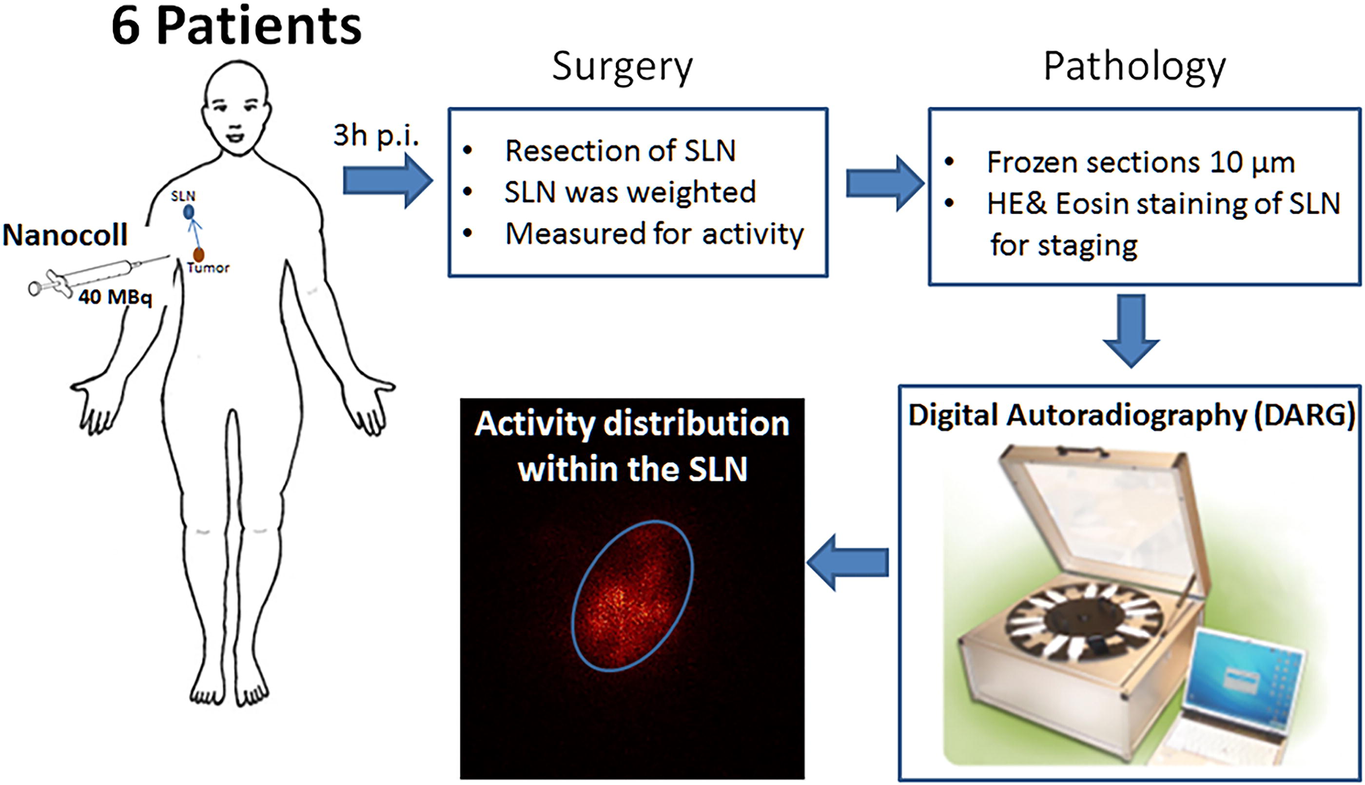
Working scheme to obtain the activity distribution images of human SLNs. SLNs, sentinel lymph nodes; SPIONs, superparamagnetic iron oxide nanoparticles.
Animal studies
Three groups of six white Wistar male rats (mean body weight, 200 g) were lightly anesthetized with a mixture of isofluran/O2 (2.5%–3%, 0.2 L/min) and injected subcutaneously with 99mTc-Nanocoll (∼7 MBq, 0.05 mL), 99mTc-SPIONs (∼24 MBq, 0.05–0.07 mL), or 68Ga-SPIONs (∼7.4 MBq, 0.05–0.07 mL) in the right hind paw. The animals were imaged with preclinical dual modality single-photon emission computed tomography (SPECT)/CT or positron emission tomography (PET)/CT (NanoSPECT-CT or NanoPET-CT, Bioscan, USA) 1, 2, 3, and 5 h postinjection. These images were only used for localization of the SLNs. Animals from the first group (injected with 99mTc-Nanocoll and 99mTc-SPIONs) were euthanized: one after the first imaging session (1 h p.i.), one after the second imaging session (2 h p.i.), two after the third imaging session (3 h p.i.), and two after the last imaging session (5 h p.i.). Animals injected with 68Ga -SPIONs were due to the short half-life of 68Ga, euthanized after 3 h p.i.
The dissected SLN were weighed and measured for activity using an NaI(Tl) well-type detector (1282 CompuGamma CS; LKB Wallac, Turku, Finland). Frozen sections (100 µm thick) of popliteal (SLN) and iliac lymph nodes were prepared, mounted on sample slides, and imaged for activity distribution with autoradiography.
Autoradiography
To determine the 99mTc-Nanocoll and 99mTc-SPIONs intranodal activity distribution in human and animal SLNs, microscope slides containing 1–3 SLN sections were imaged using autoradiography. An imaging system (Biomolex 700 Imager; Biomolex AS, Oslo, Norway) based on a double-sided silicon strip detector with an intrinsic 50 µm spatial resolution was used. 33,34 Each slide was imaged for 10–12 h. The acquired images were then assembled from list-mode data, using an in-hose developed code (IDL 6.4, L3 Harris Technologies, Melbourne, FL, USA), corrected for dead pixels by interpolation and for radioactive decay. No images were acquired for samples containing 68Ga-SPIONs due to the short half-life of 68Ga.
Dosimetry
The absorbed dose rates in SLNs were calculated from the activity image of autoradiography convolved with a point-dose kernel. The point-dose kernels for 99mTc and 68Ga were simulated with the Monte Carlo code MCNP5 1.60 (Los Alamos National Laboratory, New Mexico), using the new electron sampling logic option and a voxel size of 50 µm, corresponding to the voxel size of the autoradiography system. 34,35 Because data from adjacent tissue were missing, the same image was stacked to get the absorbed dose rate contribution from adjacent tissue. To determine the absorbed dose, the time-integrated activity coefficient, TIAc, (residence time) was determined from the dissected SLNs in rats (n = 6) at 0, 1, 2, 3, and 5 h. Activities were then decay corrected to the time of injection. Exponential fitting was performed on the first 5 h of the uptake curve, during which the maximum specific uptake was reached. For the time beyond 5 h, the uptake was considered constant, with no particles being released from the SLN. After the fitting, the physical decay was added, and the TIAc was calculated. Absorbed-dose images were obtained by multiplying the TIAc with the dose-rate images. These were then filtered using a Gaussian smoothing filter (σ = 1 voxel). Three thresholds (ratio of maximum)-based region of interests (ROIs) were derived, Region1 [0.12–0.25], Region 2 [0.25–0.4], and Region 3 [0.4–1.0]. These ROIs were then applied to the dose-rate images, and the absorbed dose in these regions was obtained. The calculated small-scale absorbed dose distribution depends on the assumption that the activity does not relocate inside the lymph node during the 5h experiment. In addition, dose–volume histograms (DVH), cumulative DVH (cDVH), and maximum to mean absorbed dose ratios were derived (Fig. 5). The short half-life of 68Ga (T1/2 = 68 min) resulted in that tissue samples containing 68Ga-SPIONs that could not be imaged using autoradiography. Therefore, we assumed similar activity distribution for 68Ga-SPIONs and 99mTc-Nanocoll in patients as well as 99mTc-SPIONs in rats.
Results
Labeling and quality control of tracers
The radiolabeling efficiency for 99mTc-Nanocoll was >95% for both patients and animals. 99mTc-SPIONs and 68Ga-SPIONs were labeled with an efficiency of 99% and 97%, respectively. The radiolabeled nanoparticles remained at the origin of the ITLC strips, whereas the free 99mTcO4 − or free 68Ga migrated with the solvent front with Rf values of 0.7 and 0.9, respectively. On analyzing the colloidal stability of 99mTc-SPIONs and 68Ga-SPIONs in human serum, the amount of free activity was <1% after 24 h (99mTc-SPIONs) and 4 h (68Ga-SPIONs).
Assessment of the SLN of patients
Patient SLN, resected for biopsy, weighed between 1 and 3 g with an average of 1.52 ± 0.94 g and 10–20 mm in size, measured along the longest axis. The mean activity accumulated in the lymph nodes was 1.31 ± 0.5 MBq (3.31% ± 1.44% IA), 3–5 h postinjection. The maximum and mean uptake in SLN is presented in Table 1.
The Maximum and Mean Uptake of Different Tracers in Patient (p) and Animal (a) Sentinel Lymph Nodes, Respectively
Assessment of the SLN of animals
The small animal SPECT-CT and PET-CT images clearly visualized popliteal lymph nodes in rats injected with 99mTc-Nanocoll, 99mTc-SPIONs, or 68Ga-SPIONs.
Rat SLNs had a mean weight of 0.01 ± 0.001 g, a size of 2–5 mm, and were bean-shaped with an off-white appearance, almost transparent color. The highest uptake in rat SLNs was 7.39% injected activity (IA) for 99mTc-SPIONs and 6.47% IA for 68Ga-SPIONs, 5 h respectively 3 h postinjection. For 99mTc-Nanocoll, the maxim uptake was 0.64% IA in rats and 6.02% IA in patients, 3–5 h postinjection. The maximum and mean uptake in SLN for each agent is presented in Table 1.
Autoradiography
Autoradiography imaging revealed a heterogeneous distribution of activity related to the anatomy, in both patient and rat lymph node sections. Despite higher uptake (activity content) in lymph nodes from rats, the activity distribution patterns were very similar when comparing the different agents for patient and rat lymph nodes.
Representative images are presented in Figure 2. The activity was primarily observed most proximal to afferent lymphatic vessels, in subcapsular and medullary sinuses, in both human and animal SLNs.

Autoradiography images presenting a comparison of activity distribution in human and rat lymph nodes.
Dosimetry
Despite the fact that 68Ga emits much more energy from charged particles than 99mTc, thus having a higher S-factor, the long accumulation time and the shorter half-life result in lower TIA and, subsequently, also a lower absorbed dose. The maximum absorbed-dose rate in the SLN of patients and rat lymph nodes was 6.4 ± 4.2 mGy/h and 774 ± 830 mGy/h, respectively, using 99mTc-Nanocoll. Assuming the same biodistribution as 99mTc-Nanocoll in patients for 68Ga-SPIONs, the maximum absorbed dose rate was 136 ± 83 mGy/h in SLNs. Representative absorbed dose images for patients are presented in Figure 3 and for animals are presented in Figure 4. The mean absorbed dose for 99mTc-Nanocoll in SLNs of patients was 1.8 ± 1.6 mGy.

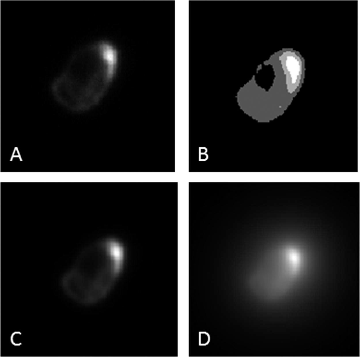
The absorbed dose calculated from the activity distribution in the autoradiography images for the different regions (Region 1 [0.12–0.25], Region 2 [0.25–0.4], and Region 3 [0.4–1.0]) is presented in Table 2.
The Mean Absorbed Dose in the Different Activity Regions in Rat and Patient Lymph Nodes
If 68Ga-SPIONs would be used instead of the 99mTc-Nanocoll, the mean absorbed dose would be as presented in the table, based on the nanocoll biodistribution. SLN, sentinel lymph node; SPIONs, superparamagnetic iron oxide nanoparticles.
Examples of DVH and cDVH for human and rat lymph nodes using 99mTc-Nanocoll, 99mTc-SPIONs, and 68Ga-SPIONs are presented in Figure 5. For comparison, the DVHs obtained for 68Ga-Nanocoll (Fig. 5B and D) are presented together with 99mTc-Nanocoll and 68Ga-SPIONs.

The figure shows the dose–volume histograms (DVH) and cumulative dose–volume histograms (cDVH) from single-section autoradiography dosimetry. The DVH has been normalized to the highest bin, and the x-axis is displayed as the absorbed dose to the mean absorbed dose ratio, where the red line visualizes the mean absorbed dose. The y-axis of the cDVH shows the percent of the volume having at least the absorbed dose ratio on the x-axis. The two columns represent 99mTc and 68Ga, respectively, and the upper row represents Nanocoll human
Absorbed dose heterogeneity differed between 99mTc- and 68Ga-SPIONs, with a maximum-to-mean absorbed dose ratio of 2.7 ± 0.3 and 1.6 ± 0.1. This difference originates from the different decay characteristics of the radionuclides. Compared with 99mTc, 68Ga has a shorter half-life and higher energy of the emitted particles, resulting in energy deposition over a larger volume.
Discussion
γ-camera or PET images have a low spatial resolution, making it difficult to obtain detailed activity distribution within organs such as, e.g., lymph nodes. Therefore, dosimetry calculations based on these imaging modalities may overestimate or underestimate the absorbed doses in organ subregions.
In the present study, autoradiography images made it possible to measure heterogeneous activity distribution and calculate the corresponding absorbed dose distribution of radiolabeled colloids/nanoparticles in lymph nodes.
99mTc-SPIONs, 68Ga-SPIONs, and 99mTc-Nanocoll present a similar pattern of activity distribution within normal lymph nodes because it primarily depends on SLN anatomy. Animal and human lymph nodes differ in size, number of afferent lymphatic vessels, and lymphatic flow rate. The radiolabeled colloids and SPIONs enter through the afferent lymphatic vessels, often located at the tip of the semiovoid-shaped nodes, and accumulate in the subcapsular and medullary sinuses. Certain particles flow through efferent vessels into the subsequent node in the chain, which can result in hot regions, as shown in autoradiography images in Figure 2. These observations indicate that small-scale dosimetry can reveal the absorbed dose distribution.
The radiolabeled colloids and SPIONs were not homogenously distributed through the entire SLN, and the mean activity concentration was 2–3 times higher in some regions compared with other regions (see Figs. 3 and 4). The mean absorbed dose calculated for the entire patient lymph node was concurrent with those reported in the product specifications for 99mTc-Nanocoll. The absorbed doses were in accordance with those for individual lymph nodes (0.49 mGy/MBq) after subcutaneous injection of 99mTc-SbSC (antimony sulfide colloid; 40 MBq) by Bergqvist et al. 12
The absorbed dose depends on the radionuclide emission properties. The conversion electrons from 99mTc deposit 90% of energy within 0.15 mm in SLNs in comparison with high-energy positrons emitted from 68Ga, having a mean range of 2.6 mm. Therefore, in human lymph nodes, the absorbed dose per IA is slightly higher if 68Ga-SPIONs were to be used instead of 99mTc-SPIONs or 99mTc-Nanocoll (Table 2), and less heterogeneous. To our knowledge, no other studies have attempted to estimate the absorbed dose using small-scale dosimetry of 99mTc-Nanocoll or 68Ga-labeled SPIONs for SLNs. In animal experiments, the absorbed dose per IA was higher than that in patients, due to the smaller size of the SLNs (Table 2).
The results of the heterogenous absorbed dose distribution in SLNs with up to a factor of 3 (maximum/mean absorbed dose) emphasize the importance of small-scale dosimetry, when a comprehensive radiobiological evaluation is needed.
As has been outlined in several publications, the radiobiological response in a tissue is dependent on the homogeneity of the absorbed dose. For radiopharmaceuticals, the uptake in a tissue is dependent on its cellular components. A recent publication by Pouget et al. states that the mean absorbed dose alone is insufficient to predict treatment response or toxicity. 36 One such factor to consider is the absorbed dose heterogeneity, and we have in this study shown a heterogeneity of absorbed dose in SLN and a deviation from mean absorbed dose. Future studies of the radiobiological response from radionuclides should, therefore, focus on small-scale dosimetry.
Conclusions
The DVH of SLNs clearly show that large volumes of the tissue will experience either a higher or lower absorbed dose than the reported mean, and therefore different biological effects. Our study also clearly shows the importance of dosimetry evaluation in organs both on a macro and small scale. When evaluating dose effects for optimization studies, small-scale dosimetry can ensure optimal dose while minimizing risks.
Footnotes
Acknowledgments
This study was performed with generous support from the Swedish Cancer Foundation, the Swedish Science Council, Mrs. Berta Kamprad’s Foundation, Gunnar Nilsson’s Foundation, and the ALF Foundation of the Medical Faculty of Lund University. The SPIONs were provided as a generous gift from Genovis AB, Sweden. The authors also want to thank Lund University Bioimaging Center (LBIC), Lund University, for providing experimental resources.
Authors’ Contributions
R.M.: Conceptualization, patient and animal data curation, formal analysis, methodology, project administration, and writing original draft preparation. E.L.: Conceptualization, data curation, formal analysis, software (dosimetry), and writing—reviewing. A.Ö.: Conceptualization, data curation, formal analysis, software (autoradiography), and writing—reviewing. C.I.: Conceptualization, patient data curation, project administration, resources, and writing and reviewing. D.G.: Conceptualization, formal analysis, investigation, methodology, and visualization (microscopy). L.K.: Conceptualization, supervising, and writing—reviewing. S-E.S.: Conceptualization, funding acquisition, methodology, supervising, and writing—reviewing.
Author Disclosure Statement
No competing financial interests exist.
Funding Information
No funding was received for this article.