
Abstract
Aim:
This review examined multiple deep learning (DL) methods, including artificial neural networks (ANNs), convolutional neural networks (CNNs), k-nearest neighbors (KNNs), as well as generative adversarial networks (GANs), relying on their abilities to differentially extract key features for the identification and classification of skin lesions.
Background:
Skin cancer is among the most prevalent cancer types in humans and is associated with tremendous socioeconomic and psychological burdens for patients and caregivers alike. Incidences of skin cancers have progressively increased during the last decades. Early diagnoses of skin cancers may aid in the implementation of more effective treatment and therapeutic regimens. Indeed, several recent studies have focused on early detection strategies for skin cancer. Among the lesion features that can aid the recognition and characterization of skin cancers are symmetry, color, size, and shape.
Results:
Our assessment indicates that CNNs delivered maximum accuracy in visual lesion recognition, yet GANs have surfaced as a strong tool for training augmentation through simulated image creation. However, there were significant limitations associated with existing datasets, such as provision of insufficient skin tone variability, demanding computational needs, and unequal lesion representations, which may hamper efficiency, inclusivity, and generalizability of DL models. Researchers must combine diverse high-resolution datasets within a structural framework to develop efficient computational models with unsupervised learning methods to enhance noninvasive and precise skin cancer detection.
Conclusion:
The breakthroughs in image-based computational skin cancer detection may be crucial in reducing the requirement of invasive diagnostic tests and expanding the scope of skin cancer screening toward broad demographics, thereby aiding early cancer detection in a time- and cost-efficient manner.
Introduction
The prevalence of skin cancers has increased during the previous years, and they are thought to be among the most frequently diagnosed cancer types in humans. Relative deficiencies of skin pigmentation in Caucasian populations make them particularly vulnerable to skin cancers. In addition, ozone depletion is one of the major causes of increased incidences of skin cancers. Based upon cell type affected, skin cancers are classified as nonmelanoma and malignant melanoma. 1 Nonmelanomas are basal cell or squamous cell carcinomas, while melanomas are associated with metastases of melanin-producing melanocytes. 2 Melanomas have reduced prevalence (approximately 1% of all cases) but are deadlier with significantly enhanced mortality rates. Skin cancer lesions are often found on sun-exposed surfaces of the hands, faces, necks, and lips. Unfortunately, if not diagnosed and/or treated properly, melanoma tumors are known to easily spread to other regions of the body, ultimately resulting in mortality. 3 Regrettably, melanomas have fewer treatment choices than other cancer types. Early identification of skin cancers, particularly melanomas, is essential for evaluating and establishing more effective therapies/treatments. 4 The most common diagnostic procedure is a biopsy of the suspicious skin lesion. However, this has time- and cost-efficiency constraints, and is painful for the subjects. Computer-based technological developments have reduced the cost, complexity, and time involved in identifying whether a person has skin cancer. 5 Furthermore, many of these regimens involve minimally invasive or noninvasive techniques for identification of melanomas.
In recent years, several databases have been created to accommodate skin lesion images. Despite their limitations in diversity, image quality, and lesion representations, these can potentially be explored for computational analyses. The standard procedure for identifying skin cancer includes image acquisition, preprocessing, segmentation, and extraction of the desired features and outcome assessment. 6 Figure 1 represents the basic scheme of deep learning (DL) methods, which involves augmentation of training and test data, followed by feature extraction. The extracted features train a classifier model, which allows the trained model to classify inputs as either benign or malignant, thereby detecting the presence of skin cancer. Color-coded blocks and arrows have been used in the flowchart to represent the sequential process from data preparation to final classification.

The process of skin cancer detection using deep learning (DL) algorithms.
DL algorithms outperform more conventional machine learning (ML) methods and have improved the pattern recognition of ML in multiple arenas, such as industries, bioinformatics, medical, and clinical fields. 7 One such tool is the artificial neural network (ANN), which recapitulates the functions of the human brain network. 8 The applications of DL algorithms have also been extended to identification and characterization of skin cancers. 9 This review attempts to compile relevant research evidence supporting the utilities of deep neural network (DNN)-based categorization in detecting, classifying, and characterizing skin cancers. This study summarized the current findings of early detection and classification of skin cancer based on DL techniques with non-invasive diagnostic procedures. The algorithms such as ANN, convolutional neural network (CNN), k-nearest neighbors (KNN), and generative adversarial network (GAN) used for identification and classification purposes are tested in the study. This study also refers to the widely used datasets: International Skin Imaging Collaboration (ISIC) and PH2 skin cancer datasets. By summarizing recent advances, it highlights opportunities to improve the accuracy of diagnoses, develop strong models, and fill the gap in data representation and training methodologies.
Research Methodology
This research identified and summarized the most recent and relevant skin cancer diagnosis methods based on neural networks (NNs). Literature search was conducted to collect and evaluate previously published research according to predetermined standards. This research study was guided through the deployment of criteria for selection to ensure relevance and quality in the choice of studies. Only those studies published in English from recognized journals, conferences, and other reputable platforms were considered. Given the dynamic nature of the area of research, more recent publications were prioritized, as they propose/evaluate/discuss the most recent developments related to NN-based skin cancer detection methods. Most of the selected studies were topical interest-based and included those that contained experimental detail and methodological robustness directly and appropriately related to the research objectives/questions (see Research questions). Literature references within, and studies citing the selected studies were included if found necessary and suitable. Other studies, which include case reports, conference proceedings, or lack proper documentation, were excluded.
Research questions
The overarching aim was to search and critically analyze the scholarly databases for articles focused on DL-mediated diagnoses of skin cancer. The authors developed the following research questions before starting the review of the literature. What are the most common algorithms used? Which skin cancer databases are typically used? What are the challenges in this field?
Search strategy
To obtain data for the above questions and summarize the relevant information, the authors conducted a thorough literature search of previously published primary studies. Relevant search terms and keywords used for literature search are summarized in Table 1. The selection criteria for primary studies were the works’ language, publication year, and topical value. All potentially interesting studies, including case studies, reports, conference proceedings, and research articles, were initially selected. Later, studies from unrecognized journals/conferences and those lacking extensive reporting were removed. The citations in these primarily selected studies were also examined carefully for pertinent researchers, which might have been overlooked during the initial search.
Primary and Secondary Keywords Used for Retrieval of Pertinent Literature
Lesion Detection
A structured framework can be applied to assess the effectiveness of algorithms, including ANN, CNN, KNN, and GAN, in skin cancer detection. The evaluation method relies on accuracy and sensitivity alongside specificity and precision metrics to determine the algorithms’ ability to diagnose cancerous and noncancerous lesions correctly. The matrices, F1-score, receiver operating characteristic (ROC) curves, and area under the ROC curve (AUC) score, enhance evaluation of lesion categorization reliably. Evaluation of generalizability focused on diverse input data collection from databases such as ISIC, PH2, HAM10000, and DermNet NZ (the Databases section) helps reduce overfitting by providing varied information about lesion types and skin tones accompanied by demographic diversity. Many of the algorithms show robust performances during tests, affirming their ability to process noisy data and heterogeneous lesions and images of varying quality. However, these testing techniques are required to be subjected to both cross-validation and clinical dataset examination to show their utilities for advancing the development of skin cancer diagnostic tools in practical real-world settings.
DL Methods
As indicated, DNNs have increasingly been used to detect and classify skin cancer lesions. These DNNs can be divided into a group of unique nodes communicating with one another. This nerve cell network is remarkably similar to that found in the human brain. To solve a specific problem, each node adds to the effectiveness of the network as a whole. 9 An NN can become a reliable source of knowledge about a set of tasks once trained to perform them. With regard to skin cancer detection, skin lesion images can be characterized and categorized using NNs for the presence/absence of skin cancers and their different characteristics. A variety of skin lesion images may be conveniently acquired from databases such as the ISIC dataset. 10 Various algorithms, including ANN, CNN, KNN, and GAN, can be used for the prediction of skin cancer using the pictorial characterization of lesion images. 11 This section is dedicated to the usage of these different DNN approaches for the prediction of skin cancer.
Table 2 concisely compares the methods, highlighting their strengths and limitations for skin lesion detection in skin cancer using advanced NN-based approaches that may increase the efficiency of diagnoses. ANNs use layered structures and back-propagation for features and classification of data, while CNN is more suitable for image-based assessment through convolution and pooling layers, which are very accurate with lesion identification. KNN is also classified by proximity, making it much more effective as it can better handle nonlinear relations. GANs, by generating synthetic images, aid overcoming the issue of data scarcity, thereby improving the model’s training and overall accuracy of detection. The workings of these still developing technologies, computationally expensive and requiring a quality dataset, high image diversity, and better lesion detection with clinical applicability, are individually discussed in subsequent subsections.
Comparison of the Different Deep Learning-Based Skin Lesion Detection Methods
ANN-based algorithms
An ANN generates predictions using statistical learning and nonlinear modeling. In ANN, there are three layers of neurons. Information is sent from the first “input” layer neurons to the second “intermediate” layer neurons. 12 Conventional ANNs are more complicated than they appear, with hidden intermediate layers beneath their simple surface. 13 As illustrated in Figure 2, an NN architecture is highlighted by input layers, hidden layers, and output layers, in which the input layer has five nodes representing variables such as RPM, slope, and their interactions. The hidden layer has several interconnected neurons processing the inputs, while the output layer consists of a single neuron predicting efficiency of lesion categorization. Furthermore, the network structure depicts a classical feedforward NN typically used for regression or classification tasks. Intermediate neurons pass the information to the third “output” layer neurons. 14 Back-propagation exposes the complex interconnections between the input and output layers, allowing refinement of predictions. 15 Computations carried out by each network level are important to achieve reliable output. 16

Basic artificial neural network (ANN) structure.
ANN is used in skin cancer detection systems to categorize the detected properties into groups (Table 3). The input photographs are split into two categories after training/classification of the training set: those with melanoma and those without. The number of photographs an ANN processes determines the thickness of its hidden layers. The input dataset links the first layer to the second layer when building an ANN. 17 Both supervised and unsupervised learning techniques can be used on labeled or unlabeled data. Semisupervised learning, the workflow of which is depicted in Figure 3, includes human annotation and task-clustering classifiers. This process allows manually annotating a human annotator on unlabeled data, which then converts the unlabeled data to labeled data. Once the data are labeled, they are fed into the task-clustering classifiers to further perfect the labeling process, which can also handle new incoming unlabeled data efficiently. Such a system forms a feedback loop that increases labeling accuracy over time. 18 In an NN, each link or connection has a “weight” learned either through back-propagation or feed-forward. Each system has a unique organizational structure for the underlying data source. Every communication takes place in the space between the input and output layers, a method for evaluating whether a skin lesion is benign or metastatic.
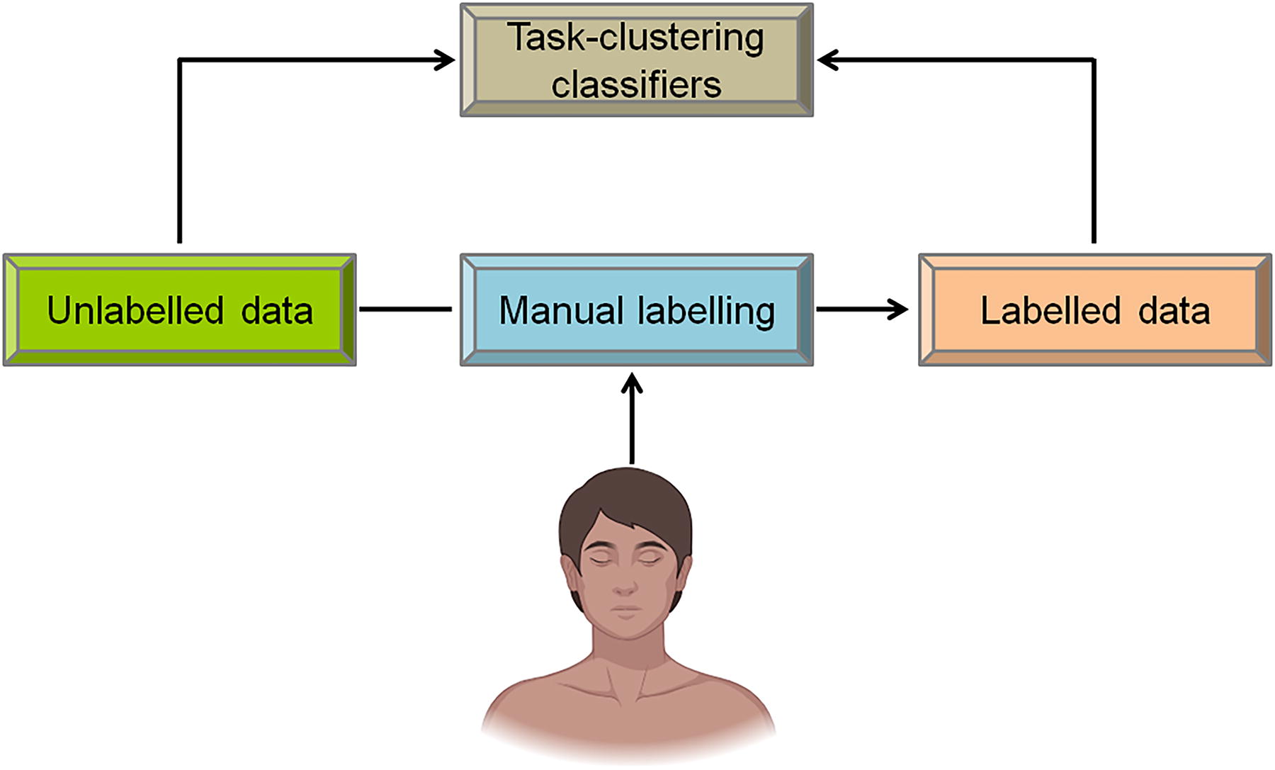
Basic scheme of semisupervised learning.
Artificial Neural Network-Based Skin Cancer Detection Paradigms
A self-generating NN can be used to initially filter out photographs with lesions. 19 Tumor margins, texture, and color are characterized in the second stage. Principal component analysis, which reduces the number of feature dimensions, can make it possible to choose the best features. In skin cancer detection, the effectiveness of the three ANN learning algorithms, Levenberg–Marquardt, robust back-propagation, and scaled conjugate gradient, was also investigated recently. 20 Levenberg–Marquardt’s learning algorithm successfully identified benign lesions even as the epoch number increased. With a sensitivity of 92.6%, the scaled conjugate gradient learning algorithm generated better outcomes in the interim. Furthermore, developing a mole categorization system increased the feasibility of detecting melanoma skin cancer early. 21 The asymmetrical border, color, and diameter (ABCD) rule can be applied to determine how the lesions appeared. Indeed, asymmetry and edges of a lesion can be identified using the Mumford–Shah algorithm and the Harris–Stephen method. 22 Such analyses can be performed on moles to determine their benignity. Thus, it may be benign if a mole is black, cinnamon, or brown. As melanoma moles are often bigger, the diameter threshold for melanoma detection may be set at 6 mm. Back-propagation feed-forward ANN can divide moles into three classes as follows: common, unusual, and melanoma moles. Such automated skin cancer diagnosis systems have been reported. 23 A “maximum entropy thresholding” technique has also been used to classify lesions into carcinogenic and noncancerous using a gray-level co-occurrence matrix for extraction of visual traits. 24 Similarly, the ANN-based classifier resulted in an accuracy rate of about 88%. 25
CNN methods
CNN is frequently used to identify, gather, and organize images as it can generate more complex features such as corners using information pertaining to angles from curves and edges. 26 Before the completely integrated layers in a CNN, there may be numerous convolutional, pooling, and fully connected hidden layers. Figure 4 depicts the working scheme of the CNN architecture for image classification, in which the process begins with a raw image input, preprocessing, augmentation, and normalization. Thereafter, convolutional layers of the CNN framework extract image features, while the pooling layers are used for reducing the dimensionality, and the fully connected layers are involved in the final classification of images. 27 Therefore, CNN brings about effective feature learning and accurate classification in computer vision tasks. 28 Its architecture for image classification includes the input first, followed by several convolutional layers with ReLU activation, pool layers for dimensionality reduction, full convolution layers, followed by fully connected layers with softmax activation and classification labels (Fig. 5). This structure efficiently extracts hierarchical features from images and can be used for accurate classification in DL-based skin cancer diagnoses. In recent years, several studies have used CNNs to identify skin cancer (Table 4). The data can be grouped using techniques such as support vector machine (SVM) and softmax classifiers. Without segmentation, melanoma can be correctly grouped with an accuracy of 82.8% and 85.5% with segmentation. A multiscale CNN from an inception v3 DNN was trained on ImageNet to further aid in the detection of melanoma. Classification of skin cancer was significantly improved using coarse-scale (lesion environment and form as a whole) and finer scale (lesion textual descriptions) parameters for high-resolution images of skin lesions. 29 Diagnosis of skin lesions involves extracting properties from deep CNNs that have already received training. Some of the trained versions used as deep feature generators include Alex Net, ResNet-18, and VGG16. 30 For culminating image sorting, combining all the classifiers’ outputs is required. 31 Such protocols are effective in classifying seborrheic keratosis (SK) and melanoma images with high accuracy. 30 A pretrained version of ResNet-152 was used as the foundation for a deep CNN architecture for grouping 12 different types of skin lesions. Similarly, after extracting features from a trained deep CNN termed AlexNet, an error-correcting output coding SVM has been used for skin lesion classification, resulting in scores for sensitivity and specificity. 32 Identification of squamous cell carcinoma, actinic keratosis, and basal cell carcinoma can have high accuracy rates (of > 85%) using CNN designs. 3
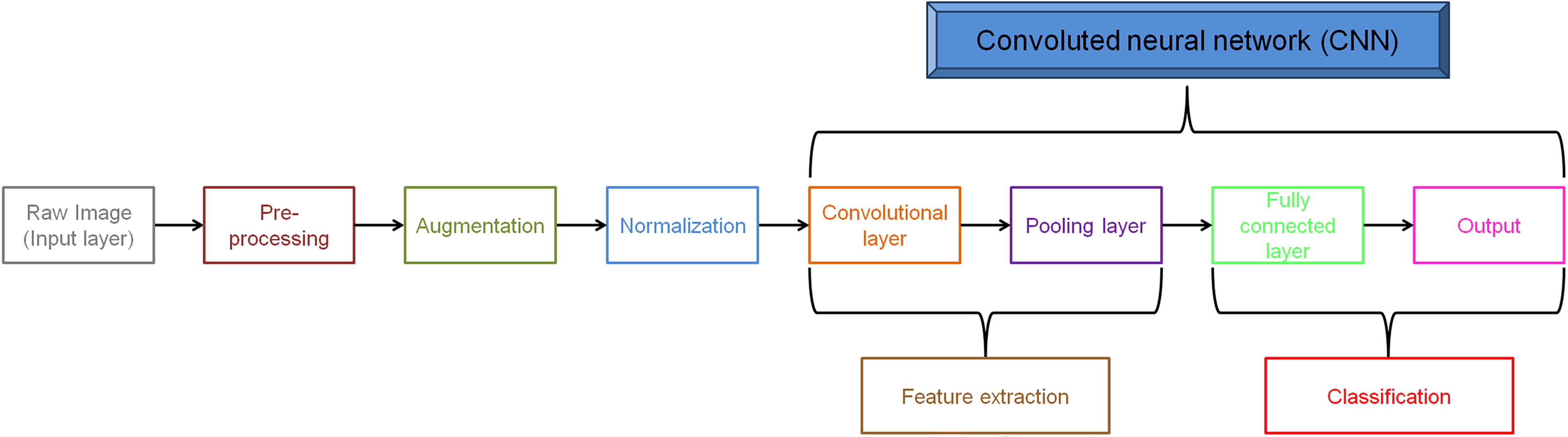
Basic scheme of convolutional neural network (CNN) architecture.
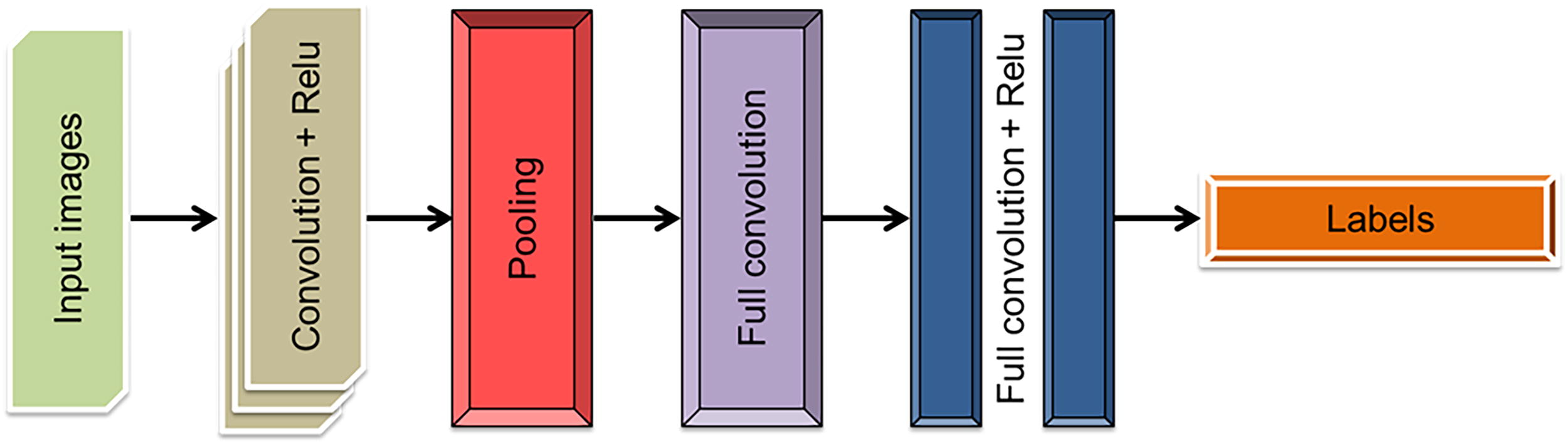
Basic convolutional neural network (CNN) architecture.
A Comparative Analysis of Skin Cancer Detection Using Convolutional Neural Network-Based Approaches
CNN, convolutional neural network; SVM, support vector machine; AUC, area under the ROC curve.
KNN models
Similar to CNNs, KNNs do not rely on external aid because they learn via unsupervised learning, requiring only a basic understanding of the properties of the input data. 33 The two-dimensional plane of KNN typically has two levels, an input layer and an output layer, connected via bridges. 34 Figure 6 illustrates a decision-making process based on KNN. A message is encoded into several embeddings, which are stored in different storages. Each store retrieves a set of nearest neighbors, and the scoring function processes them to decide the final outcome. This structure is typically used in recommendation systems, anomaly detection, and natural language processing applications. Without knowing the relationships between the individuals in the input data set, a KNN can be used to cluster data. 35 Each node in the competitive layer must act as both an input and an output since a KNN lacks a specific output layer. A KNN preserves the topological structure of the initial data space as it transitions from high to low dimensions. The “preservation” distance calculates how evenly distributed the information is in a given area. 36 By mapping the data points farther apart, this method considers the distance between data points in the input data space. Hence, KNN is the best method for handling enormous amounts of data. The generalizability of KNN is another important feature. Unknown incoming data can be recognized and categorized by the network as KKN can map complex, nonlinear connections between data points effectively. 37
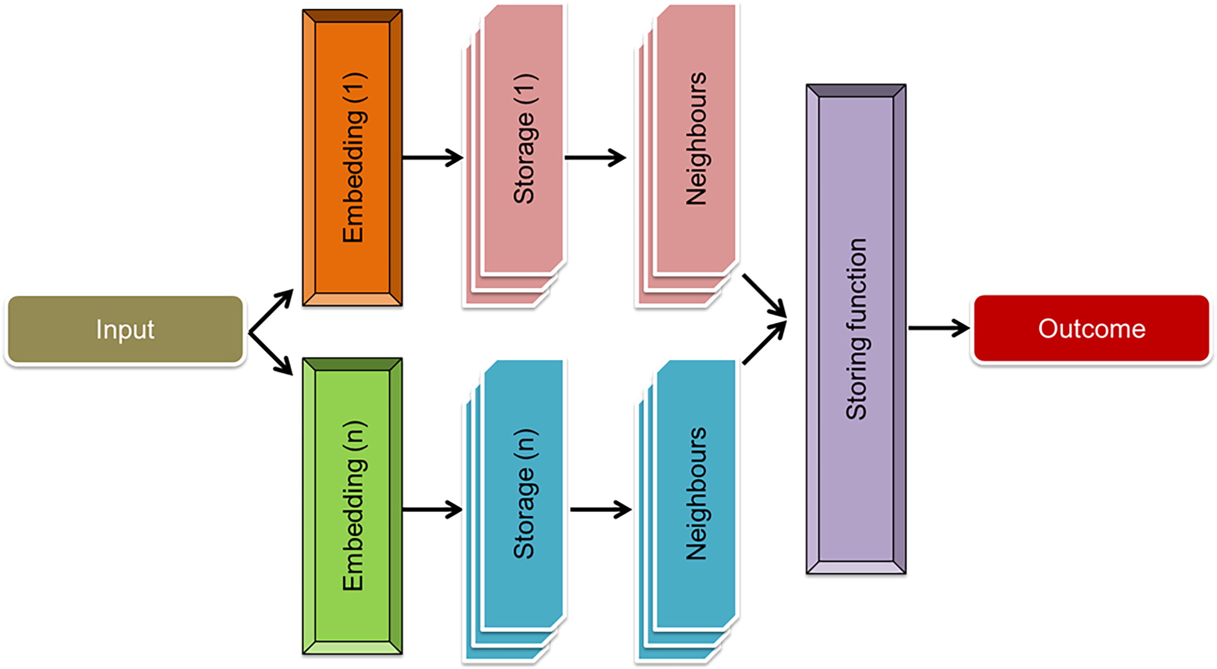
Basic k-nearest neighbor (KNN) architecture.
Due to these advantages, KNNs have been used in many skin cancer detection systems (Table 5). A KNN-based detection was used for synchronized fluorescence spectra from melanoma, nevus, and healthy skin samples to train an NN. The spectra’s high dimensionality was significantly reduced via principal component analysis (PCA).36 Similarly, KNN and ANN have been used to identify melanomas, with the error rate being 3%–4% on the test dataset. 38 Basal cell carcinoma, melanoma, and squamous cell carcinoma may be identified using a self-organizing NN and a radial basis function NN. 39 Skin lesion images have been categorized using color, GLCM, and shape attributes, using KNN, ANN, and Naive–Bayes classifiers. 40 An automated method based on KNN to lessen ambient noise in the filtered images was used to recognize skin cancer with an exceptionally high accuracy. 41 A similar KNN-based method has been compared with SVM, back propagation neural network (BPN), and 3-layer NN algorithms for its utilities in lesion classification performance and found to be highly accurate. 42
A Comparative Analysis of Skin Cancer Detection Using k-Nearest Neighbor-Based Approaches
GAN-based techniques
Deep CNNs such as GAN are powerful DNNs built on the idea of negligible games. Two NNs, discriminator and generator, compete to learn and record available data. 43 The generator module creates highly biased data, and the discriminator component differentiates between fake and real image. 44 Both networks will carry out these tasks repeatedly during training. 45 Figure 7 shows a GAN comprising two networks as follows: the generator network, which takes real images and creates fake images, and the discriminator network, which tries to determine whether an image is real or fake. Each evaluates images from the other, for concerted improvement of the capabilities of image generation and distinguishment of both networks. Overall, the generator becomes better at fooling the discriminator, and the discriminator becomes better at telling the real from the fake. The main benefit of GAN is its capacity to produce fakes, including photorealistic images, with the same data distribution as real samples. 6 It can aid DL in overcoming the deficiency of good training examples. Figure 8 highlights working of a GAN consisting of two NNs, the generator that generates fake samples from noise, and the discriminator that can differentiate real from fake samples. The discriminator is trained using real data as well as generated samples, which is fed back into the system via back-propagation. Both the networks are controlled by loss functions, so that the generator keeps improving the capability of generating real samples with time.

Basic generative adversarial network (GAN) architecture.
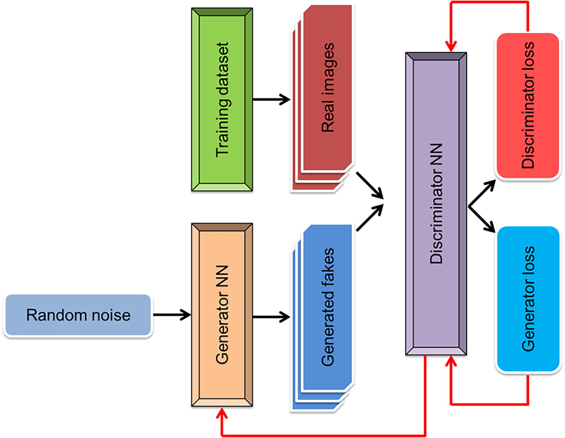
Working scheme of the generative adversarial network (GAN)-based image differentiation.
Studies have evaluated the effectiveness of GAN models in skin lesion detection (Table 6). For example, a GAN-based technique was used for training to generate fake images resembling real skin lesions. The CNN-based discriminator module was then used as a classifier for categorizing skin lesions into seven different categories. 46 GAN models have also been compared with benchmarks such as ResNet-50 and DenseNet for their effectiveness in the detection of skin cancer and found to result in increased accuracy of classifying dermoscopy images as melanoma, SK, or nevus. 47 Ahmad et al. have suggested a new approach for characterizing skin lesions using self-attention progressive GAN with significantly increased accuracy of detection. 48
A Comparative Analysis of Skin Cancer Detection Using Generative Adversarial Network-Based Approaches
Databases
Reliable image collection is indispensable for evaluating skin cancer diagnostic effectiveness of ML/DL techniques. 49 However, insufficiency of data types and numbers make this difficult. This section discusses the datasets of lesion images, which can be used for skin cancer detection. Research incorporating new datasets should fill important gaps regarding dataset variability and patient representation to enhance technological reliability, clinical applicability, and predictive accuracy for DL models that detect skin cancer.
PH2
Dermoscopic images (∼200 typical and atypical nevi, and melanoma) of PH2 dataset 49 are based in Pedro Hispano Hospital Dermatology Center, Portugal. The pictures are taken by Tuebinger-Mole-Analyzer at 20 × magnification under similar illumination. The images in PH2 are 768 × 560 pixels with 8 bit of each RGB color. Images can be evaluated for features such as stripes, colors, melanin networks, regression zones, and blue-white veil globules. 50
International Skin Imaging Collaboration
The ISIC repository is another database initiated in 2016 at the International Symposium on Biomedical Imaging. 42 Training and testing images of benign nevi, SH, and malignant melanomas are organized separately. 51 ISIC regularly updates its image library, and includes additional information such as patient demographic data.
Dermatology Information System
Dermatology information system (DermIS) 52 was created as a collaborative effort of the Universities of Erlangen and Heidelberg. It contains >6500 lesion images and has been used to create two new online image atlases for dermatology and pediatric dermatology.
DermQuest
DermQuest dataset 53 harbors images in excess of 22,000. It has lesion tags for images; however, it was transferred to Derm101 in 2018, and since December 31, 2019, it is no longer in use.
Ham10000
The HAM10000 dataset 42 includes >10,000 dermatoscopic images of numerous lesion types, enabling researchers to create durable predictive models to properly identify disorders in underrepresented groups, including people with dark skin.
DermNet NZ
The DermNet NZ database 54 expands DL model diagnostic abilities through its extensive coverage of dermatological conditions, including complex and rare lesions, even though skin cancer remains its secondary focus.
MED-NODE
The MED-NODE dataset 55 functions as an essential resource for algorithm testing under controlled conditions because of its precise melanoma focus and its emphasis on high-quality data curation.
PAD-UFES-20
PAD-UFES-20 dataset 56 that includes clinical images taken from smartphones, alongside dermoscopic photographs of multiple skin conditions, may boost model generalization by presenting algorithms to different lesion characteristics among varied patient groups.
XiangyaDerm
XiangyaDerm database 57 stores >150,000 skin images, mainly from individuals of Asian ethnicity. In addition to adding racial diversity for skin cancer diagnoses, the repository in XiangyaDerm encompasses >570 skin diseases and has been shown to efficiently aid in the identification of multiple dermal conditions, including psoriasis. 58
Discussion
Recent progresses in the applications of DL algorithms for skin lesion detection/categorization are commendable. The DL methodologies achieved exceptional diagnostic effectiveness in detecting skin lesions through automated scalable and noninvasive solutions. CNNs have demonstrated exceptional performance in handling high-resolution pictures while finding intricate features that particularly suit image-based classification needs. GANs resolve the elimination of medical data by creating artificial images to develop stronger training abilities as well as exemplary model results. Overall, DL models eliminate human dependency in diagnostic work, which may enhance error-free standardized outcomes that match data from different sources. Early skin cancer detection benefits substantially from these techniques because they excel at recognizing minor skin lesion patterns to enhance patient treatment results.
However, there are significant obstacles that must be addressed before widespread clinical implementation of DL methods for skin cancer diagnoses. A notable hurdle is the requirement for time- and cost-intensive extensive training, which in turn relies on powerful software, extensive hardware, and computing facilities. 59 These are all challenging for clinicians and hospital staff, particularly considering their cost-ineffectiveness, hindering their widespread use under clinical settings. Computational needs are among the major drawbacks of DL models because effective deployment and technical know-how require advanced hardware and software systems, which limit their accessibility, especially in resource-limited areas. Another critical aspect is that these models suffer from the absence of diverse and high-resolution images that can severely hamper the generalizability of the models. Lesions can be quite heterogeneous and come have different geometries and sizes, and many images lack diagnostic clarity and resolution. This is a significant challenge for detecting small lesions (1–2 mm). 60 In fact, lesion heterogeneity with regard to their geometries, sizes, and background is a significant hurdle that compromises the accuracy of detection/prediction results. Identification of lesion shapes is equally critical. 61 Similarly, skin color is a critical issue as most databases are overrepresented with fair-skinned subjects. Indeed, databases lean toward a more favorable complexion population and neglect the darker complexion population, affecting the diagnostic precision widespread use across different populations. Hence, accurate training of detection systems and identification of lesions in individuals of color need recognition of background skin color. 62 This, in turn, requires larger samples of both dark- and light-skinned subjects to be included in the databases. In addition, available dermoscopy images of these rare types of skin cancer and age group information are not completely available. Most of these limitations must be addressed while taking DL-based diagnostic systems ahead. Likewise, the insufficiency of dermoscopy images of rare lesion types in databases is another limitation. 51 Another point of concern is the age of subjects; several skin cancer types, such as Merkel, basal, and squamous cell carcinomas, are more prevalent in people older than 65 years. 63 An adequate number of lesion images from this age group is required for effective NN-based training and detection. Similarly, skin cancer types are strongly influenced by a plethora of environmental and genetic factors, 64,65 which must be represented and taken into account while using DL/ML models. Finally, patient data privacy and unconsented sharing of lesion images are another issue that must be addressed for implementation of DL methods for skin cancer diagnoses.
Many NNs exist as uninterpretable “black boxes” that prevent clinicians from understanding the basis of their diagnostic decisions. Model robustness faces challenges because data reliability problems lead to degraded performance, while encounters with uncommon lesion presentations present algorithm difficulties. The evaluation of DL techniques shows their ability to reinvent skin cancer diagnosis. Yet, additional work on dataset biases alongside computational hurdles and interpretability barriers is required to achieve broad clinical acceptance and equitable health care results.
Conclusion and Future Perspectives
The review discusses the ability of various DL-based NN algorithms, mainly ANN, CNN, KNN, and GAN, to identify/categorize skin cancer lesions. Such characterizations are particularly interesting as they are noninvasive and do not require surgery or biopsy. The benefits are accuracy in diagnosis, auto-categorization of lesions, and minimization of reliance on interventional procedures and human subjection. Using preprocessing, image segmentation, feature extraction, and classification techniques of ML/DL, it is possible to identify and characterize skin cancers with high accuracy. While each algorithm has benefits and drawbacks, it is crucial to choose the best classification strategy for optimum performance, and develop it further. Because CNN is more closely related to computer vision than other NN types, it is more effective at classifying images and predicting whether a lesion is cancerous or not. While CNNs are highly effective for image-based lesion classification, GANs can be beneficial with a dearth of data by generating synthetic images. Hence, DL models using CNNs alongside GANs may demonstrate a stronger potential for delivering precise noninvasive skin cancer diagnoses. DL method advancement must include work on diverse datasets that use high-resolution inputs while implementing unsupervised learning methods to maximize feature detection ability and overall model consistency. Unsupervised ML techniques such as auto-organization may be used to find features, connections, or patterns in image datasets to enhance the accuracy of their feature representations. 66 Such image processing systems could be improved through research so that even minute features may be identified. In addition, it is essential to improve computational efficiency and minimize dataset biases to achieve broader clinical deployment. Developing interpretable automated models may enhance acceptance of the prediction results by medical practitioners and clinicians, thereby extending the use of computerized systems for actual clinical implementation. In conclusion, despite the significant progresses, further steps to address the challenges associated with diversity in the datasets, computational complexity, and underrepresentation of some populations, are warranted to diversify image acquisition and improve the protocols.
Footnotes
Acknowledgments
The authors gratefully acknowledge the funding of the
Authors’ Contributions
S.H.: Conceptualization, methodology, formal analysis, investigation, resources, writing—original draft, writing—review and editing, visualization, supervision, and project administration. F.A.: Conceptualization, methodology, formal analysis, investigation, writing—original draft, writing—review and editing, and visualization. V.S.: Conceptualization, methodology, formal analysis, investigation, writing—original draft, writing—review and editing, and visualization. D.M.M.: Conceptualization, methodology, formal analysis, investigation, writing—original draft, writing—review and editing, and visualization. A.B.: Conceptualization, methodology, formal analysis, investigation, writing—original draft, writing—review and editing, and visualization.
Disclosure Statement
The authors declare no conflicts of interests.
Funding Information
Deanship of Graduate Studies and Scientific Research, Jazan University, Saudi Arabia – Project No. RG24-L06.