
Abstract
Objectives:
To prepare a novel 68Ga-labeled pH (low) insertion peptide-like peptide, YJL-11, and study its ability to be used as a probe for the diagnosis of triple-negative breast cancer (TNBC) via in vivo imaging of tumor-bearing nude mice.
Methods:
Circular dichroism (CD) analysis of YJL-11 was performed to assess its secondary structure. YJL-11 was labeled with 68Ga, and the in vivo biodistribution of 68Ga-YJL-11 in MDA-MB-231 xenograft mice was evaluated. This probe was then applied for small animal positron emission tomography (PET) imaging of tumor-bearing nude mice.
Results:
CD analysis of YJL-11 confirmed a typical pH-dependent transition in its secondary structure. The radiochemical yield of 68Ga-YJL-11 was 75.5 ± 0.25%, and the radiochemical purity was 95.75 ± 0.15%. Biodistribution studies showed that the tumor uptake of 68Ga-YJL-11 was significantly higher than in the control group, 1 and 2 h after injection. Small animal PET imaging results were consistent with the biodistribution data, showing clear images of the tumors and livers 1 and 2 h after injection of 68Ga-YJL-11, whereas tumors were not detected in the control group.
Conclusion:
68Ga-YJL-11 was prepared with high radiochemical yield and can target TNBC tissues, indicating that it has great potential in the diagnosis of TNBC.
Introduction
The incidence of breast cancer has continued to rise, surpassing the incidence of lung cancer to become the number one cancer worldwide in some recent years. 1 Triple-negative breast cancer (TNBC) is a high-risk breast cancer subtype. Compared with other types of breast cancer, TNBC is more likely to metastasize and has a higher risk of recurrence along with a poorer prognosis. 2 Thus, diagnosing TNBC early is critical to reduce the difficulties with its treatment and improve both the cure rate and prognosis. TNBC is likely to occur in young women, 3 as the breast tissue in this population is usually denser. The diagnostic performance of traditional imaging examinations for TNBC is not adequate, which results in missed diagnoses and delayed patient treatment. Despite the widespread use of fluorine-18 fluorodeoxyglucose (18F-FDG) positron emission tomography/computed tomography (PET/CT) in tumor imaging, there is heterogeneity in the uptake of 18F-FDG in breast cancer. Studies have shown that the uptake of 18F-FDG in MDA-MB-231 TNBC tumors is only 2.3 ± 0.0%ID/g 1 h after injection. 4 Additionally, some inflammatory lesions may also result in high uptake of 18F-FDG, leading to false positive results. 5 Radiolabeled antibodies such as bevacizumab demonstrate effective tumor targeting; however, their clinical translation is hampered by high production costs, potential immunogenicity, and extended blood circulation leading to elevated background signals. 6,7 Similarly, while prostate-specific membrane antigen (PSMA) tracers show promise for TNBC imaging, their utility is challenged by heterogeneous target expression and off-target accumulation. 8 Therefore, there is an urgent need to develop an examination method that can diagnose TNBC.
The pH (low) insertion peptide (pHLIP) family is derived from the C-helix of the protein bacteriorhodopsin, 9 it is a novel carrier capable of targeting the acidic tumor microenvironment (TME). The molecular mechanism of targeting the TME relies on the negatively charged pHLIP becoming electrically neutral in the low pH environment, leading to the formation of an α-helical secondary structure, which subsequently allows its C-terminus to insert into the tumor cell membrane. 10 –17 The pHLIP family has been applied in targeted research focusing on breast cancer, prostate cancer, melanoma, and pancreatic cancer. 18 –23 The TME of TNBC is acidic, with a pH range of 6.2–7.0. 24 In our previous studies, 25 –30 the pHLIP family was used as vectors to target MDA-MB-231 TNBC cells. We showed that this family of peptides can target tumor cells, but their affinity for tumor cells needed further improvement.
Therefore, to enhance the tumor-targeting abilities of these peptides, we performed an in-depth analysis of pHLIP variant 3 (var3), a peptide that shows relatively good tumor-targeting ability, via a template-assisted method and modified the amino acid sequence of its α-helical region to obtain the novel pHLIP-like peptide YJL-11. 31 In this study, 68Ga-labeled YJL-11 was used for PET/CT imaging to investigate its ability to detect TNBC.
Materials and Methods
Main instruments
The following instruments were utilized in this study: Shimadzu LCMS-2020 system (Shimadzu Corporation, Japan), Chirascan plus ACD spectropolarimeter (Applied Photophysics Ltd., UK), CRC-55tR radiopharmaceutical dose calibrator (Capintec Inc., USA), Wizard 1480 automatic gamma counter (PerkinElmer Life Sciences Inc., USA), 20 mCi 68Ge/68Ga generator (Obninsk Cyclotron Co., Ltd., Russia), Scan-RAM+PS Radio-TLC scanner (LabLogic Systems Ltd., UK), and Inveon small animal PET scanner (Siemens Medical Solutions USA Inc., USA).
Major reagents
The primary reagents employed in this study included isoflurane (Shenzhen RWD Life Technology Co., Ltd., China) and hydrochloric acid (Merck KGaA, Germany).
Cell lines and experimental animals
Human breast cancer MDA-MB-231 cells, obtained from the Cell Bank of the Chinese Academy of Sciences, were cultivated in high-glucose Dulbecco’s modified eagle medium supplemented with 10% fetal bovine serum and a 1% penicillin-streptomycin mixture. The cells were incubated at 37°C in an atmosphere containing 5% CO2. For the establishment of the MDA-MB-231 tumor model, 4–5-week-old female specific pathogen-free BALB/c nude mice, obtained from Saiye Biological Technology Co., Ltd., China [license number: SCXK (Su) 2018-0003], were utilized. A suspension containing 1 × 10⁶ MDA-MB-231 cells (composed of 50 µL of phosphate-buffered saline and 50 µL of Matrigel, totaling 0.1 mL) was administered subcutaneously into the right axilla of each mouse. The animal studies were conducted with the approval of the ethics committee of Qingdao University and the management of the animals in compliance with ethical guidelines.
Design and characterization of the peptide YJL-11
The α-helical region of the template peptide pHLIP (var3) was analyzed with the PEP-FOLD server (https://mobyle.rpbs.univ-paris-diderot.fr/). 32,33 The analysis revealed that the helical region of pHLIP (var3) can be divided into two parts (Table 1): the helix region 1 and the helix region 2. The amino acid composition of the helix region 1 was as follows: positively charged amino acid R2; negatively charged amino acid D6; and nonpolar amino acids W1, A3, Y4, L5, and L7. Additionally, the amino acid composition of the helix region 2 was as follows: negatively charged amino acids D12 and D17; nonionized polar amino acid T13; and nonpolar amino acids L14, L15, L16, and L18. There are many types of nonpolar amino acids in the helix region 1 of pHLIP (var3); therefore, we simplified the types of nonpolar amino acids in this region and replaced both A3 and Y4 with a more hydrophobic amino acid (L) to obtain the novel peptide sequence YJL-11.
Basic Bioinformatics Parameters of the Peptides
pHLIP, pH (low) insertion peptide; sOPEP, the optimized potential for efficient structure prediction.
The secondary structure of YJL-11 was predicted by the PEP-FOLD server. The online tool HeliQuest (http://heliquest.ipmc.cnrs.fr/) was used to analyze the hydrophobic moment of the α-helical region of this peptide. The LogP values of the peptides were assessed using Chem3D software.
Synthesis and purification of the peptides
The peptides YJL-11 (WRLLLDLLFPTDTLLLDL-NH2) and the control peptide kVar7 34 (WARYLKWLFPTKTLLLKL-NH2) were synthesized using standard Fmoc solid-phase peptide synthesis (SPPS) methodology at Shanghai Science Peptide Biological Technology Co., Ltd. The synthesis was performed on Rink amide MBHA resin (loading capacity: 0.45–0.55 mmol/g) using N,N-dimethylformamide (DMF) as the primary solvent. Fmoc-protected amino acids were coupled using N,N’-diisopropylcarbodiimide and hydroxybenzotriazole as coupling reagents. Each coupling reaction was conducted for 2 h at room temperature. Fmoc deprotection was achieved using 20% piperidine in DMF (v/v) for 20 min. After synthesis completion, the crude peptides were cleaved from the resin using a mixture of trifluoroacetic acid (TFA)/triisopropylsilane/H2O (95:2.5:2.5, v/v/v) for 2 h at room temperature.
Additionally, the N-terminal amino acids of YJL-11 and kVar7 were modified with the chelator 1,4,7-triazacyclononane-N,N′,N″-triacetic acid (NOTA) (Supplementary Data S1) via a condensation reaction, and the C-terminal amino acids were subjected to amidation. Specifically, the peptides were connected to paraisothiocyanatobenzyl NOTA via a 6-aminohexanoic acid linker.
NOTA-YJL-11 and NOTA-kVar7 were purified via high-performance liquid chromatography (HPLC) on a Shimadzu LC-20AT system (Shimadzu Corporation, Japan) at room temperature. The chromatographic conditions were as follows: column, Agela C18 analytical column (250 × 4.6 mm, 5 μm); mobile phase, solvent A (0.05% TFA + 2% acetonitrile) and solvent B (0.05% TFA + 90% acetonitrile); gradient elution was performed from 0∼18 min with 42%–60% B; flow rate, 1.0 mL/minute; and detection wavelength, 220 nm. The peptides were analyzed using a Shimadzu LCMS-2020 system (Shimadzu Corporation, Japan) for low resolution mass spectrometry (LRMS) analysis. The observed [M + H]+ peaks confirmed the identity of the target compounds.
CD spectroscopy
YJL-11 (0.1 mg/mL) and 1-palmitoyl-2-oleoyl-sn-glycero-3-phosphocholine (POPC; diameter ≤50 nm, 2 mmol/L) were dissolved in 10 mmol/L phosphate buffer (pH = 8) and equilibrated at room temperature for at least 10 h. The pH value of the solution was adjusted to 4.0 with 0.1 mol/L HCl.
The circular dichroism (CD) spectrum of YJL-11 was measured with a Chirascan plus ACD spectropolarimeter at pH values 8.0 and 4.0. The determination conditions were as follows: quartz sample cell optical path length, 1 mm; scanning wavelength range, 190–260 nm; wavelength step size, 1 nm; time per point, 0.5 s; and temperature, 25°C.
Preparation and purification of the 68Ga -labeled peptides
The 68Ge/68Ga generator was eluted with 5 mL of 0.1M HCl, the eluent was collected as fractions with a volume of 0.5 mL per fraction, and the fraction with the highest radioactivity was selected for subsequent labeling. First, 500 μL of 0.1 M sodium acetate buffer (pH = 4.5), 190.5 MBq/500 μL of 68Ga eluent, and 80 µL of 0.1 mg/mL NOTA
Measurement of radiochemical purity was performed by an Agilent 1200 radio-HPLC system (Agilent Technologies Inc., USA) with mobile phase A (0.1% aqueous TFA) and mobile phase B (acetonitrile) with gradient elution from 0 to 15 min with 95%–5% A (5%–95% B) at a flow rate of 1 mL/min. The retention time of the radioactive peak was noted. All radiochemical terminology and calculations strictly adhere to the Consensus Nomenclature Rules for Radiopharmaceutical Chemistry. 35
Serum stability and protein binding
Two hundred microliters of the 68Ga-labeled peptide (18.5 MBq) was mixed with 1.0 mL of fresh mouse serum for incubation at 37°C, and the radiochemical purity was determined by Scan-RAM+PS TLC scanner (LabLogic Systems Ltd., UK) after 30, 60, 120, and 240 min.
The radiotracer, with an activity of 0.37 MBq in a volume of 100 µL, was incubated in 1.0 mL of mouse serum at 37°C under constant stirring at 600 rpm. Aliquots of 250 µL of the serum were taken at 30, 60, 120, and 240 min, and each was transferred to a 1.7 mL Eppendorf tube containing 250 µL of ice-cold acetonitrile. The resultant mixture was vortexed for 1 min, followed by centrifugation at 13,000 rpm for 5 min. The supernatant was then pipetted into another 1.7 mL Eppendorf tube and centrifuged again at 13,000 rpm for 2 min. Both the supernatant and sediment were collected individually, and their radioactivity was measured using a gamma counter to ascertain the radiotracer’s binding to serum proteins.
In vivo biodistribution evaluation
When their tumor diameters reached approximately 1.0 cm, 12 tumor-bearing nude mice were selected for in vivo biodistribution experiments. Each nude mouse was injected with approximately 1.85 MBq/200 μL 68Ga-labeled YJL-11 or kVar7 via the tail vein. The mice were humanely sacrificed 1 or 2 h after injection, and tissues and organs, such as brain, heart, lungs, liver, kidney, stomach, small intestine, blood, muscle, bone, and tumors, were collected and weighed, and their radioactive counts were determined. The results are expressed as a percentage of the injected dose per gram of tissue (%ID/g).
Small animal PET imaging
When their tumor diameters reached approximately 1.0 cm, 6 tumor-bearing nude mice were selected for imaging. Each nude mouse was injected with approximately 7.4 MBq/200 μL 68Ga-labeled YJL-11 or kVar7 via the tail vein. The tumor-bearing mice were anesthetized with 2% isoflurane 1 and 2 h after injection, and static 10-min PET and whole-body CT with moderate resolution were performed.
Statistical analysis
SPSS 24.0.0.0 software (IBM) was used to process the data. Quantitative data are expressed as the mean ± standard deviation. Variables were compared via one-way analysis of variance. A p-value <0.05 was considered to indicate statistical significance.
Results
Bioinformatics analysis of the novel peptide
Upon analysis by the PEP-FOLD server, it was revealed that the novel peptide YJL-11 (Fig. 1) could be divided into a helix region 1, a hinge region, and a helix region 2. The amino acid composition of helix region 1 was as follows: a positively charged amino acid R2; a negatively charged amino acid D6; and nonpolar amino acids W1, L3, L4, L5, and L7. Additionally, the amino acid composition of the hinge region was as follows: nonpolar amino acids L8, F9, and P10; and the nonionized polar amino acid T11. Moreover, the amino acid composition of helix region 2 was as follows: negatively charged amino acids D12 and D17; the nonionized polar amino acid T13; and nonpolar amino acids L14, L15, L16, and L18. The energy of the coarse-grained model of YJL-11 (the optimized potential for efficient structure prediction [sOPEP] energy) was −40.4108, which was lower than the energy values of pHLIP (var3) (−36.2973) and the peptide YJL-4 (−32.2099; Table 1). 25
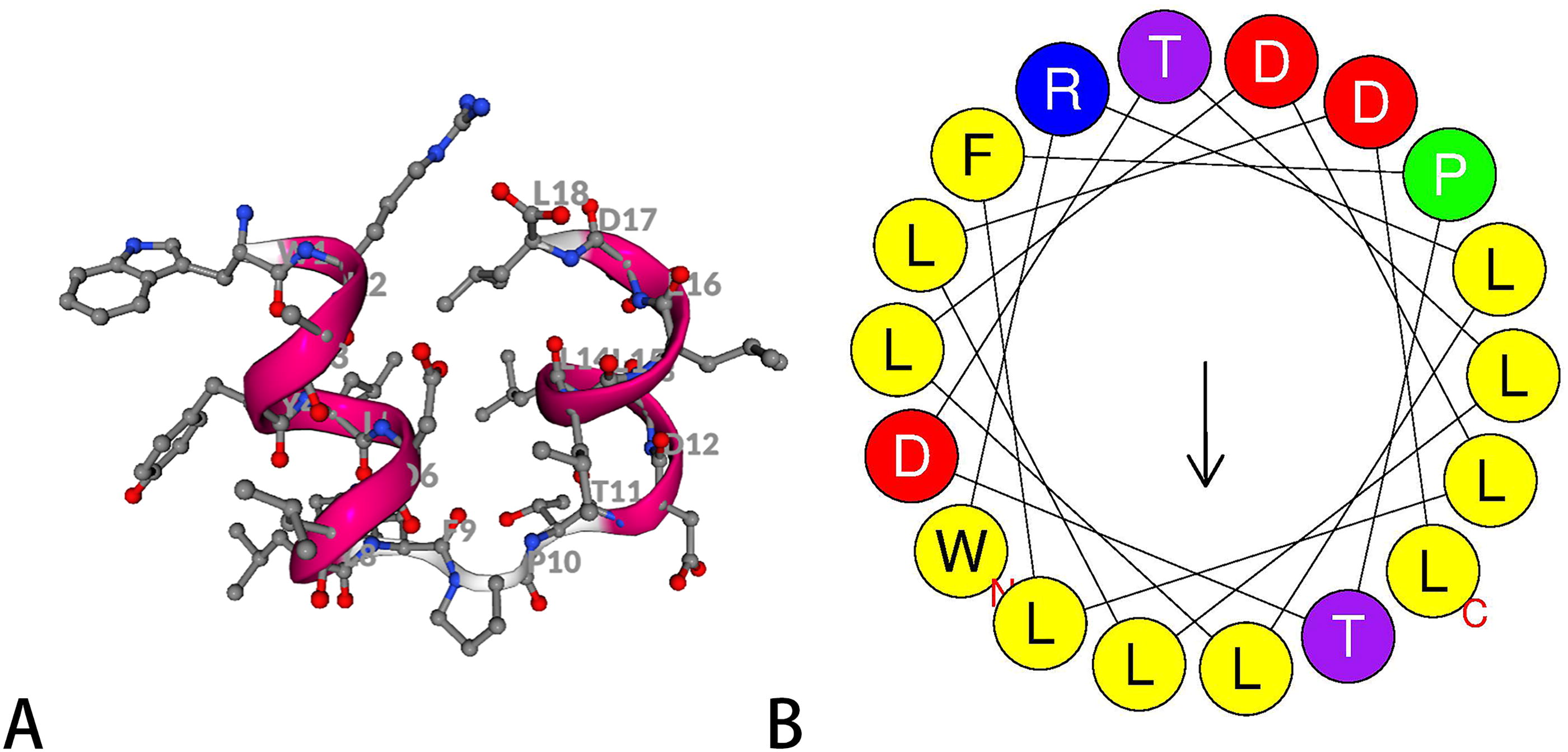
Analysis by the HeliQuest online tool revealed that the hydrophobic moment of the YJL-11 α-helix region was 0.399 µH, which was higher than the hydrophobic moments of pHLIP (var3) (0.389 µH) and YJL-4 (0.358 µH; Table 1). The Chem3D analysis indicates that the LogP values for YJL-11, YJL-4, and pHLIP (var3) are 3.8709, 1.2979, and 2.5031, respectively (Table 1).
Synthesis and purification of the peptides
NOTA-YJL-11 and the control peptide NOTA-kVar7 were successfully synthesized via SPPS. HPLC analysis of NOTA-YJL-11 revealed one main peak (95.24%, 13.08 min) and three smaller impurity peaks, and mass spectrometry analysis of the product revealed one peak at m/z 2421.45 (Supplementary Data S2). The molecular mass was confirmed by LRMS (ESI+): m/z [M + H]+ calcd. for C111H194N26O33: 2420.92; found: 2421.45 (Shimadzu LCMS-2020).
RP-HPLC analysis of the control peptide NOTA-kVar7 revealed one main peak (99.07%, 10.12 min) and three smaller impurity peaks, and mass spectrometry analysis of the product revealed one peak at m/z 2441.01 (Supplementary Data S3).
CD spectroscopy
The secondary structure of YJL-11 was evaluated by CD spectroscopy at pH values of 8.0 and 4.0. In the presence of POPC, when the pH was decreased from 8.0 (blue line) to 4.0 (red line), YJL-11 exhibited a typical pH-dependent transition from structureless to adopting an α-helical conformation (Fig. 2).
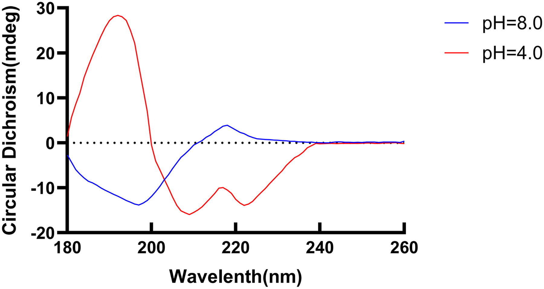
CD spectrum of YJL-11 at pH values of 4.0 and 8.0. CD, circular dichroism.
Labeling the peptides with 68Ga
Following purification, the mean activity of 68Ga-YJL-11 was determined to be 143.86 ± 0.48 MBq (n = 3), while 68Ga-kVar7 exhibited an average activity of 138.15 ± 0.86 MBq (n = 3). The radiochemical yields of 68Ga-YJL-11 and 68Ga-kVar7 were 75.5 ± 0.25% (n = 3) and 72.5 ± 0.45%(n = 3), respectively. Radio-HPLC analysis of the purified products revealed that the radiochemical purity of 68Ga-YJL-11 was 95.75 ± 0.15%, which appeared with a retention time of 11.51 min (Supplementary Data S4). Moreover, the radiochemical purity of 68Ga-kVar7 was 96.1 ± 0.5%, with a retention time of 7.03 min. The specific activities of 68Ga-YJL-11 and 68Ga-kVar7 were 17.98 ± 0.06 MBq/μg and 17.27 ± 0.11 MBq/μg, respectively.
Serum stability and protein binding
68Ga-YJL-11 and 68Ga-kVar7 showed reliable stability. After incubation in mouse serum at 37°C for 30, 60, 120, and 240 min, the radiochemical purity of 68Ga-YJL-11 was 98.63 ± 0.25%, 97.5 ± 0.27%, 96.51 ± 0.21%, and 96.18 ± 0.19%, respectively (Fig. 3; Supplementary Data S5). Similarly, the radiochemical purity of 68Ga-kVar7 was 97.8 ± 0.31%, 96.9 ± 0.26%, 95.8 ± 0.21%, and 95.04 ± 0.23%, respectively (Fig. 3).
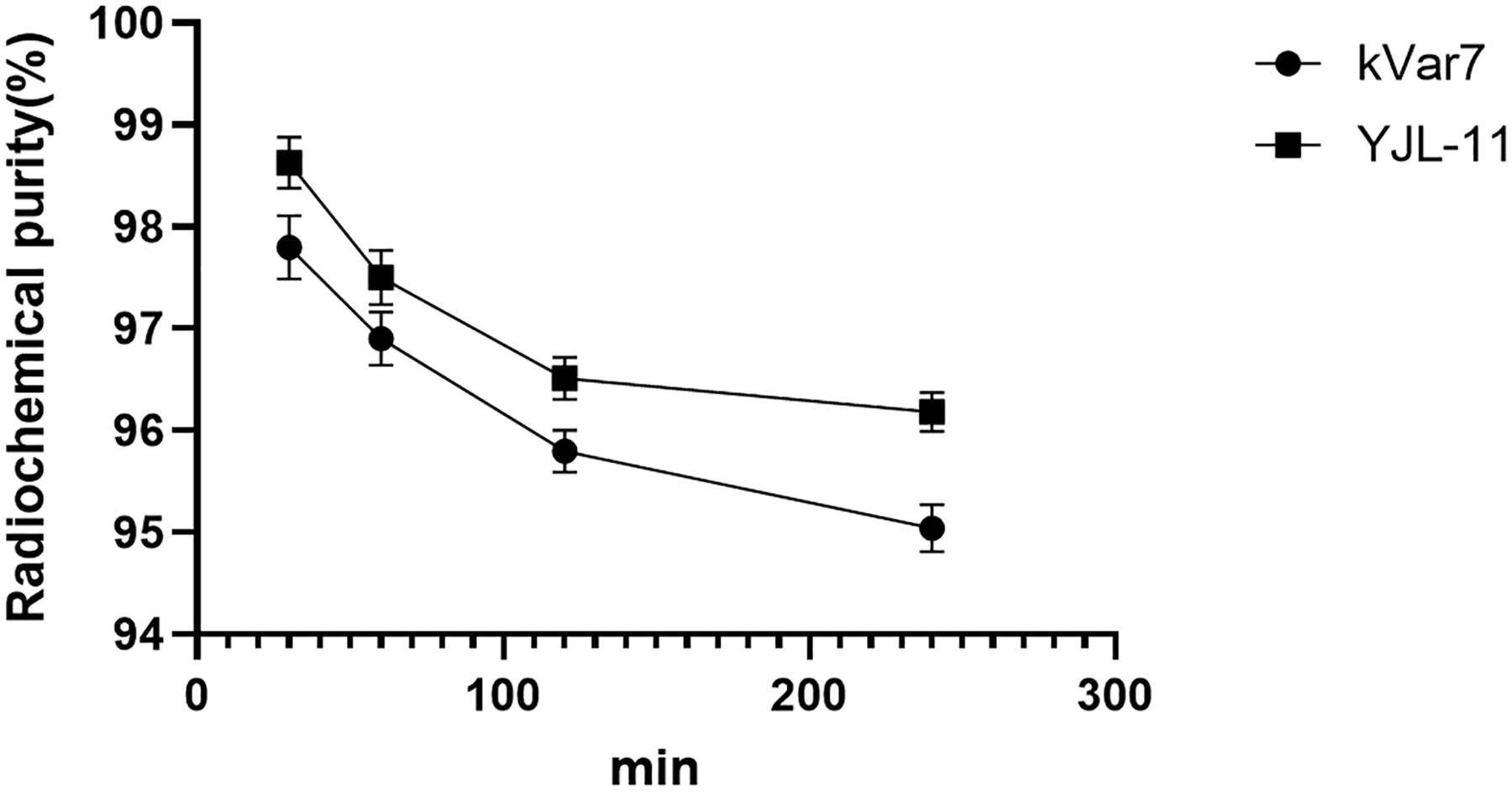
In vitro stability of 68Ga−YJL-11 and 68Ga-kVar7.
Figure 4 displays the serum protein binding rates for both 68Ga-YJL-11 and 68Ga-kVar7. The binding rates for 68Ga-YJL-11 were found to be 22.95 ± 1.85% at 30 min, 22.65 ± 1.25% at 60 min, 22.61 ± 1.30% at 120 min, and 22.87 ± 1.75% at 240 min. In addition, the binding rates for 68Ga-kVar7 were 26.28 ± 2.18%, 26.21 ± 1.65%, 25.79 ± 1.47%, and 26.16 ± 1.36% at the respective time points.
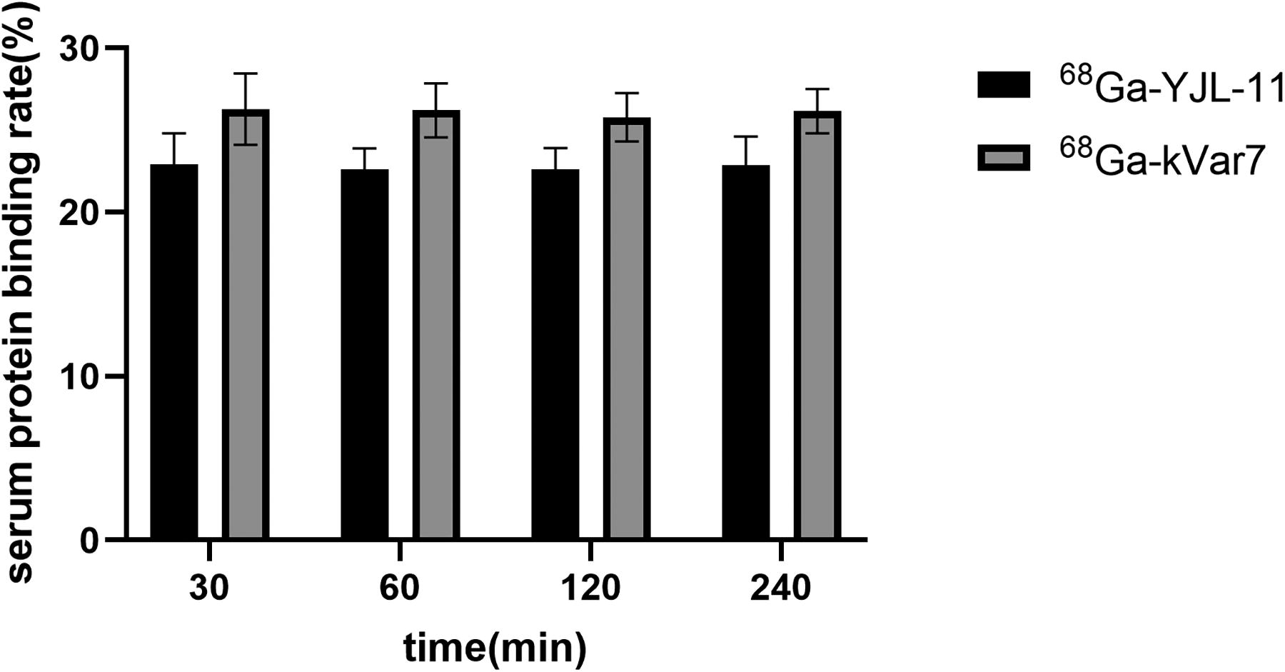
Serum protein binding rates for both 68Ga-YJL-11 and 68Ga-kVar7.
In vivo biodistribution
One and two hours after the 68Ga-labeled peptide (1.85 MBq/200 μL) was injected into tumor-bearing nude mice via the tail vein, the distributions of 68Ga-YJL-11 in the tumors were 8.94 ± 2.2%ID/g and 6.65 ± 1.75%ID/g, respectively, which were significantly higher than the distributions of 68Ga-kVar7 in the tumors (p < 0.01; Figs. 5 and 6). The tumor/nontumor ratio in the 68Ga-YJL-11 group was higher than in the 68Ga-kVar7 group at each time point (p < 0.01; Fig. 7; n = 3). In addition, the distribution of 68Ga-YJL-11 in the liver was relatively high, with values of 7.49 ± 1.64%ID/g and 5.46 ± 1.98%ID/g at 1 and 2 h after injection, respectively. Moreover, 68Ga-kVar7 was very highly distributed in the liver 1 and 2 h after injection (14.31 ± 2.93 and 9.77 ± 2.87%ID/g, respectively). Compared with 68Ga-YJL-4, 68Ga-YJL-11 exhibited no statistically significant difference in tumor distribution (p > 0.05); however, its distribution in the liver was significantly decreased (p < 0.05; Table 2).
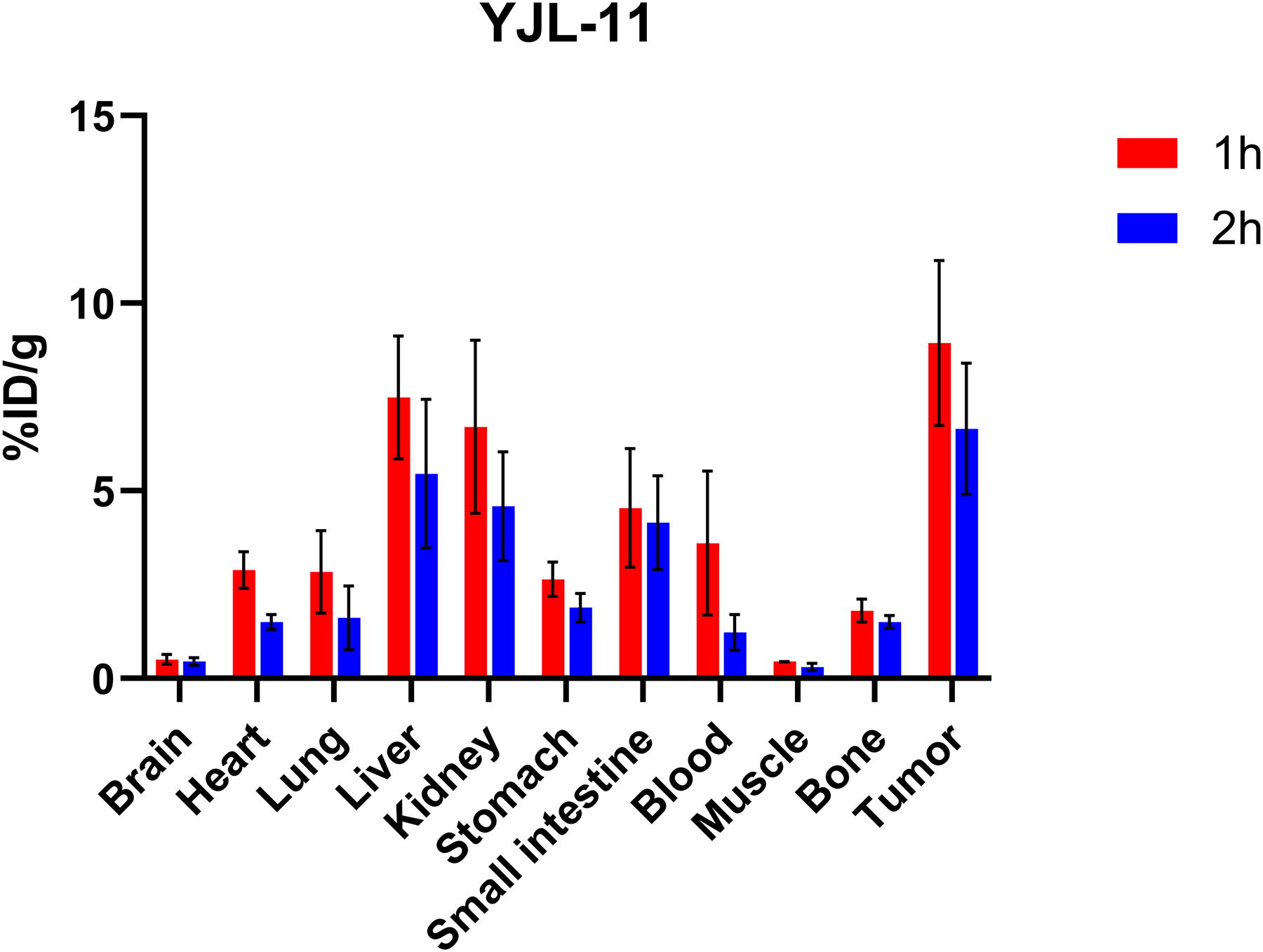
Biodistribution of 68Ga-YJL-11 in mice with MDA-MB-231 tumors (n = 3).

Biodistribution of 68Ga-kVar7 in mice with MDA-MB-231 tumors (n = 3).
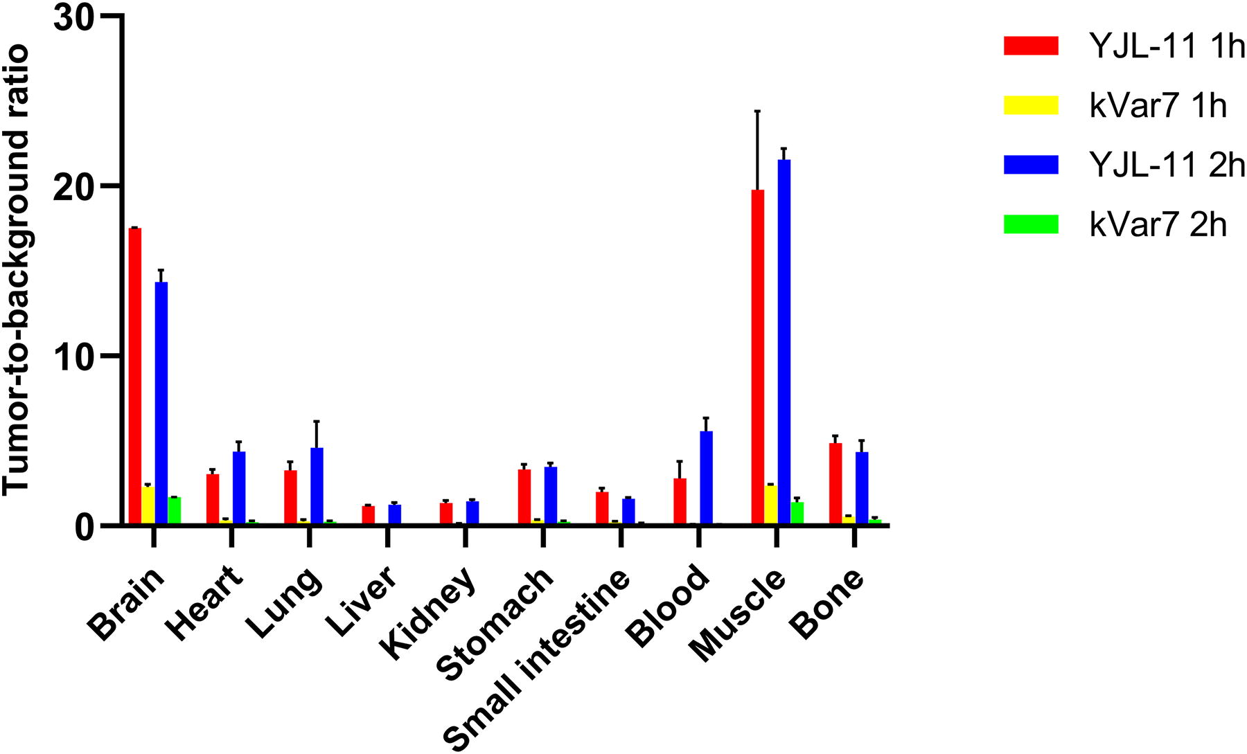
The T/NT ratio in MDA-MB-231 tumor-bearing mice after 68Ga-YJL-11 and 68Ga-kVar7 administration. T/NT, tumor/nontumor.
Biodistribution of Peptides
Small animal PET imaging
Tumors were clearly observed 1 and 2 h after injection in the 68Ga-YJL-11 group but not in the control group. Moreover, the liver was clearly seen at all time points after injection of 68Ga-YJL-11 and 68Ga-kVar7 (Fig. 8, n = 3).

Small animal PET imaging with 68Ga-YJL-11 and 68Ga-kVar7 in MDA-MB-231 tumor-bearing model mice. PET, positron emission tomography.
Discussion
The novel peptide YJL-11 was generated by replacing the alanine and tyrosine amino acid residues on the hydrophobic side of the α-helix region of the pHLIP (var3) sequence with the more hydrophobic amino acid leucine. The peptide bioinformatics analysis revealed the followings: First, using the PEP-FOLD server to analyze the peptides, we found that the energy of the coarse-grained model of the novel peptide YJL-11 was lower than those of pHLIP (var3) and YJL-4 from our previous study, 25 suggesting that YJL-11 may be more structurally stable than pHLIP (var3) and YJL-4. The energy of the coarse-grained model reflects the stability of the peptide, with a lower value suggesting a more stable structure. Second, heliQuest analysis revealed that the hydrophobic moment of the α-helix region of YJL-11 was higher than those of pHLIP (var3) and YJL-4, indicating that YJL-11 is more amphipathic than pHLIP (var3) and YJL-4. The hydrophobic moment reflects the amphipathicity of α-helical peptides, and a higher hydrophobic moment indicates that the peptide is more amphipathic. High amphipathicity makes peptide insertion into the tumor cell membrane easier, and these data suggest that YJL-11 may more easily insert itself into such a membrane. Chem3D analysis revealed that compared with YJL-4 and pHLIP(var3), YJL-11 has a higher LogP value, indicating its stronger liposolubility and tends to be metabolized primarily through the liver. YJL-11 is a peptide derived from pHLIP(var3) with only a few amino acid modifications. Predictive analysis by the PEP-FOLD server suggests that YJL-11 and pHLIP(var3) share similar secondary structures. In addition, CD analysis of YJL-11 suggested that YJL-11 can go from a structureless conformation to an α-helical structure in an acidic environment, confirming that the novel peptide YJL-11 still has this basic pH-dependent characteristic of the pHLIP family. Based on these findings, it is hypothesized that the mechanism of YJL-11 binding to TNBC is the same as that of the pHLIP family.
68Ga can be generated by a 68Ge/68Ga generator and used in areas without cyclotrons; notably, 68Ga has a very short half-life of only 68 min, which makes it an important option to ensure the acquisition of high-quality, high-resolution images. Moreover, its application can effectively reduce the patient’s radiation dose, making 68Ga very suitable for imaging small molecule peptides. Studies have shown that NOTA is a reliable chelator for 68Ga labeling, when labeling 68Ga with NOTA-Peptide, higher radiochemical yields can be obtained at lower temperatures, and the generated markers exhibit better stability in vivo. 36 However, the connection between 68Ga and the NOTA ring requires the formation of six covalent bonds with three N atoms and three carboxyl groups on the NOTA ring to remain stable. This means that the peptide cannot occupy the carboxyl groups of the NOTA ring; otherwise, 68Ga labeling will be hindered. 37 In this study, the peptide was linked to the carbon atom in the NOTA ring through indirect means. This design retained the three carboxyl groups in the NOTA ring and provided a strong guarantee for 68Ga labeling. The labeling of 68Ga in this study exhibited high radiochemical yield and radiochemical purity, which fully meet the requirements of imaging. The specific activities of 68Ga-YJL-11 (17.98 ± 0.06 MBq/μg) and 68Ga-kVar7 (17.27 ± 0.11 MBq/μg) are slightly lower than the typical range reported for peptide-based imaging agents (21.30–28.79 MBq/μg). 38 This is due to using an increased amount of precursor (8 μg) during the radiolabeling process to ensure high radiochemical yields, which naturally results in lower specific activities. Despite being below the theoretical maximum, these values are adequate for preclinical applications due to the desired level of imaging contrast achieved. In future studies, we aim to optimize precursor usage to enhance specific activities without compromising the radiochemical yield.
The biodistribution study revealed that 68Ga-YJL-11 was highly distributed in the tumors 1 and 2 h after injection, suggesting that 68Ga-YJL-11 has tumor-targeting effects and can rapidly accumulate in tumors. In addition to its accumulation in tumors, YJL-11 was also highly distributed in the liver, suggesting that this probe may be primarily metabolized in the liver, which is consistent with its high liposolubility revealed by Chem3D analysis. Compared with the distribution of the peptide YJL-4 in our previous study, 25 the distribution of 68Ga-YJL-11 in the liver and blood was significantly reduced. This difference may be related to the following factors: (1) The energy prediction from the coarse-grained model for YJL-11 was lower than that for YJL-4, suggesting that the structure of YJL-11 is more stable than that of YJL-4; (2) The hydrophobic moment of YJL-11 is higher than that of YJL-4, suggesting that YJL-11 is more amphipathic, which may improve its tumor targeting.
The PET imaging results with 68Ga-YJL-11 were consistent with the biodistribution trends. The probe accumulated at the tumor site quickly and could be used to obtain clear images, indicating that it has an efficient tumor-targeting ability. These characteristics indicate that this probe has great potential in the diagnosis of tumors. Research conducted by other team on 68Ga-labeled pHLIP demonstrated poor imaging quality. In a study by Wei X et al., 39 Fe3O4-PEG-[68Ga] DOTA/pHLIP nanoparticles were utilized to image 4T1 tumor-bearing mice and found only mild uptake in the tumor area, with abnormal uptake in the liver and spleen. The imaging performance could be influenced by multiple factors, including the hydrodynamic size and surface charges of the nanoprobe, which affect its interaction with blood proteins and clearance by the mononuclear phagocyte system. In comparison, our 68Ga-YJL-11, a small molecule probe labeled with NOTA, demonstrated more favorable biodistribution profiles and higher tumor uptake, which might be attributed to its smaller size and different physicochemical properties. These findings suggest that both chelator type and probe physicochemical properties play important roles in determining imaging outcomes. Compared with other PET imaging agents targeting TNBC, although 89Zr-bevacizumab and 68Ga-PSMA-11 exhibited superior tumor uptake, they demonstrated considerable nonspecific accumulation in the abdomen. 6,40 Such distribution patterns may compromise the detection and assessment of abdominal metastases. The uptake of 68Ga-NOTA-YJL-11 in MDA-MB-231 tumors was lower than that of 64Cu- and 18F-labeled pHLIP in 4T1 tumors, as reported by Demoin et al. 41 This difference is likely due to variations in tumor models. Specifically, 4T1 tumors exhibit a more acidic TME compared with MDA-MB-231 tumors. Previous studies have shown that the extracellular pH of MDA-MB-231 tumors ranges from 6.9 to 7.0, 42,43 whereas 4T1 tumors have a lower extracellular pH of 6.5–6.8. 44 In subsequent experiments, we plan to directly measure pH to verify these differences in the acidic microenvironment between MDA-MB-231 and 4T1 tumors. We will also systematically investigate the influence of pH on 68Ga-NOTA-YJL-11 uptake through both in vitro and in vivo studies. Compared with that of 68Ga-YJL-4, 25 the image quality with 68Ga-YJL-11 was significantly improved, and although the liver was still clearly seen, the extent of this effect was significantly reduced. These data suggest that, owing to the optimization of the peptide structure, 68Ga-YJL-11 has higher tumor affinity and lower nonspecific binding, which thereby improved the contrast and clarity of the image.
Conclusion
The novel peptide YJL-11 has the unique property of the pHLIP family to target acidic environments. 68Ga-YJL-11 was produced with high radiochemical yield, showed good stability, can target TNBC tissues, and has broad application prospects in the diagnosis of TNBC.
Footnotes
Availability of Data and Material
The datasets used or analyzed during the current study are available from the corresponding author on reasonable request.
Authors’ Contributions
Conception and design: M.Y. and Y.C. Collection of data: S.S. and F.W. Data analysis and interpretation: Y.S. and S.S.; Article writing: M.Y. Article revising: M.Y. and Y.C.
Disclosure Statement
No existing financial conflicts.
Funding Information
This study was funded by the Natural Science Foundation of Shandong Province (grant no. ZR2021MH038).
Supplementary Material
Supplementary File S1
Supplementary File S2
Supplementary File S3
Supplementary File S4
Supplementary File S5
Supplementary File S6
References
Supplementary Material
Please find the following supplemental material available below.
For Open Access articles published under a Creative Commons License, all supplemental material carries the same license as the article it is associated with.
For non-Open Access articles published, all supplemental material carries a non-exclusive license, and permission requests for re-use of supplemental material or any part of supplemental material shall be sent directly to the copyright owner as specified in the copyright notice associated with the article.