
Abstract
Abstract
Three viable female dogs, which have the same genotype, have been successfully produced by somatic cell nuclear transfer (SCNT); however, data on the growth pattern of cloned dogs are lacking. Thus, the aim of this study was (1) to assess growth parameters among those cloned dogs with measurement of body weight, height, and radiographic analysis of skull size and bone plate, and (2) to compare hematologic characteristics among the donor dog, cloned dogs, and age-matched control dogs. The cloned dogs were kept in the same environmental conditions. The body weight increased from 0.52, 0.46, and 0.52 kg at birth to 21.9, 22.9, and 20.4 kg at 68 weeks of age for individual cloned dogs, respectively. The withers height increased from 34.5, 32.6, and 35.2 cm at 8 weeks of age to 67.1 cm at 68 weeks of age in the three clones. The radiographic data demonstrated that patterns of bone growth were similar among cloned dogs, and all measured parameters of matured cloned dogs were similar with that of the fully grown donor dog. An age-specific pattern was identified on hematologic and serum biochemical measurements in both cloned dogs and age-matched controls. The parameters examined were within the normal reference ranges for healthy dogs. In conclusion, three genetically identical cloned dogs showed similar growth characteristics and had normal hematological and serum biochemical parameters.
Introduction
At the same time, there is growing concern about unpredictable or unknown effects of cloning. For example, the question of whether cloned animals will grow normally as naturally propagated animals or not, remains to be determined. Accordingly, the comprehensive studies concerning the health status and the reproductive performances of cloned domestic and laboratory animals were conducted (Chavatte-Palmer et al., 2002; Kasai et al., 2007; Landry et al., 2005). In cloned cattle, no remarkable differences in health status or similarities in the growth curves have been reported among conventionally bred cattle, or nuclear donor and clones originating from the same nuclear donor (Watanabe and Nagai, 2008). Likewise, cloned mice attained a comparable level of development with controls when parameters such as body weight gain, motor ability, and learning and memory skills were assessed (Tamashiro et al., 2000).
Since the first cloned dog, “Snuppy,” was born, it has now been established that normal viable cloned dogs can be produced by nuclear transfer (Jang et al., 2007, 2008; Lee et al., 2005). Unlike other cloned animals, the effects of SCNT on postnatal development have not been well studied in cloned dogs, mainly because of the low number of cloned dogs. In view of this, the aim of the present study was to assess growth parameters among cloned dogs generated by SCNT and compare the data between adult cloned and donor dogs. Also, we compared the hematologic characteristics between cloned dogs and age-matched dogs produced by natural fertilization.
Materials and Methods
Animals
Three genetically identical cloned female dogs produced by SCNT as previously described by Jang et al. (2007) were used in this study. Briefly, adult fibroblasts isolated from a 2-month-old female Afghan hound were used as donor cells. For SCNT, in vivo matured canine oocytes collected surgically from the oviducts of oocyte donor dogs were used as recipient oocytes. After SCNT, the reconstructed embryos were surgically transferred into the oviducts of recipient female dogs. Three healthy female puppies were delivered by Caesarean section on day 60.
These cloned female dogs (clones A, B, and C) were raised under the care of their mother. After weaning, all cloned dogs were fed three meals per day with commercial dry dog food until 5 months of age followed by two meals per day afterward, depending on their body weight. Every dog was fed individually, and was given water ad libitum at all times except when it was fasted prior to blood collection. The food rations sufficient to meet the energy requirements of each puppy were adjusted daily according to its growth level (Niblock, 1980; Swanson et al., 2004).
Healthy age-matched female Afghan hound (n = 8 for 7 to 12 weeks of age, n = 4 for 20 to 30 week of age, n = 7 for adults) were used in this experiment. Somatic cell donor dog and unrelated control dogs were privately owned with provision of individualized nutrition and environment. Cloned animals were kept in conventional conditions (open system) on the Seoul National University campus under the following environmental conditions; temperature 25–27°C, relative humidity 50–70%, 12-h light:12-h dark cycle. All puppies received standard vaccinations (Vanguard Plus 5CV®, Pfizer Animal Health Korea ud., Korea), and received annual booster injections throughout their lives. None of the dogs were used for breeding during the study. Dog care facilities and the procedures performed met or exceeded the standards established by the Committee for Accreditation of Laboratory Animal Care at Seoul National University. The study was conducted in accordance with recommendations described in “The Guide for the Care and Use of Laboratory Animals” published by the Institutional Animal Care and Use Committee (IACUC) of Seoul National University.
Measurement of body weight and height
Body weights and withers heights were recorded weekly from 4 to 16 weeks of age, then at 2-week intervals until 1 year of age for evaluation of growth in the three cloned dogs. Body heights were measured from the ground in a straight line up to the highest point of the withers. To obtain the most accurate measurements, we made the dog stand on firm, level ground with its front feet together. Growth data were compared among cloned dogs, the data from donor dog after reaching the fully grown stage and the reference ranges (Niblock, 1980; Race, 1999).
Radiological monitoring
Radiographic assessments were performed at 8, 12, 16, 26, and 68 weeks of age to compare skeletal growth among the cloned animals with three unrelated control dogs and the donor dog after reaching the fully grown stage using techniques described by others (Schoenmakers et al., 2000; Voorhout et al., 1994). All radiographs were obtained with a standard 100-cm beam source distance.
Skeletal growth of the dogs was assessed using images derived from dorsoventral radiographs of the skull, left–right lateral radiographs of lumbar vertebrae, and the ventro–dorsal image of pelvic region. The selected values of length and width were measured and the parameters of bone length and width were defined as follows:
Longitudinal length of skull (Sk 1) as a straight line between bone–rim of the upper incisor and the end of the external occipital protuberance in a dorsoventral skull radiograph. Transverse length of skull (Sk 2) as a straight line between the left lateral border of the zygomatic arch and the right lateral border of the zygomatic arch in a dorsoventral skull radiograph. Width of skull (Sk 3) as a straight line between the left and right lateral borders of the calcarium in a dorsoventral skull radiograph. Posterior vertebral body height of the L4 (PH) is the distance between the uppermost and lowest points on the posterior border of the body of L4 in a left–right lateral radiograph of the lumbar vertebrae. Upper body length of the L4 (UL) is the distance between the most anterior and most posterior point on the upper border of the body of L4 in a left–right lateral radiograph of the lumbar vertebrae. Lower body length of the L4 (LL) is the distance between the most anterior and most posterior point on the lower border of the body of L4 in a left–right lateral radiograph of the lumbar vertebrae. Longitudinal length of the pelvic bone (Pe 1) as a straight line between the tip of the cone-shaped upper end of the iliac crest and the lower border of the pelvic bone in a ventrodorsal radiograph of the pelvic region. Transverse length of the pelvic bone (Pe 2) as a straight line between the left and right lateral borders of the ischiatic tuberosity in a ventrodorsal radiograph of the pelvic region.
For the comparison of dental development, oblique radiographic views of the maxilla and mandible were also obtained. Deciduous teeth were always allowed to exfoliate naturally and the eruption patterns at 8, 12, 16, 26, and 68 weeks of age were radiographically evaluated. Identification of teeth was according to the following dental formulas and the first premolar was considered to erupt without a deciduous precursor (Kremenak, 1967, 1969; Shabestari et al., 1967).
Deciduous: incisor (I) 3/3, canine (C) 1/1, molar (M) 3/3 Permanent: incisor (I) 3/3, canine (C) 1/1, premolar (PM) 4/4, molar (M) 2/3
Blood typing by dog erythrocyte antigen
Blood collection was performed at 5 months of age. Approximately 2 mL of blood were collected from the jugular vein into an evacuated tube containing EDTA (ethylenediaminetetraacetic acid) as anticoagulant. A sample was submitted to a commercial laboratory kit (Antech Diagnostics, Phoenix, AZ, USA) for a blood typing, and the results were compared between the cloned dogs and the donor dog.
Hematologic and serologic profiles
Blood samples were collected via jugular or cephalic venipuncture for complete blood counts (CBC) and serum biochemistry from cloned dogs and normal age-matched dogs at 7 to 12 weeks of age and fully grown stage. From the donor dog, blood sample collection was performed only at the fully grown stage. To avoid diurnal variations, all blood samplings were performed at approximately the same time of day. At each collection time, 2 mL of blood was collected into an evacuated tube containing EDTA to measure red blood cell (RBC) count, white blood cell (WBC) count, hemoglobin concentration (Hb), packed cell volume (PCV), mean corpuscular volume (MCV), mean corpuscular hemoglobin (MCH), mean corpuscular hemoglobin concentration (MCHC), and platelet count (PLT). For serological analyses, 4 mL of blood was collected and transferred into a serum-separating tube (Becton Dickinson, Franklin Lakes, NJ, USA) For serum biochemistries, alanine aminotransferase, aspartate aminotransferase (AST), alkaline phosphatase (ALP), blood urea nitrogen (BUN), creatine, glucose, total bilirubin, albumin, total protein, phosphorus, and calcium were examined. For the determination of complete blood count, Celltac α hematology analyzer (Nihon Kohden, Tokyo, Japan) was used. The serum biochemistry profile was determined using a Selectra 2 chemistry analyzer (Merck, Dieren, The Netherlands) according to the manufacturer's protocols.
Statistical analysis
As this was an observational study, the measured parameters in growth were compared with reference data (Niblock, 1980; Race, 1999). The hematologic and serologic profiles were compared to normal age-matched dogs and reference data (Ikeuchi et al., 1991; Meyer and Harvey, 1992; Shifrine et al., 1973). Values were expressed as the mean ± SD for each group of dogs, and statistical analysis was performed using Student's t-test (SAS Institute, version 9.1, Cary, NC, USA). Differences among the groups were considered statistically significant when the p-values were less than 0.05.
Results
Measurement of body weight and height
Comparisons of the growth parameters among cloned dogs are shown in Figure 1. All three cloned dogs were born from different recipients, with an average birth weight of 500 g (520, 460, and 520 g, respectively). These cloned dogs were raised under the care of their mother, and each of these animals was weaned at around 30 days of age. As they grew, the estimated body weights and heights increased rapidly during the first 16 weeks after birth and then plateaued, reaching maturity between 40 and 60 weeks of age. The cloned dogs demonstrated that the body weights at 8 weeks of age were 5.7, 5.8, and 5.5 kg, and body weights at 68 weeks of age were 21.9, 22.9, and 20.4 kg, respectively. The withers heights increased from 34.5, 32.6, and 35.2 cm at 8 weeks of age to 67.1, 67.1, and 67.1 cm at 68 weeks of age, respectively. The body weight and height of donor dog at fully grown stage was 22.7 kg and 68 cm, respectively.

Comparisons of (
Radiological monitoring
As shown in Figures 2, 3, and 4, parameters were measured on dorsoventral radiographs of the skull, on lateral radiographs of the lumbar vertebrae, and on ventrodorsal radiographs of the pelvic region. All measured parameters increased in an accelerated manner from 8 to 16 weeks of age in the three cloned dogs, and the patterns of bone growth were similar each other. Also, measured parameters of three cloned dogs and donor dog at the fully grown stage were more comparable to each other than those from unrelated control dogs.
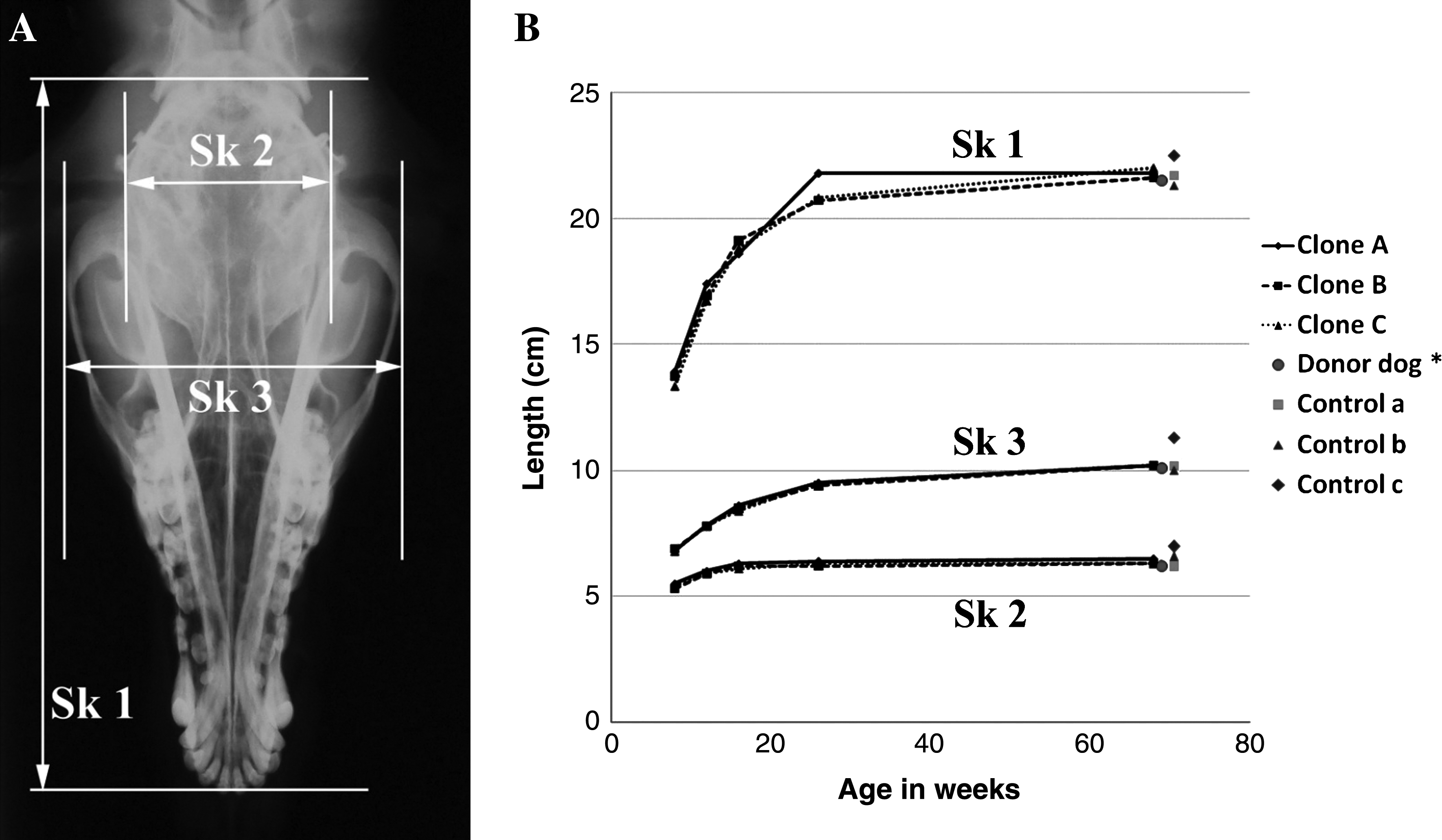
Change in each part of the skull measured by dorsoventral skull radiograph in cloned dogs. (

Change in each part of the lumbar vertebrae measured by lateral lumbar vertebrae radiography in cloned dogs. (
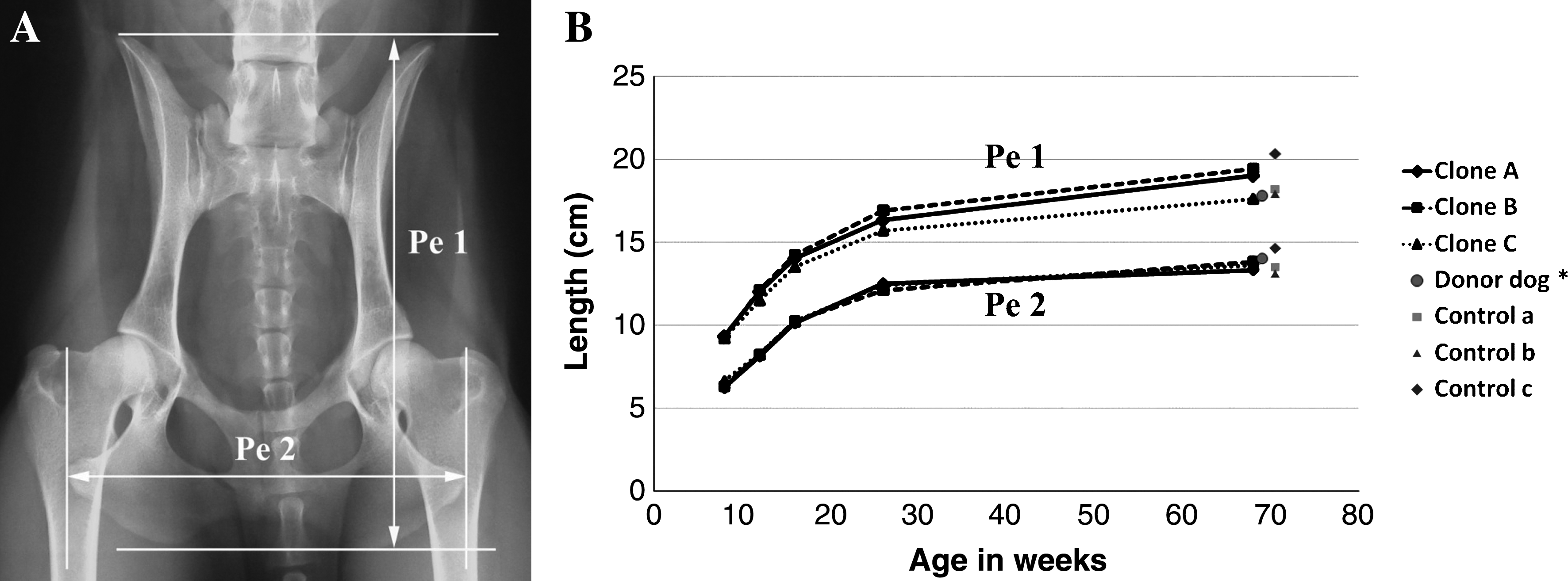
Change in each part of the pelvic bone measured by ventrodorsal pelvic region radiograph in cloned dogs. (
As shown in Table 1, Figures 5 and 6, each cloned dog's deciduous tooth exfoliation and permanent tooth emergence were observed at 8, 12, 16, 26, and 68 weeks of age by radiologic assessments. The data revealed that the developmental pattern of molar teeth was different among the cloned dogs. The number of permanent molars on the left side of the maxilla was one and on the right side was two in clone B, whereas the other dogs and the donor dog had two molar teeth on both sides of the maxilla. Also, there were two permanent molars on the left side of the mandible and three on the right side in clone C, whereas the other dogs and the donor dog had three molar teeth in both sides of their mandibles. In Figure 7, one of three control dogs demonstrated reduced number of molar teeth on the right side and premolar teeth on the left side of the maxilla, but other control dogs had normal dental formula.

Comparisons of dental development of molar teeth were measured by oblique view of maxilla radiograph in the donor dog (

Comparisons of dental development of molar teeth were measured by oblique view of mandible radiograph in the donor dog (

Oblique radiographic views of the maxilla (
The number of permanent molar on the left side is one, and on the right side is two in the maxillary.
The number of permanent molar on in the left side is two, and on the right side is three in the mandible.
d, deciduous tooth; p, permanent tooth; nd, no data. Data presented between parenthesis is the number of nonerupted permanent teeth.
Identity of blood type
The blood samples from the three cloned dogs and the donor dog had a negative agglutination reaction with DEA (dog erythrocyte antigen) 1.2 reagent, and had a positive agglutination reaction with DEA-1, 1.1, 3, 4, and 7 reagent. As shown in Table 2, the blood type was the same in all three cloned dogs and in the donor dog.
The DEA-1, 1.1, 1.2, 3, 4, and 7 were monoclonal antibodies against canine RBCs.
Hematologic and serologic profiles
The values of the mean, SDs, and the results of the statistical analysis of the hematologic and serum biochemical parameters analyzed are listed in Tables 3 and 4. Table 3 shows data on the comparisons of the hematologic parameters as a function of age in age-matched control dog and cloned dog groups. In age-matched control dogs, there was a significant effect of age on all hematologic tests (p < 0.05) except MCHC values. Values of RBC counts, Hb, and PCV gradually increased during the first year of life in controls, whereas the values of WBC, MCV, MCH, and platelet counts showed gradual decrease as age increased. On the other hand, only RBC counts, Hb, PCV, and MCH values were significantly different (p < 0.05) between different ages in the cloned dog group, which showed a gradual increase as also seen in the control group. The differences observed (p < 0.05) between control and cloned dogs were in the values of RBC counts, Hb, PCV, MCV, MCHC, and platelet counts during the ages of 7 to 12 weeks.
Means with different superscripts in the same line are significantly different (p < 0.05).
Cloned dog values with significant difference from age-matched control dogs (p < 0.05).
WBC, white blood cell count; RBC, red blood cell count; Hb, hemoglobin concentration; PCV, packed cell volume; MCV, mean corpuscular volume; MCH, mean corpuscular hemoglobin; MCHC, mean corpuscular hemoglobin concentration; PLT, platelet count.
Means with different superscripts in the same line are significantly different (p < 0.05).
Cloned dog values with significant difference from age-matched control dogs (p < 0.05).
AST, aspartate aminotranferase; ALP, alkaline phosphatase; ALT, alanine aminotransferase; BUN, blood urea nitrogen.
Table 4 shows the values of the serum biochemical parameters of the animals as a function of age. In both groups, all biochemical parameters demonstrated significant differences (p < 0.05) between ages except in AST, BUN, bilirubin, and calcium concentrations. Also, the levels of ALT, creatinine, albumin, and total protein were increased, whereas ALP, glucose, and phosphorus decreased with age in both groups. Differences were observed (p < 0.05) between control dogs and cloned dogs in the concentrations of glucose, calcium, and phosphorus during the period 7 to 12 weeks, and in the concentrations of ALP and creatinine during the 35 weeks to 1 year old. However, all estimated values of the donor dog, control, and cloned dog groups at the fully grown stage were within the normal reference ranges for healthy dogs.
Discussion
In the present study, we evaluated several key aspects of growth including overall measurements of body weight and height, radiographic assessment, blood type, and measurements of hematology and serum biochemistry to examine the effect of cloning in a canine species. Because the DNA donor dog was privately owned and had individualized nutrition and environment with no maintenance protocol required by the project leaders, there was no recorded growth or hematologic data during the growth stages for evaluating growth performance. The number of animals used in this study was small because of very a few number of canine cloned to other cloned livestock. Therefore, it was not possible to use statistical analysis in this study. With this reason, we assessed growth data among the cloned dogs to describe if all clones share similar characteristics, and compared the data with the cloned dogs, the donor dog after reaching the fully grown stage, and the reference data (Niblock, 1980; Race, 1999).
The increase in body weight and height of the cloned dogs was similar to each other, as well as mature body weight and height at 68 weeks of age were similar to each cloned dog and the donor dog (Fig. 1). As shown in Figure 1, the estimated variances of the body weight were greater than those of the body height. It is widely assumed that not only genetic factors but also other factors such as physical activities could be related with body weights in adult animals (West and York, 1998), and this suggestion also can be applied to cloned dogs. Other researchers reported maternal influences on growth patterns as well as environmental and genetic factors (Landry et al., 2005). A study of swine revealed that litter, environmental, and maternal effects were important for body weight at 100 days of age (Johnson et al., 2002). Because the three cloned dogs used in this study were delivered from different surrogate mothers (Jang et al., 2007), the maternal effect should be considered in studying growth and development in cloned dogs.
Skeletal maturation staging by radiographic analysis have been widely used for assessing maturational status and predicting growth potential. The method of evaluating skeletal maturity is radiographic examination of the developing bones, which include various areas of the skeleton (Dobenecker et al., 2006; Krailassiri et al., 2002; Kremenak, 1967; Sato et al., 2001; Shabestari et al., 1967). In this study, we performed radiographic assessment by X-ray images derived from the skull, the lumbar vertebrae, and the pelvic region. The radiographic monitoring results demonstrated that the bone growth patterns were similar among cloned dogs and the donor dog at the fully grown stage (Figs. 2, 3, and 4). All measured parameters increased rapidly during the 8 to 16 weeks of age and then plateaued, which is similar to the increasing patterns of the body weight and height. However, some differences in dental development among the cloned dogs and the donor dog were observed. As shown in Table 1, Figures 5, and 6, although the donor dog and clone A had normal dental formulas (Kremenak, 1967; Kremenak, 1969; Shabestari et al., 1967), clone B was missing one permanent molar tooth on the left side of the maxillary bone, and clone C was missing one permanent molar tooth on the right side of its mandible. In Figure 7, the permanent dentition of an unrelated control Afghan hound dog also demonstrated variance in the number of premolar and molar teeth. It has been reported that dogs of several breeds showed variation in the dentition (Arnall, 2008). These findings suggest that further studies to increase knowledge about genetic effects on dental growth should be encouraged. In canines, generally the lower cheek teeth erupt earlier than their upper counterparts (Shabestari et al., 1967). In accordance with the previous studies (Shabestari et al., 1967), the first permanent premolar in the mandible also appeared at 16 weeks of age, before the upper permanent premolar eruption occurred in cloned dogs.
To investigate the similarity in cloned dogs, the blood typing by dog erythrocyte antigen were performed. All blood groups are known to be decided via an autosomal dominant genetic mechanism (Hohenhaus, 2004; Ogasawara et al., 1996). As shown in Table 2, the blood phenotype of cloned dogs was identical to each other, which may be due to equal genetic information of the cloned dogs and the donor dog. The results obtained on our study indicate that the investigation of genetically and phenotypically identical animals could have a wide application to research related with genetic relationship of blood group, transfusion medicine, and transplantation immunology.
Measured hematologic and serum biochemical values provide fundamental biological data, and variation from these reference values often provide helpful diagnostic information (Kimura et al., 1992; Kley et al., 2003). In the present study, data were collected from 7-week to 1-year-old female cloned dogs and age-matched control dogs to evaluate if apparently normal cloned dogs share similar clinical characteristics with normal controls produced by natural breeding. Many researchers have reported that most hematologic and serum biochemical measurements are affected by age (Earl et al., 1973; Harper et al., 2003; Ikeuchi et al., 1991; Kimura et al., 1992; Mundim et al., 2007; Shifrine et al., 1973; Swanson et al., 2004). Measurement of RBC counts, Hb, and PCV are known to be interrelated and, in the absence of significant alterations of red cell size and Hb concentration, these values tend to be parallel with each other (Feldman et al., 2000). It has been documented that low RBC and hemoglobin concentrations are common in young dogs because RBC life span is shorter in young dogs and young RBCs contain less hemoglobin than old RBCs (Bush, 1992). In line with earlier reports, results of the present study demonstrated age-specific patterns in the control and cloned dog groups. Differences were observed between control and cloned dogs during weeks 7 to 12; however, there was no evidence of health problems because the hematologic values of the donor dog, cloned dogs, and control dogs were within laboratory reference ranges of young dogs (Shifrine et al., 1973).
Results of the serum biochemical analysis in this study also identified age-specific patterns in the control and cloned dog groups. Serum ALP and phosphate level were increased during the growing stage of life in both groups. The influence of age on serum ALP level is well documented that the serum ALP levels are very high in young dogs and decrease slowly with age, which reflects the slowdown of bone growth and activity of osteoblasts (Harper et al., 2003; Ikeuchi et al., 1991; Mundim et al., 2007; Swanson et al., 2004). As well, it has been reported that the high level of inorganic phosphate is maintained in the immature animal and a high plasma level of phosphate is essential for both cellular growth and mineralization of the skeleton (Caverzasio and Bonjour, 1993). Thus, higher levels of serum ALP and phosphate in immature control and cloned dogs are a consequence of the high demand in response to rapid rate of growth.
Moreover, serum protein and albumin concentrations and metabolites of protein metabolism, that is, urea and creatinine, were all increased during the first year of life in both groups. These data agree with previous observations that progressive increase in these values occur in normal growing puppies with advancing age (Mundim et al., 2007; Nap et al., 1991; Swanson et al., 2004). It is probably, in part, due to an increase in gammaglobulins induced by vaccinations (Mundim et al., 2007). Dogs in the growth phase also present special physiological characteristics, such as high protein metabolism (Gessert and Phillips, 1956; Mundim et al., 2007), which can be a consequence of the lower levels of serum protein, albumin, urea, and creatinine in the cloned and control groups at less than 16 weeks old. Notably, there was no evidence of progressive impairment of renal function among dogs, because those values in the donor dog, cloned dogs, and controls were still within published reference ranges. In addition, one consideration in determining the cause of different glucose, calcium, and phosphorus values in the growth phase of cloned dogs and ALP and creatinine values in the adult phase of cloned dogs with the age-matched controls may be the genetic and environmental difference between the two groups in this experiment rather than the possible confounding factors involved in the particular effect of cloning.
In conclusion, cloned dogs derived from SCNT showed similar growth characteristics, and we found no evidence of serious adverse effect of cloning using adult somatic cells. We believe that this study will be invaluable for risk assessment of dog cloning and beneficial to reassure the public of the social advantages of applying animal cloning to natural resources. However, further studies over their entire life span should be done to examine the effect of the aging process in cloned animals; because dogs have a long life span and the long-term consequences of cloning will not be known for years.
Footnotes
Acknowledgments
We thank Dr. Barry D. Bavister (Wayne State University) for his valuable editing of the manuscript. This study was financially supported by KOSEF (grant #M10625030005-09N250300510, approval number by ILAR (Institute of Laboratory Animal Resources) in SNU; snu-090508-5), TS Corporation, Nestlé Purina PetCare, Natural Balance Korea, and the Korean MEST, through the BK21 program for Veterinary Science.
Author Disclosure Statement
The authors declare that no conflicting financial interests exist.
Current address for Min Kyu Kim: Department of Animal Science and Biotechnology, College of Agriculture and Life Sciences, Chungnam National University, Daejeon 305-764, Republic of Korea.