
Abstract
Abstract
An innovative technique called high hydrostatic pressure (HHP) treatment has recently been reported to improve the cryosurvival of gametes and embryos in certain mammalian species, including the mouse, pig, and cattle. In the present study the parthenogenetic activation (PA) of pig oocytes caused by HHP treatment was investigated in different holding media with or without Ca2+. The efficiency of activation was tested at different pressure levels and media including T2 (HEPES-buffered TCM-199 containing 2% cattle serum), and mannitol-PVA fusion medium with (MPVA + Ca2+) or without Ca2+ and Mg2+(MPVA). The results showed that HHP cannot induce PA in T2, but only in MPVA + Ca2+ with low Ca2+ concentration and MPVA without Ca2+. The highest activation efficiency was achieved with 10 min HHP treatment using 100 or 200 bars for oocytes in MPVA + Ca2+ or MPVA, respectively. In the light of these results, the possible source of Ca2+ during activation was investigated. It was found that even after a total of 30-min wash with TL-HEPES-PVA buffer without Ca2+ before HHP treatment in MPVA, the oocytes could still be activated, indicating the possibility of an intracellular Ca2+ source caused cytoplasmic free Ca2+ elevation. In conclusion, parthenogenetic activation could be induced by HHP in certain holding media with low or zero Ca2+ content. Further experiments are needed to identify the exact mechanisms of activation.
Introduction
High hydrostatic pressure (HHP) treatment was initially reported by Pribenszky et al. (2005a, 2005b, 2005c) to increase the cryotolerance of bovine and murine blastocyst as well as boar spermatozoa, and porcine oocytes (Du et al., 2008a; Pribenszky et al., 2008), which are highly sensitive to low temperatures, partly because of the high lipid content in the cytoplasm (Isachenko et al., 2001; McEvoy et al., 2000). Recently, HHP treatment was reported to support somatic cell nuclear transfer by increasing the developmental competence of the reconstructed embryos both in vitro and in vivo (Du et al., 2008b).
Parthenogenetic activation could be triggered with various physical (mechanical, electronic, osmotic), chemical, and other treatments (Machaty and Prather, 1998; Machaty et al., 1999; Somfai et al., 2007; Tian et al., 2007). Osmotic stress and hydrostatic pressure in Drosophila melanogaster has also induced parthenogenetic activation by causing a rise of intercellular Ca2+ (Horner and Wolfner, 2008). The purpose of the present study was (1) to investigate the possibility that HHP treatment on activation of porcine oocytes in different holding media with different pressure levels, and (2) to test the effect of Ca2+ concentrations of holding media on activation efficiency with HHP treatment.
Materials and Methods
Except where otherwise indicated, all chemicals were obtained from Sigma Chemical Co. (St. Louis, MO, USA); all manipulations were performed on a heated stage adjusted to 39°C, and all drops used for handling oocytes were 20 μL covered with mineral oil to avoid evaporation.
Oocyte collection and in vitro maturation (IVM)
Cumulus-oocyte complexes (COCs) were aspirated from 2 to 6 mm follicles from slaughterhouse-derived sow ovaries and matured in groups of 50 in 400 μL in vitro maturation (IVM) medium consisting of bicarbonate-buffered TCM-199 (GIBCO BRL, Grand Island, NY, USA) supplemented with 10% (v/v) cattle serum (CS), 10% (v/v) pig follicular fluid, 10 IU/mL eCG, 5 IU/mL hCG (Suigonan Vet; Skovlunde, Denmark) in the Submarine Incubation System (SIS) (Vajta et al., 1997) for 41–44 h.
High hydrostatic pressure treatment
Matured oocytes were denuded with 1 mg/mL hyaluronidase by vigorous pipetting, and randomly distributed into 18 (Experiment 1) or 8 (Experiment 2) groups. Oocytes in each group were washed twice in holding medium and then loaded into a 0.25 mL plastic straw with holding medium without air bubbles (see details in Experimental Design). The straw was sealed by heat and placed into a prewarmed pressure machine (Cryo-Innovation Inc.; Budapest, Hungary) filled with water as pressure medium. Oocytes were exposed to different pressures for 10 min at 37°C.
Embryo culture and cell number counting
Embryos were subsequently cultured in groups of 20 to 30 in wells of four-well dishes (Nunc, Skovlunde, Denmark) containing 400 μL PZM-3 medium (Yoshioka et al., 2002) covered with 400 μL mineral oil at 38.5°C in 5% O2, 5% CO2, and 90% N2 with maximum humidity. Cleavage rates were determined on Day 1. Blastocyst rates were determined on Days 6 and 7.
For determination of cell numbers, Day 7 blastocysts were individually fixed and mounted on a glass microscopic slide in an approx. A total of 3–4 μL drops of glycerol supplemented with 20 μg/mL Hoechst 33342 fluorochrome. After staining for 24 h, embryos were observed under a Diaphot 200 inverted microscope with epifluorescence attachment and UV-2A filter (Nikon, Tokyo, Japan). Cell nuclei were counted manually.
Statistical analysis
Statistical analysis was performed using SPSS 13.0 (SPSS, Chicago, IL, USA). Analysis of variance (ANOVA) was used to analyze differences in cleavage rates, blastocyst rates, and cell numbers. A probability of p < 0.05 was considered to be statistically significant.
Experimental Design
Experiment 1: Effect of HHP treatment on parthenogenetic activation of porcine oocytes with different pressure level in different holding media
Denuded oocytes were observed under a stereomicroscope, and those without detectable signs of degeneration were randomly distributed into 18 groups, according to six different pressure levels (0, 100, 200, 400, 600, and 800 bar) and three different holding media (T2, MPVA + Ca2+, and MPVA). Concentrations of Ca2+, Mg2+, mannitol, and polyvinyl alcohol (PVA) of the three media are shown in Table 1. After 10 min HHP treatment, oocytes were cultured in PZM-3 medium under conditions described above.
Experiment 2: Effect of Ca2+ concentration of holding media on activation efficiency with HHP treatment
Selected denuded oocytes with appropriate morphological appearance were randomly distributed into eight groups. In this experiment, only two different pressure levels were used: 100 bar (Groups 1–4) and 200 bar (Groups 5–8). To to test the effect of Ca2+ concentration on the efficiency of activation, besides the three different holding media (with two quick prewashes of 5–10 sec for each), one more group was added where the wash was performed twice for 15 min with Ca2+ free TL-HEPES-PVA buffer composed of 10 mM HEPES, 137 mM NaCl, 2.68 mM KCl, 0.42 mM NaH2PO4, 1.7 mM MgCl2, 11.9 mM NaHCO3, 5 mM glucose, 0.1% PVA before using MPVA as holding medium to make sure there was no Ca2+ outsite the oocytes during the HHP treatment. After 10 min HHP treatment in different holding media with different pressure levels, the oocytes were cultured in PZM-3 medium as described above.
Results
Experiment 1: Effect of HHP treatment on parthenogenetic activation of porcine oocytes with different pressure level in different holding media
Denuded oocytes were distributed in 18 groups as described in Materials and Methods. Six replicates with a total of 1993 oocytes were analyzed.
Microscopic view of oocytes after HHP treatment: after 10 min HHP treatment in MPVA + Ca2+ and MPVA, the perivitelline space was considerably enlarged in all treated groups, regardless of the intensity of the pressure treatment. The average diameter of oocytes had decreased approximately by 20%. However, no such changes were observed in untreated groups, or treated groups in T2 holding medium (Fig. 1).
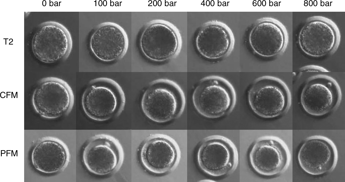
The morphology of oocytes after HHP treatment for 10 min in different solutions at different pressure levels.
Developmental rates
Low cleavage rates were found in all T2 groups and control groups in MPVA + Ca2+ and MPVA medium. However, high cleavage rates after HHP treatment in MPVA + Ca2+ and MPVA medium were achieved, with 55 ± 11% and 51 ± 12% for MPVA + Ca2+ group treated with 100 bar, and MPVA group treated with 200 bar, respectively (Fig. 2a). After 6 days of culture, very low blastocyst rates were found in all T2 medium groups and all control groups. HHP treatment in MPVA + Ca2+ and MPVA media resulted in significant improvement in embryo development to the blastocyst stage, with rates of 17 ± 3% and 19 ± 6% for MPVA + Ca2+ group treated with 100 bar, and MPVA group treated with 200 bar, respectively (Fig. 2b). The same trend was observed on Day 7 (Fig. 2c).

(
Experiment 2: Effect of Ca2+ concentration of holding media on activation efficiency with HHP treatment
Denuded oocytes were distributed into eight groups and subjected to treatments described in Materials and Methods. Six replicates with a total of 1351 oocytes were analyzed. After treatment with 100 bar for 10 min, very low development was observed when T2 was used as holding medium compared with the other three groups, whereas no difference on development was found among the other three groups (Table 2). When 200 bar was applied for HHP treatment, the T2 group still showed practically no activation. Higher cleavage rates on Day 1 were found in groups with MPVA + Ca2+ and MPVA as holding media compared with the long prewash group (Table 3). However, the differences in blastocyst rates between the long prewash group and any of the other groups were not significant. No difference in blastocyst cell numbers was observed between the MPVA + Ca2+, MPVA and long prewash groups.
Data are the mean ± SEM of six replicates. Values in the same column with different superscripts differ significantly (p < 0.05).
N.A. stands for data not available (no blastocyst).
Data are the mean ± SEM of six replicates. Values in the same column with different superscripts differ significantly (p < 0.05).
N.A. stands for data not available (too few blastocysts).
Discussion
PA can be induced by either physical stimuli such as temperature and electric treatment, or by chemicals such as Ca2+ ionophores. It was reported that pressure and osmotic stress may cause parthenogenetic activation of Drosophila eggs (Horner and Wolfner, 2008). In the present study similar results were observed on porcine oocytes. After 10 min HHP treatment in both MPVA + Ca2+ and MPVA media, porcine oocytes were activated. Only slight differences between these two media were observed when optimal activation parameters were used. In MPVA + Ca2+ medium the peak developmental competence of oocytes was induced by 100 bar, whereas in MPVA medium it was 200 bar. In contrast to the HHP treatment in MPVA + Ca2+ and MPVA media, similar HHP parameters resulted in a negligible activation level in T2 medium.
It should be mentioned that HHP treatment of porcine oocytes in MPVA without Ca2+ could cause activation with almost the same blastocyst rate as in MPVA + Ca2+. It is well known that Ca2+ signaling plays an important role in PA, somatic cell nuclear transfer (SCNT), and IVF. In natural fertilization, the oscillation in the cytoplasmic free Ca2+ levels is induced by the fertilizing sperm and the origin of the increased Ca2+ levels is the intracellular stores (Ducibella and Fissore, 2008). In certain in vitro activation approaches, this process is mimicked by inducing elevation of intracellular Ca2+ either from external or internal resources (Machaty et al., 1999).
At least three possible mechanisms may explain the activation in MPVA. The first mechanism is that two quick washes (3–5 sec each) in MPVA are not enough to completely remove Ca2+ from the extracellular medium, and it was demonstrated previously that even a very low amount of Ca2+ contamination may be able to trigger oocyte activation (Machaty et al., 1995). The second possible mechanism is that there is a release of Ca2+ from internal reserves, because HHP treatment might “squeeze” Ca2+ out of the intracellular stores such as the endoplasmic reticulum (ER). The third possibility is that the activation has no relationship with Ca2+ but it is triggered by events downstream of the Ca2+ signal.
To test the first possibility, a washing experiment was performed. To avoid Ca2+ contamination, two quick and two long (15 min) washes with Ca2+-free TL-HEPES-PVA buffer were performed before HHP treatment of oocytes in Ca2+-free medium (MPVA). The wash with extended duration did not significantly reduce the HHP-induced blastocyst development. HHP treatment could still activate the oocytes; thus, a role for external Ca2+ influx during HHP-induced oocyte activation is not likely. The similar cell numbers of the resultant blastocysts in the three treatment groups (MPVA + Ca2+, MPVA, prewashed MPVA) also suggest that extracellular Ca2+ plays a very limited role in the activation with HHP.
The morphology of oocytes in MPVA + Ca2+ and MPVA groups was changed after HHP treatment. The oocytes were more compact, with enlarged perivitelline space in the MPVA and MPVA + Ca2+ groups, but not in the T2 group (Fig. 1). This enlarged perivitelline space was never observed in untreated groups, indicating that the enlarged perivitelline space was caused by HHP treatment. Because the three holding media have almost the same osmolarity, the possibility that the enlarged perivitelline space was induced by a combination of HHP and osmotic pressure was excluded. These changes were only induced by HHP treatment in MPVA + Ca2+ and MPVA medium, but not in T2, and were thus correlated to the activation efficiency. The functional relationship between the enlarged perivitelline space and activation is not clear. More studies are needed to answer these questions.
In the second experiment, lower mean cell numbers of Day 7 blastocysts were observed after treated with 200 bar, indicating that 100 bar was more appropriate for activation purpose. Not only HHP, but also both osmotic (Lin et al., 2009) and oxidative (Vandaele et al., 2010) stresses were found supportive for embryos developmental competence if they were properly optimized, while higher osmotic (Hay-Schmidt, 1993) and oxidative (Dennery, 2007) stresses could have completely opposite effect.
In conclusion, HHP treatment could activate porcine oocytes in certain media such as MPVA + Ca2+ and MPVA, but not in T2. The Ca2+ concentration of holding media did not seem to affect the blastocyst development and quality after activation induced by HHP treatment. A total of 100 bar HHP for 10 min seems to be the appropriate parameters to induce parthenogenesis. As far as is known, this report is the first to describe the parthenogenetic activation induced by HHP treatment in mammals.
Author Disclosure Statement
The authors declare that no conflicting financial interests exist.