
Abstract
Abstract
Trichostatin A (TSA) has previously been used in somatic cell nuclear transfer (SCNT) to improve the cloning efficiency in several species, which led our team to investigate the effects of TSA on the full-term development of bovine SCNT and gaur–bovine interspecies SCNT (gaur iSCNT; gaur somatic cells as donors and bovine oocytes as recipients) embryos. Treatment with 50 nM TSA for 10 h after fusion had no positive effects on the rates of fusion, cleavage, or the development to eight-cell or morula stages in both bovine SCNT and gaur iSCNT embryos. However, TSA treatment significantly enhanced the blastocyst formation rate in bovine SCNT embryos (44 vs. 32–34% in the TSA-treated and TSA-untreated groups, respectively), but had no effects on gaur iSCNT embryos. The fresh blastocysts derived from bovine SCNT and gaur iSCNT embryos (fresh groups), as well as vitrified bovine SCNT blastocysts (vitrified group), were transferred to bovine recipients. We found that TSA treatment increased the pregnancy rates only in recipients receiving fresh bovine SCNT embryos. In recipients receiving TSA-treated bovine SCNT embryos, three cloned calves from the fresh group and twin cloned calves from the vitrified group were delivered; however, no calf was born from the TSA-untreated bovine SCNT embryos. In contrast, one gaur iSCNT calf was born from a recipient receiving blastocysts from the TSA-untreated group. In summary, TSA improved the preimplantation development and pregnancy rates of bovine SCNT embryos, but did not have any beneficial effect on gaur iSCNT embryos. However, one gaur iSCNT calf reached full-term development.
Introduction
It was hypothesized that successful live offspring production from iSCNT embryos depends on the complete reprogramming of the donor nucleus, resulting in proper embryonic initiation and fetal gene expression (Mastromonaco and King, 2007). Incomplete reprogramming involves abnormal epigenetic remodeling, such as DNA methylation and histone modifications (Wee et al., 2006). In theory, assisted reprogramming of the donor nucleus through SCNT might improve embryo development (Wilmut et al., 2002). Previous studies have indicated that Trichostatin A (TSA) treatment of cloned embryos improved in vitro embryo development in many species, including mice (Kishigami et al., 2006, 2007; Rybouchkin et al., 2006; Tsuji et al., 2009; Costa-Borges et al., 2010; Dai et al., 2010; Hai et al., 2011; Kang and Roh, 2011), cattle (Lee et al., 2011; Akagi et al., 2011), pigs (Zhang et al., 2007; Li et al., 2008, Beebe et al., 2009; Martinez-Diaz et al., 2010; Himaki et al., 2010; Kim et al., 2011), and rabbits (Shi et al., 2008). However, TSA treatment had no positive effect on preimplantation development in cloned rats (Mizumoto et al., 2008). Although TSA can enhance the live birth rate in cloned mice (Kishigami et al., 2006; Rybouchkin et al., 2006; Maalouf et al., 2009; Tsuji et al., 2009), there was no positive effect on pregnancy and birth rates in cloned rabbit embryos (Meng et al., 2009). For iSCNT using human somatic cells and enucleated rabbit oocytes, TSA treatment did not improve the in vitro development of iSCNT embryos (Shi et al., 2008). In addition, no positive effect was found on the full-term development of black-footed cat iSCNT embryos (Gómez et al., 2011). Taken together, these previous studies have indicated that the effects of TSA treatment on cloning efficiency have shown no consistent pattern. Therefore, in this study, we assessed the effects of TSA treatment on the full-term development of bovine SCNT and gaur iSCNT embryos.
Materials and methods
Animals
Animal care and procedures were conducted according to the guidelines of the Ethics Committee of the Laboratory Animal Care of Suranaree University of Technology.
Chemicals and media
All reagents were purchased from Sigma-Aldrich Chemical (St. Louis, MO, USA) unless otherwise specified.
Donor cell preparation
The donor cells were prepared as previously described (Suteevun et al., 2006). Briefly, skin tissue was biopsied from male and female bovines and gaurs. The tissue was then cut into small pieces and cultured in α-mimimum essential medium (αMEM) supplemented with 10% fetal bovine serum (FBS; Gibco, MD, USA) in a humidified atmosphere of 5% CO2 at 37°C for 8–10 days. At subconfluence, the fibroblasts were harvested using 0.25% trypsin/EDTA and cultured in αMEM supplemented with 10% FBS for expansion. The fibroblasts were frozen in αMEM supplemented with 20% FBS and 10% dimethyl sulfoxide (DMSO; Merck, Darmstadt, Germany) and stored in liquid nitrogen. Frozen–thawed donor cells were cultured for 2–3 days, and only the fourth passage of cultured cells from all cell types was used as the donor cells. Several minutes before injection, the proliferating donor cells were harvested and resuspended in Emcare® holding medium (ICPBio, Glenfield, New Zealand).
Bovine SCNT and gaur iSCNT embryo production
The oocyte preparation, nuclear transfer procedure, and in vitro embryo culture were performed as previously described (Srirattana et al., 2010). Briefly, bovine oocytes were obtained from local abattoir-derived ovaries and cultured in in vitro maturation (IVM) medium for 21 h. The cumulus cells were then removed by repeat pipetting in 0.2% hyaluronidase. Metaphase II (MII) oocytes with the first polar body were selected for enucleation. The MII oocytes were placed in 5 μg/mL cytochalasin B for 5 min. The zona pellucida above the first polar body was cut with a glass needle, and a small volume (approximately 5–10%) of the cytoplasm beneath the first polar body was then squeezed out. Completed enucleation was confirmed by staining the squeezed-out cytoplasm with 5 μg/mL Hoechst 33342 and visualizing the stained nucleus in the cytoplasm under a fluorescence microscope (IX71, Olympus, Tokyo, Japan). An individual donor fibroblast from a male and female bovine or gaur was inserted into the perivitelline space of the successfully enucleated bovine oocyte. The donor cell–cytoplast couplet (DCCC) was fused in Zimmermann fusion medium (Zimmermann and Vienken, 1982) between the tips of the fusion electrodes and electrostimulated by two direct current pulses (24 V, 15 μsec). The DCCCs were subsequently washed six times in Emcare® holding medium. The success of the fusion was examined 1 h after electrostimulation. The fused DCCCs were activated with 7% ethanol (Carlo Erba, Val de Reuil, France) in Emcare® holding medium for 5 min at room temperature and cultured in modified oviduct synthetic fluid with amino acid medium (mSOFaa; Gardner et al., 1994) supplemented with 1.25 μg/mL cytochalasin D and 10 μg/mL cycloheximide at 38.5°C in a humidified atmosphere of 5% CO2 for 5 h.
The embryos were then cultured in fresh mSOFaa medium at 38.5°C in a humidified atmosphere of 5% CO2, 5% O2, and 90% N2 for 2 days. The eight-cell stage embryos were selected and cocultured with bovine oviductal epithelial cells in mSOFaa medium at 38.5°C in a humidified atmosphere of 5% CO2 for 5 days. Half of the volume of mSOFaa medium was replaced daily and the development of embryos was also recorded.
TSA treatment
After fusion, the DCCCs were separated into two groups (TSA-treated and TSA-untreated). For the TSA-treated group, DCCCs were placed in Emcare® holding medium supplemented with 50 nM TSA for 1 h. The fused DCCCs were then activated and cultured in medium supplemented with 50 nM TSA for an additional 9 h (Kishigami et al., 2006). For the TSA-untreated group, fused DCCCs were activated and cultured with the same conditions except without TSA supplementation.
Embryo staining
Blastocysts from all treatment groups at 7 days after culture were counterstained to distinguish trophectoderm (TE) cells and the inner cell mass (ICM), as described by Suteevun et al. (2006) with some modifications. Briefly, the zona pellucida of blastocysts was removed with 0.5% protease and washed in modified Dulbecco's phosphate-buffered saline (mDPBS) supplemented with 0.1% polyvinylpyrrolidone. The zona pellucida-free blastocysts were incubated for 30 min with rabbit anti-bovine splenocyte antibodies for bovine SCNT embryos or rabbit anti-gaur fibroblast antibodies for gaur iSCNT embryos. Both antibodies were generated in our laboratory according to the protocol described by Iwasaki et al. (1990). After incubation, the blastocysts were transferred into a mixture of 10% guinea pig complement, 10 μg/mL propidium iodide, and 10 μg/mL Hoechst 33258 for 30 min. The blastocysts were mounted with glycerol (Merck) on glass slides and covered with cover slips. The ICM cells (blue) and TE cells (red) were counted under ultraviolet light using IX71 fluorescence microscopy.
Embryo vitrification and warming
Male bovine SCNT embryos from TSA-treated and TSA-untreated groups were vitrified using the microdrop technique. Briefly, after 7 days of culture, the hatching blastocysts were washed in TCM199-HEPES supplemented with 20% FBS, 7.5% (v/v) ethylene glycol (EG), and 7.5% (v/v) DMSO for 3 min and then transferred to vitrification medium [TCM199-HEPES supplemented with 20% FBS, 0.5 M sucrose, 16.5% (v/v) EG, and 16.5% (v/v) DMSO] for 30 sec. Small drops of the vitrification solution (1–2 μL) containing 5 embryos were directly dropped into liquid nitrogen and then transferred into cryotubes (Nunc, Roskilde, Denmark) and stored in liquid nitrogen (Papis et al., 2000). The embryos were thawed by immersing the vitrified drops in 0.6 M sucrose in TCM199-HEPES supplemented with 20% FBS for 5 min at 39°C followed by a stepwise dilution in 0.4, 0.2, and 0 M sucrose for 5 min each. The embryos were subsequently washed three times in mSOFaa medium and cocultured with bovine oviductal epithelial cells in a humidified atmosphere of 5% CO2 at 38.5°C for 12 h before the embryo transfer.
Embryo transfer
The recipients (2–3 years old) were synchronized to estrus and used for embryo transfer. Briefly, an intravaginal progesterone-releasing device (CIDR; Pharmacia & Upjohn Company, Hamilton, New Zealand) was inserted into the vagina of each recipient for 7 days. Two days before CIDR removal, an intramuscular (i.m.) injection of 500 IU equine chorionic serum gonadotrophin (eCG; Folligon®, Intervet International B.V., Boxmeer, The Netherlands) was administered. At the time of CIDR removal, each recipient received an i.m. injection of 0.75 mg of synthetic prostaglandin (PGF2α; Iliren®, Intervet International GmbH, Unterschleissheim, Germany). At 15 h after standing estrus, each recipient received an i.m. injection of 0.25 mg of gonadotropin-releasing hormone (GnRH; Fertagyl®, Intervet International B.V.). On day 8 postestrus, 2–3 bovine SCNT (fresh and vitrified groups) and gaur iSCNT (fresh group) embryos from TSA-treated and TSA-untreated groups were transferred nonsurgically into the recipients. Pregnancy diagnosis was performed at day 45 after embryo transfer by transrectal ultrasound scanning. The pregnancy status was rechecked by rectal palpation on days 60, 90, 120, 150, 180, 210, 240, and 270 of gestation.
DNA microsatellite analysis
The genotyping of clones and donor cells was performed by polymerase chain reaction (PCR) using bovine DNA microsatellite markers (Table 1). The reaction mixture contained 100 ng of DNA sample, 1× PCR buffer (Promega, Madison, USA), 2 mM MgCl2 (Promega), 0.2 mM deoxyribonucleotide triphosphates (dNTPs) mix (Fermentas, Burlington, Canada), 0.5 μM of each primer (Table 1), and 0.625 U Taq polymerase (Promega) in a total of 20 μL. The amplification conditions were as follows: 95°C for 2 min, followed by 45 cycles at 95°C for 20 s, an annealing temperature (Table 1) for 20 s, and 72°C for 30 s. The final elongation was performed at 72°C for 5 min. The PCR products were resolved through a 5% polyacrylamide gel, stained with 1 mg/mL ethidium bromide, and visualized under ultraviolet light (ChemiDoc XRS System, Bio Rad, Hercules, USA).
Statistical analysis
Data were evaluated using the Statistical Analysis System (SAS Inst. INC., Cary, USA). Data of fusion rates, embryo development, and cell numbers were analyzed using a completely randomized design. Data of pregnancy and calving rates were analyzed using the chi-squared test. For all statistical analyses, p<0.05 was considered significant.
Results
The effects of TSA on preimplantation development of bovine SCNT and gaur iSCNT embryos
Fibroblasts from a male and female bovine and gaur were fused with enucleated bovine oocytes using the same parameters. After fusion, the reconstructed embryos were washed and incubated in Emcare® holding medium with or without TSA supplementation for 1 h. The fusion rates ranged between 85% and 91%, with no significant difference between treatment groups (Table 2). These results indicated that the gaur and bovine fibroblasts had a similar ability to fuse with the enucleated bovine oocytes.
−, TSA-untreated, +, TSA-treated.
Different superscripts within each column indicate significant differences (p<0.05; ANOVA).
Percentages calculated from the number of cultured embryos that cleaved and developed to each stage.
TSA, Trichostatin A; SCNT, somatic cell nuclear transfer; iSCNT, interspecies SCNT; TE, trophectroderm; ICM, inner cell mass; ANOVA, analysis of variance.
The rates of cleavage (94–98%) as well as development to the 8-cell (70–83%) and morula (42–51%) stages in bovine SCNT and gaur iSCNT embryos were similar for both the TSA-treated and TSA-untreated groups and the male and female fibroblasts. The blastocyst formation rate of TSA-treated bovine SCNT embryos (44%) was significantly higher than those of the TSA-untreated bovine SCNT embryos (32–34%; Table 2). However, TSA treatment did not increase the blastocyst rate of gaur iSCNT embryos (33–37%) when compared with TSA-untreated gaur iSCNT embryos (33–38%). These results demonstrated that there were no significant differences in the developmental potential between male and female cloned embryos in either bovine SCNT or gaur iSCNT embryos.
In terms of embryo quality, there were no significant differences in the number of TE and ICM cells or in the ICM ratio between treatment groups (Table 2). These results indicated that gaur iSCNT blastocysts were of similar quality as bovine SCNT blastocysts (Fig. S1) (Supplementary Data are at www.liebertonline.com).
The effects of TSA on postimplantation development of bovine SCNT and gaur iSCNT embryos
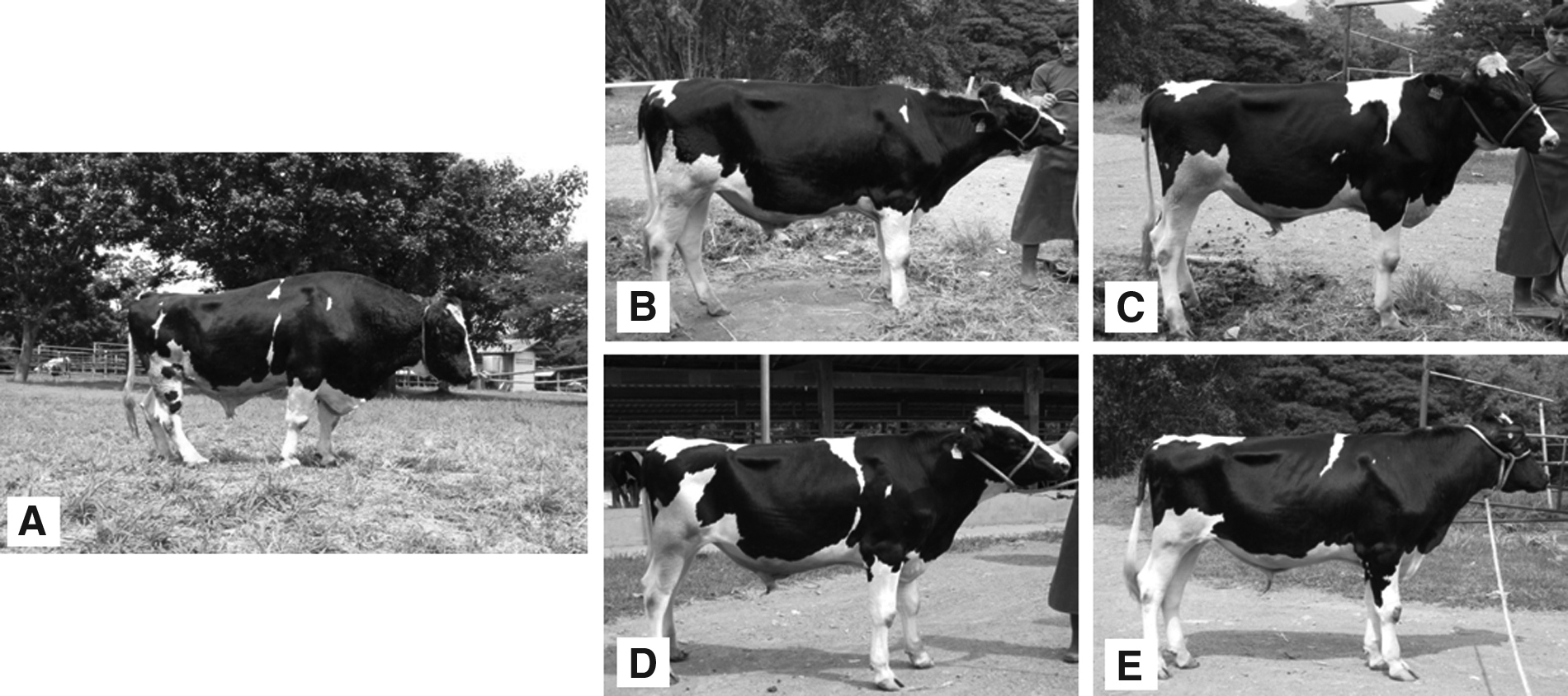
The postimplantation development of male bovine SCNT (fresh and vitrified groups), as well as male and female gaur iSCNT (fresh group) embryos, is shown in Table 3. For the fresh bovine SCNT embryos, 36 TSA-treated and 18 TSA-untreated blastocysts were transferred to 13 and 7 synchronized recipients, respectively. The pregnancy rate of recipients receiving TSA-treated blastocysts (6/13, 46.2%) was significantly higher (p<0.05) than the recipients receiving TSA-untreated blastocysts (0/7, 0%). However, there was no significant difference in postimplantation development to term between TSA-treated and TSA-untreated embryos. Only three recipients maintained pregnancy until term (3/13, 23.1%). From these pregnancies, three cloned calves were born (Fig. 1B, C); however, one calf died during veterinary-assisted delivery. In the vitrified bovine SCNT embryos, 22 TSA-treated and 14 TSA-untreated blastocysts were transferred to eight and seven synchronized recipients, respectively. The pregnancy rate was not significantly different (p>0.05) between the TSA-treated (3/8, 37.5%) and the TSA-untreated (2/7, 28.6%) groups. Only one pregnant recipient receiving a blastocyst from the TSA-treated group carried the pregnancy to term, and twin cloned calves (Fig. 1D, E) were born healthy. No offspring were born from the TSA-untreated group.
Male bovine donor (
Values with different superscripts for each donor cell were significantly different (P<0.05; ANOVA).
TSA, Trichostatin A; SCNT, somatic cell nuclear transfer; iSCNT, interspecies SCNT.
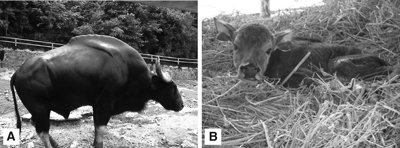
In gaur iSCNT embryos, 44 TSA-treated and 9 TSA-untreated male gaur iSCNT blastocysts were transferred to 18 and three synchronized bovine recipients, respectively. The pregnancy rate of recipients receiving blastocyst from the TSA-treated group (3/18, 16.7%) was not significantly different (p>0.05) compared to those receiving blastocysts from the TSA-untreated group (1/3, 33.3%). Only one recipient of a blastocyst from the TSA-untreated group maintained the pregnancy to term. The cloned male gaur calf was delivered by cesarean section on day 283 of gestation. The cloned gaur newborn had a normal cardiac rhythm for the first 3 h postdelivery, but signs of dypsnea subsequently appeared and the calf died 12 h after birth (Fig. 2B). A necropsy was performed and incomplete development of lung tissue was found, which may have been the cause of death. For the female gaur iSCNT embryos, 50 TSA-treated and 3 TSA-untreated gaur iSCNT blastocysts were transferred to 16 and one synchronized bovine recipients, respectively. Six out of the 16 recipients (37.5%) of the TSA-treated group blastocysts achieved pregnancy; however, most of the recipients lost the fetuses in the early stages of pregnancy (days 45–60 of gestation), with one aborted pregnancy on day 242 of gestation (Fig. 3B). Furthermore, one mummified fetus was obtained by caesarean section on day 311 of gestation (Fig. 3C). No pregnancy was observed in the recipients receiving blastocysts from the TSA-untreated group.
Male gaur donor (

Female gaur donor (
Twelve DNA microsatellite markers (MGTG4B, TGLA263, TGLA57, ETH225, INRA037, HEL9, TGLA53, CSSM66, ETH3, TGLA126, INRA063, and INRA005) were used to amplify genomic DNA (gDNA) from the bovine donor cells, the four cloned bovine calves, and the three bovine recipients. The markers used were able to distinguish PCR products of the bovine donor cell and cloned bovine calves from those of the three bovine recipients, except for the INRA063 and INRA005 markers (data not shown). The results of the 10 microsatellite analyses confirmed that all cloned calves were genetically identical to the donor cells and differed from the recipients (data not shown). For the gaur group, seven bovine DNA microsatellite markers (MGTG4B, TGLA263, TGLA57, ETH225, INRA037, HEL9, and TGLA53) were used to amplify the gDNA from the gaur donor cells, the aborted and mummified gaur fetuses, cloned gaur calf, and the three bovine recipients (data not shown). Analysis of the bovine DNA microsatellite markers confirmed that the aborted fetus, mummified gaur fetus, and cloned gaur calf were genetically identical to their donor cells. Representative results from markers TGLA263 and HEL9 are shown in Figures 4 and 5, respectively. Importantly, the four cloned bovine calves developed normally and remain alive today.

DNA microsatellite analysis using the TGLA263 marker in cloned calves. MBF, male bovine fibroblasts; R1, R2, R4, bovine recipient; CB1, CB2, CB4, CB5, cloned bovine calf.

DNA microsatellite analysis using the HEL9 marker in a cloned gaur. R5, R6, R7, bovine recipient; MCG, male cloned gaur; MGF, male gaur fibroblasts; FGF, female gaur fibroblasts; FCGa, female cloned gaur aborted; FCGm, female cloned gaur mummified.
Discussion
In this study, we found that TSA treatment of 50 nM for 10 h after fusion increased the blastocyst and pregnancy rates of bovine SCNT embryos, whereas the same treatment had no effect on gaur iSCNT embryos. Similar blastocyst qualities were found among bovine SCNT and gaur iSCNT embryos in both the TSA-treated and TSA-untreated groups. Moreover, TSA increased the pregnancy rate of the bovine SCNT embryos; however, no positive effect was observed on the gaur iSCNT and vitrified bovine SCNT embryos.
The positive effects of TSA treatment on the preimplantation development of bovine SCNT embryos were consistent with previous reports. Akagi et al. (2011) found that treatment with 5 nM TSA for 20 h increased the blastocyst formation rate but did not influence the blastocyst cell number, and that the positive effect of TSA was dependent on the individual donor cell line. Lee et al. (2011) reported that 50 nM TSA treatment for 20 h also increased the in vitro development of cloned bovine embryos to the blastocyst stage. In contrast, TSA treatment did not improve the in vitro development of cloned bovine embryos (Iager et al., 2008; Ding et al., 2008; Cui et al., 2011). However, the combined treatment of TSA and 5-aza-2′-deoxycytidine with donor cells or reconstructed embryos increased blastocyst rates (Ding et al., 2008; Wang et al., 2011), resulting in a significant increase in calving rates (Wang et al., 2011). In this study, treatment of bovine SCNT with TSA provided viable cloned bovine offspring, whereas no viable offspring were obtained from TSA-untreated SCNT embryos.
We found that TSA can also increase the pregnancy rate on day 45 after embryo transfer, and the calving rate after TSA-treated bovine SCNT embryo transfer to the recipients was higher than that of a previous report (23.1 vs. 6.8%, respectively) (Heyman et al., 2002). However, a high pregnancy rate was also reported by Kato et al. (1998) when eight cloned calves derived from a somatic cell of a single adult bovine were produced (80%). The incidence of abortion during the first 4 months of gestation observed in this study may have been caused by the aberrant placental development of cloned embryos (Hill et al., 2000; Lonergan et al., 2007; Buczinski et al., 2009). A comparison of TSA results from previous studies revealed several differences. For example, TSA was found to improve the blastocyst rate (Shao et al., 2009; Tsuji et al., 2009; Dai et al., 2010; Hai et al., 2011; Kang and Roh, 2011) and full-term development of cloned mouse embryos (Kishigami et al., 2006; Rybouchkin et al., 2006; Costa-Borges et al., 2010). TSA treatment of 50 nM for 9 h increased the percentage of cloned mice offspring and decreased the number of apoptotic cells in cloned blastocysts compared to the TSA-untreated group (Ono et al., 2010). However, Maalouf et al. (2009) reported that TSA did not increase blastocyst rate, but it did increase the blastocyst cell number and birth rate in mice. In contrast, TSA treatment had no positive effect on preimplantation development in cloned rats (Mizumoto et al., 2008), but it did have a beneficial effect in cloned pigs (Zhang et al., 2007; Li et al., 2008; Beebe et al., 2009; Yamanaka et al., 2009; Martinez-Diaz et al., 2010; Kim et al., 2011). Himaki et al. (2010) also reported that treatment with 50 nM TSA for 28 h improved the blastocyst formation rate of cloned Clawn miniature pigs.
TSA treatment can also improve embryo development to the blastocyst stage of cloned rabbit embryos (Shi et al., 2008); however, no positive effect on the pregnancy and birth rates after embryo transfer was observed (Meng et al., 2009). As described above, the effects of TSA treatment on the cloning efficiency vary depending on the species. Moreover, TSA treatment had no positive effect on postimplantation development of vitrified bovine SCNT embryos, and no differences in the pregnancy rates between fresh and vitrified groups were observed. The pregnancy rate of vitrified embryos in this study was lower than that of a previous report (67.7 vs. 33.1%, respectively) (Lonergan et al., 2007). Fetal loss during the first and third trimester of gestation was also found in vitrified embryos. However, healthy twin cloned calves were obtained from blastocysts of the TSA-treated group. Therefore, this vitrification technique had no detrimental effect on postimplantation development after embryo transfer. Several studies have reported on the effects of other histone deacetylase inhibitors on cloned embryos in several species.
Van Thuan et al. (2009) reported that 250 nM 6-(1,3-dioxo-1H, 3Hbenzo[de]isoquinolin-2-yl)-hexanoic acid hydroxyamide (Scriptaid) treatment for 10 h improved the cloning efficiency of inbred mouse strains. Scriptaid treatment of 500 nM for 14–16 h also increased the blastocyst formation rate in cloned inbred miniature pigs. However, Scriptaid treatment decreased the blastocyst rate in in vitro–fertilized pig embryos (Zhao et al., 2009). Moreover, Scriptaid treatment enhanced the blastocyst formation rate in cloned Landrace pigs and increased histone H4K8 acetylation in the cloned embryos to a level similar to the in vitro–fertilized embryos (Zhao et al., 2010). Ono et al. (2010) reported that oxamflatin treatment (1 μM for 9 h) decreased the apoptosis rate in blastocysts and improved the full-term development of cloned mice. Su et al. (2011) also found that oxamflatin treatment (1 μM for 12 h) increased the blastocyst formation rate of cloned bovine embryos, increased the number of ICM cells, and reduced apoptotic cells in blastocysts.
Costa-Borges et al. (2010) found that valproic acid treatment (2 mM for 8 h) improved the in vitro and full-term development of cloned mice embryos similar to TSA treatment. Moreover, 4 mM valproic acid treatment for 48 h increased the blastocyst formation rate in cloned miniature pig embryos (Miyoshi et al., 2010). Treatment of donor cells with sodium butyrate also improved the in vitro development of cloned bovine (Shi et al., 2003) and rabbit (Yang et al., 2007) embryos. In contrast, Das et al. (2010) found that treatment of donor cells with sodium butyrate did not improve the blastocyst formation rate of cloned pigs. However, the blastocyst formation rate increased when the cloned embryos were treated with sodium butyrate. Cloned mice treated with 1 μM suberoylanilide hydroxamic acid for 9 h demonstrated an improvement in the in vitro and in vivo development (Ono et al., 2010). Moreover, m-carboxycinnamic acid bishydroxamide treatment (1 μM for 10 h) also improved the full-term development of cloned mice (Dai et al., 2010).
No beneficial effect of TSA was found in gaur iSCNT embryos derived from male and female somatic cells. However, the blastocyst formation rate in this study was higher than those of previous reports (33–38% vs. 12–18%, respectively) (Lanza et al., 2000; Mastromonaco et al., 2007). This may have been due to the individual donor cell line, proper embryo culture system, or manipulation during the cloning process. Our previous study found that the blastocyst formation rate of cloned embryos of the domestic cat using Emcare® holding medium was significantly higher than that of embryos using 199-HEPES medium (Imsoonthornruksa et al., 2011).
TSA treatment also had no positive effect on postimplantation development of either male or female gaur iSCNT embryos, and there was no difference between the gender of the donor cell on postimplantation development in cloned gaur iSCNT embryos in this study. Similarly, Le Bourhis et al. (1998) reported that the sex of donor had no effect on the pregnancy and calving rates of cloned bovine embryos. In this study, fetal loss during the first and last trimesters of gestation was observed in gaur iSCNT embryos. Similarly, Lanza et al. (2000) found that one recipient aborted on day 202 of gestation. The low pregnancy rate and high abortion incidence could be due to several factors, such as the iSCNT procedure, abnormal placental development, fetal malnourishment, or hypoxia (Hammer et al., 2001).
In this study, a cloned gaur calf from TSA-untreated embryos showed respiratory defects and eventually died within 12 h after birth. Respiratory problems due to immature lung development in bovines have been reported in many studies (Lanza et al., 2001; Pace et al., 2002; Li et al., 2005; Panarace et al., 2007). Fetal lungs are filled and supported with amniotic fluid, and during normal development, lung tissue usually matures to a state that allows the newborn to breathe air at birth. Immature lung tissue is not prepared for breathing air at birth, which increases the chance of respiratory distress. In addition, lung tissue may collapse, which can make breathing difficult. Insufficient lung power results in low oxygen levels in the blood of premature babies and can lead to respiratory distress. From other reports, TSA (100 nM for 3 and 6 h) did not improve the in vitro development of human iSCNT embryos (Shi et al., 2008).
On the other hand, TSA treatment (5 nM for 10 h) of dog iSCNT embryos can increase the blastocyst formation rate when tail tip cells are used as the donor, but no positive effect was found when dewclaw cells are used (Sugimura et al., 2009). Moreover, TSA treatment (1 μM for 48 h) of leopard cat cells before transfer to enucleated domestic cat oocytes did not significantly increase the cleavage or blastocyst formation rates; however, the total cell number of the blastocysts was significantly increased (Lee et al., 2010). Recently, TSA treatment (50, 100, and 500 nM for 20 h) did not increase the rates of cleavage and blastocyst formation of black-footed cat iSCNT embryos, and postimplantation development was not enhanced (Gómez et al., 2011). The mechanism of nuclear reprogramming during iSCNT is still unclear. The low pregnancy rate and high mortality of fetuses of both bovine SCNT and gaur iSCNT embryos may be due to the incomplete reprogramming of important early embryonic genes or maternal–fetal incompatibility (Beyhan et al., 2007). Therefore, a better understanding of the molecular and biochemical events during nuclear reprogramming is needed to improve these SCNT and iSCNT procedures.
In conclusion, we found that TSA treatment can improve the preimplantation development and pregnancy rate of bovine SCNT embryos, resulting in viable cloned offspring, but it provides no beneficial effect on the full-term development of gaur iSCNT embryos. However, one gaur iSCNT calf was born.
Footnotes
Acknowledgments
The authors thank R. Davahude, N. Sripunya, K. Keawmungkun, W. Phewsoi, and K. Punyawai (Embryo Technology and Stem Cell Research Center) for their technical assistance and D.P.O. staff for caring for the bovine recipients and offspring. This study was supported by the National Center for Genetic Engineering and Biotechnology, the Higher Education Research Promotion and National Research University Project of Thailand, Office of the Higher Education Commission and Suranaree University of Technology. K. Srirattana and S. Imsoonthornruksa were supported by SUT postgraduate research fellowships.
Author Disclosure Statement
The authors declare that no conflicting financial interests exist.
References
Supplementary Material
Please find the following supplemental material available below.
For Open Access articles published under a Creative Commons License, all supplemental material carries the same license as the article it is associated with.
For non-Open Access articles published, all supplemental material carries a non-exclusive license, and permission requests for re-use of supplemental material or any part of supplemental material shall be sent directly to the copyright owner as specified in the copyright notice associated with the article.