
Abstract
Abstract
We have devised an embryoid body–based screening method for the selection of human embryonic stem cell (hESC) lines capable of forming functional hepatocyte-like cells (HLCs) after single-cell dissociation. The screening method highlighted one cell line from a panel of five that produced albumin-positive cells during embryoid body (EB) formation. Cell lines that did not produce albumin-positive cells during EB formation were shown to respond less well to directed differentiation following single-cell replating. Additionally, the seeding density of the pluripotent populations prior to differentiation was shown to exert a significant effect on the hepatic function of the final population of cells. In summary, we have developed a simple procedure that facilitates the identification of human hESC lines that tolerate single-cell replating and are capable of differentiating to HLCs. Although the hepatic function of cells produced by this method is ∼10-fold lower than our current gold standard stem cell–derived models, we believe that these findings represent an incremental step toward producing HLCs at scale.
Introduction
Definition of differentiation protocols is steadily advancing, although current methods still suffer from drawbacks. Initial protocols for the generation of hepatocyte-like cells (HLCs) from human embryonic stem cells (hESCs) involved the use of embryoid body (EB) formation, leading to mixed populations of cells requiring further purification (Baharvand et al., 2006; Basma et al., 2009; Imamura et al., 2004). In 2008 Hay et al (2008) developed a direct differentiation procedure based on developmental signaling that used activin A and wnt3a to prime the cells to definitive endoderm prior to further differentiation, improving efficiency levels. This system also used adherent, 2D culture conditions, making it more amenable to scale-up. A number of other protocols have also been developed for hESCs and induced pluripotent stem cells (iPSCs), demonstrating varying levels of culture definition, scalability, maturity, and function (Agarwal et al., 2008; Brolén, et al. 2010; Duan, et al. 2007; Si-Tayeb, et al. 2010; Sullivan et al., 2010; Touboul et al., 2010).
Despite some of these protocols being chemically defined, one area in which many published protocols lack definition is in the starting population of cells. Pluripotent cells are often cultured using mechanical passaging and ratio-based reseeding for scale-up and differentiation. Therefore, in our quest for a more defined automation-ready approach, we investigated the ability of hESCs to be passaged as single cells and subsequently differentiated to HLCs at high efficiency. Using an EB-based screen, we investigated the variance in hepatocyte differentiation ability of a panel of hESC lines. This led to the selection of a promising line that tolerated single-cell replating and exhibited efficient differentiation to HLCs.
While these studies are highly promising, the next challenge will be to use a scalable method to deliver cells demonstrating comparable function to our current hESC colony-derived HLCs, which exhibit similar activity to freshly isolated primary human hepatocytes replated on collagen (Szkolnicka et al., 2013,).
Materials and Methods
hESC culture
All hESCs were sourced from Roslin Cells Limited; these were RCM-1 (XX), RC-6 (XY), RC-8 (XX), RC-9 (XY), and RC-10 (XY). All lines were derived in accordance with strict United Kingdom and European Union regulations and with rigorous ethical oversight and approval. Cells were cultured using CellSTART, StemPRO medium and basic fibroblast growth factor (bFGF), all from Life Technologies. Work was performed in a six-well plate format. Passaging was performed mechanically.
Formation of EBs
Pluripotent cells were transferred into suspension culture in EB medium [knockout Dulbecco's modified Eagle medium (DMEM) supplemented with 20% fetal bovine serum (FBS), 100 μM nonessential amino acids (NEAA), 100 μM 2-mercaptoethanol, and 2 mM L-glutamine; all from Life Technologies] for 1 week until floating aggregates were visible. These were then transferred to gelatin-coated dishes for a further 2 weeks. Medium was changed every 48 h in both culture systems. Cells were then fixed with a 10-min treatment of ice-cold methanol.
Single-cell dissociation and hepatocyte differentiation.
Cells were washed with phosphate-buffered saline (PBS) before being lifted by a 2-min treatment with TrypLE Select (Life Technologies) at 37°C and dissociated using a P1000 pipette. Single cells were counted using a hemocytometer and microscope and seeded onto Growth Factor Reduced Matrigel (BD Biosciences)-coated 12-well plates at a density of 2×10⁵ cells/well. Differentiation experiments were also performed using cells seeded at 1×10⁵ and 3×10⁵ cells per well for comparison. After 24 h, the cells were treated with a three-stage, 17-day differentiation protocol as previously described (Hay et al., 2008; Medine et al., 2011). Cells were photographed throughout the protocol using phase-contrast microscopy at 10× with a Zeiss microscope and Motic imaging software. On the final day, cells were either assayed for CYP3A activity and lysed for protein quantification or fixed with a 10-min treatment of ice-cold methanol.
Immunofluorescence and quantification of HLCs
Fixed cells were stained for albumin (green) and hepatocyte nuclear factor-4α (HNF4α) (green) as described previously (Medine et al., 2012). Cells were incubated with the ZO-1 (green) antibody (Santa Cruz) at a dilution of 1:50. Vectashield with 4′,6-diamidino-2-phenylindole (DAPI) (blue) was used as a mounting agent. Cells were imaged using a Zeiss Observer fluorescent microscope at 10× for albumin and HNF4α and 20× for ZO-1. For cell quantification, four random areas of >100 cells were visualized and counted.
CYP3A activity
CYP3A activity was measured using a P450-Glo Luminescent Assay (Promega) in accordance with the manufacturer's instructions and as previously published (Zhou et al., 2012). Cells were incubated with 1 mL of medium containing luciferin–6′-pentafluoro-benzyl ether (PFBE) for 5 h. Supernatants were then collected and assayed. Activity was expressed in relative luminescence units and normalized to medium volume and protein weight.
Protein extraction and quantification
Cells were lysed in 150 μL of SUMO lysis buffer containing 2% sodium dodecyl sulfate (SDS), 50 mM Tris (pH 8), 1 mM EDTA, and 10 mM iodoacetamide (all from Sigma-Aldrich) for 5 min at room temperature. Total protein was quantified using a QuantiPro BCA Assay Kit (Sigma-Aldrich).
Statistics
Significance levels in the numerical data were measured using the Student t-test. p values of<0.05 are denoted by *, p values of <0.01 are denoted by **, and p values of <0.001 are denoted by ***.
Results and Discussion
Successful selection of a cell line with endoderm forming capability through EB screening
Five pluripotent hESC lines (RCM-1, RC-6, RC-8, RC-9, and RC-10) were assessed for their ability to form endoderm [alpha-fetoprotein (AFP)-positive cells] in a nondirected environment using immunofluorescence (Fig. 1). The three lines producing the highest abundance of AFP-positive cells (RC-6, RC-9, and RC-10) were characterized in more detail by probing for two well-established hepatocyte markers, HNF4α and albumin (Fig. 2). RC-6, RC-9, and RC-10 exhibited HNF4α gene expression; however, only RC-10-derived cells expressed albumin.
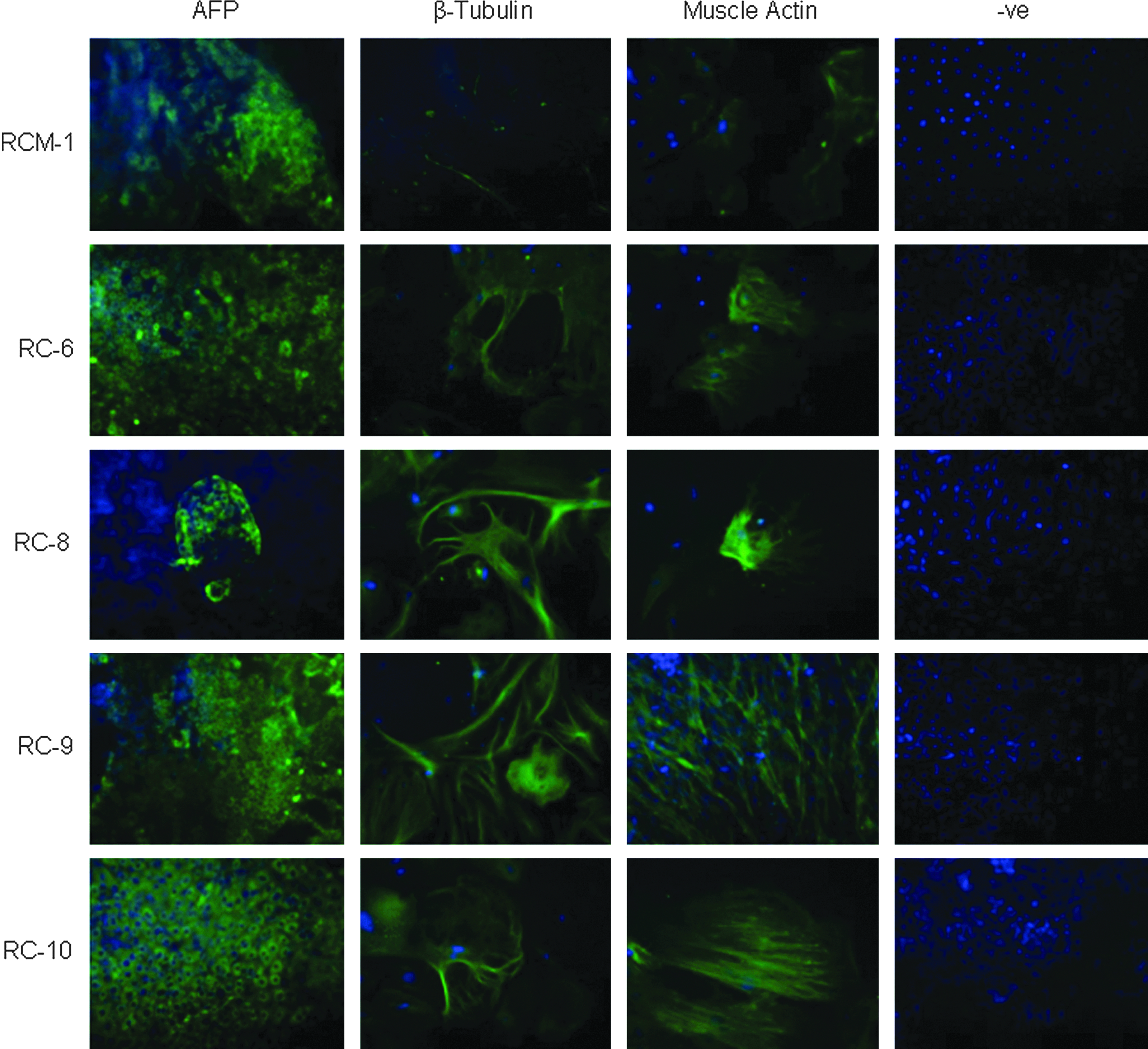
Immunofluorescence demonstrates expression of markers of ectoderm (β-tubulin), mesoderm (muscle actin), and endoderm (AFP) in EBs formed from five hESC lines. −ve, Negative. Color images available online at www.liebertpub.com/cell
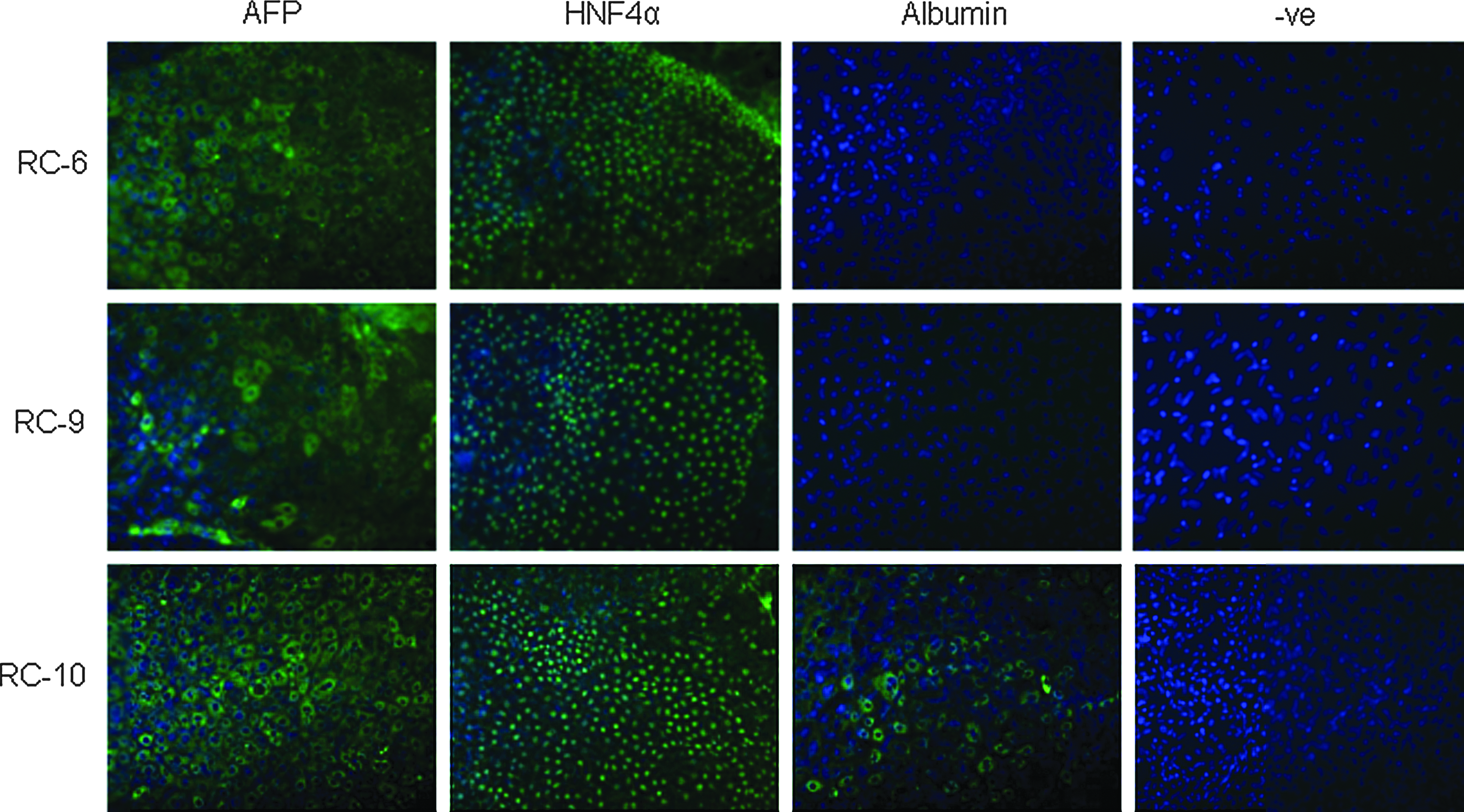
Immunofluorescence demonstrates expression of HNF4α and repeated expression of AFP in the three cell lines producing the highest abundance of AFP-positive cells in Fig. 1. Further staining indicates that the RC-10 cell line alone expressed albumin. Methods were as described for Fig. 1. −ve, Negative. Color images available online at www.liebertpub.com/cell
Significantly better response to directed hepatocyte differentiation observed in the RC-10 hESC line
Our subsequent experiment focussed on whether albumin expression from EB formation was an indicator of a cell line's ability to produce HLCs from single-cell preparations. Cells were dissociated to a single-cell suspension using TrypLE Select (Life Technologies) and seeded onto Matrigel (BD Biosciences)-coated 12-well plates at a density of 2×10⁵ cells/well, corresponding to 5.26×10⁴ cells/cm2. At 24 h after replating, hESCs were differentiated to hepatocytes using a well-established directed differentiation protocol (Hay et al., 2008; Medine et al., 2011). Following differentiation, the efficiency of hepatocyte production was examined using albumin immunostaining. RC-10 hESCs exhibited a high level of hepatocyte differentiation (93±3%) whereas RC-6 (4±3%) and RC-9 (13±5%) responded significantly less well (Fig. 3). Results are given as the mean percentage of albumin positive cells from four fields of view±standard deviation (SD).
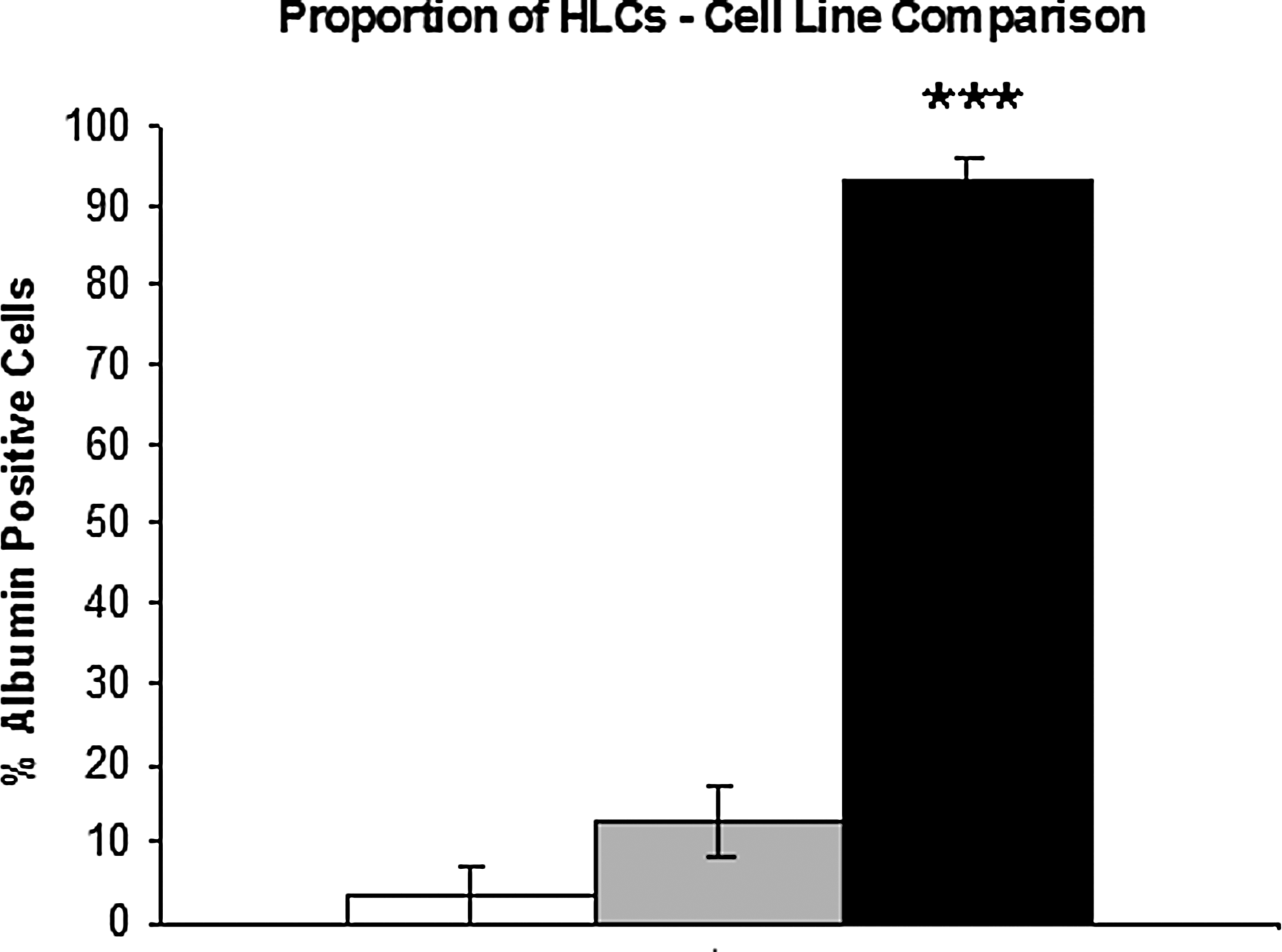
Quantification of albumin-positive cells indicates substantially higher efficiency of RC-10 to differentiate to HLCs (black bar) over RC-6 (white bar) and RC-9 (grey bar) cells. The significance levels of these comparisons were measured using the Student t-test. (***) p value of <0.001.
Single-cell seeding and directed differentiation of RC-10 cells produces HLCs displaying mature hepatocyte markers and hepatocyte function
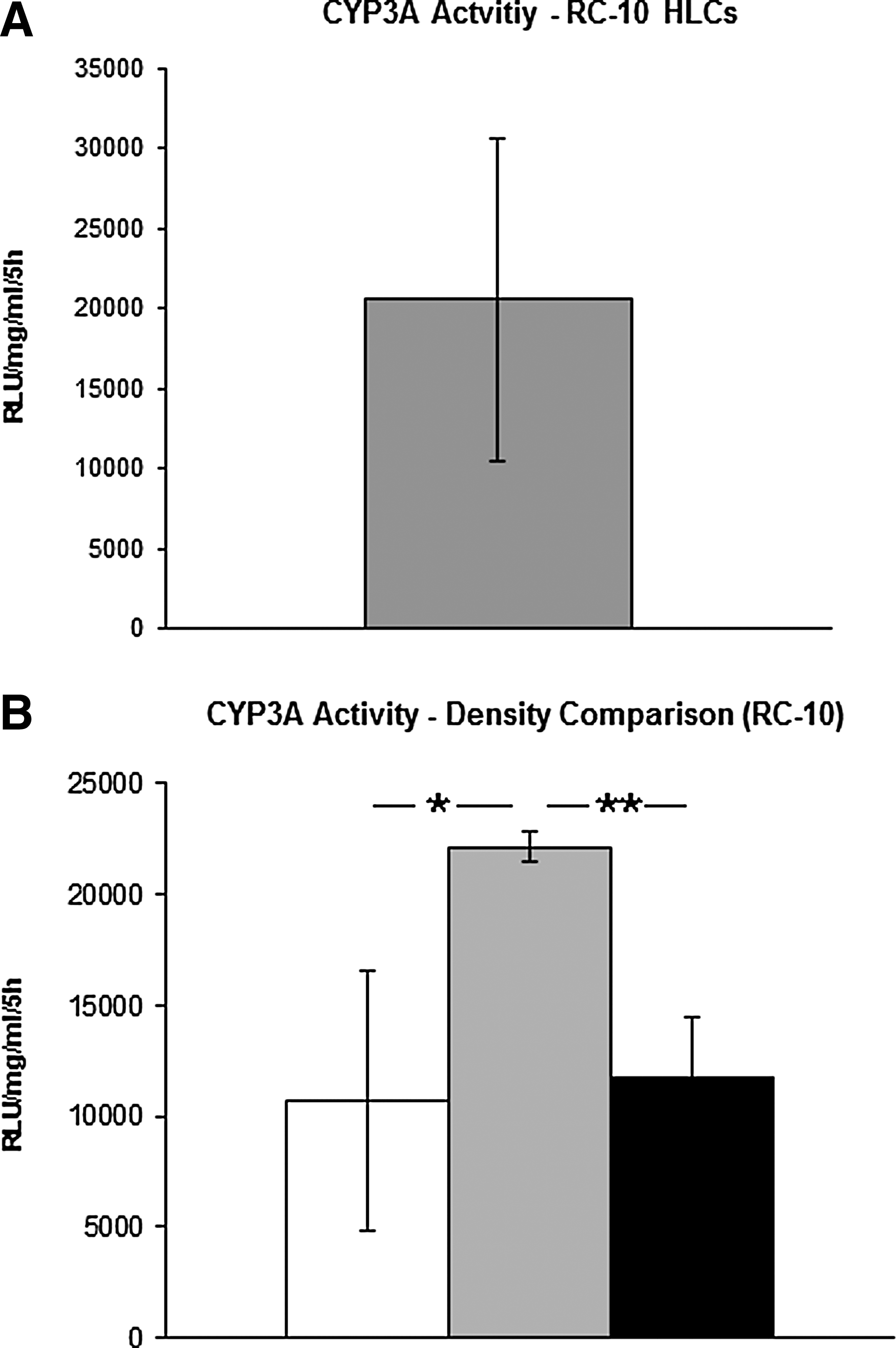
Representative phase-contrast photographs (10×) were taken throughout the directed differentiation protocol (Fig. 4). At 24 h after seeding, cells began to expand and gradually took on a hepatic cell shape. On the final day, the majority of cells demonstrated typical hepatocyte morphology—large and angular with visible tight junctions, some of which contained multiple nuclei. The differentiated HLCs uniformly displayed the hepatocyte markers albumin and HNF4α, and tight junction formation was demonstrated by zonula occludens-1 (ZO-1) expression (Fig. 5). In addition to marker expression, stem cell–derived HLCs exhibited CYP3A function (Fig. 6A). Results are representative of four experimental replicates±SD.

Morphology of RC-10 hESCs following single-cell replating and directed differentiation to hepatocyte-like cells. On day 0, cells displayed elongation and began to cluster together. During the protocol, the cells took on a hepatic shape, with the final population demonstrating large, angular morphology, tight junction formation, and some instances of multiple nuclei. Photographs were taken using phase-contrast microscopy at 10× magnification.
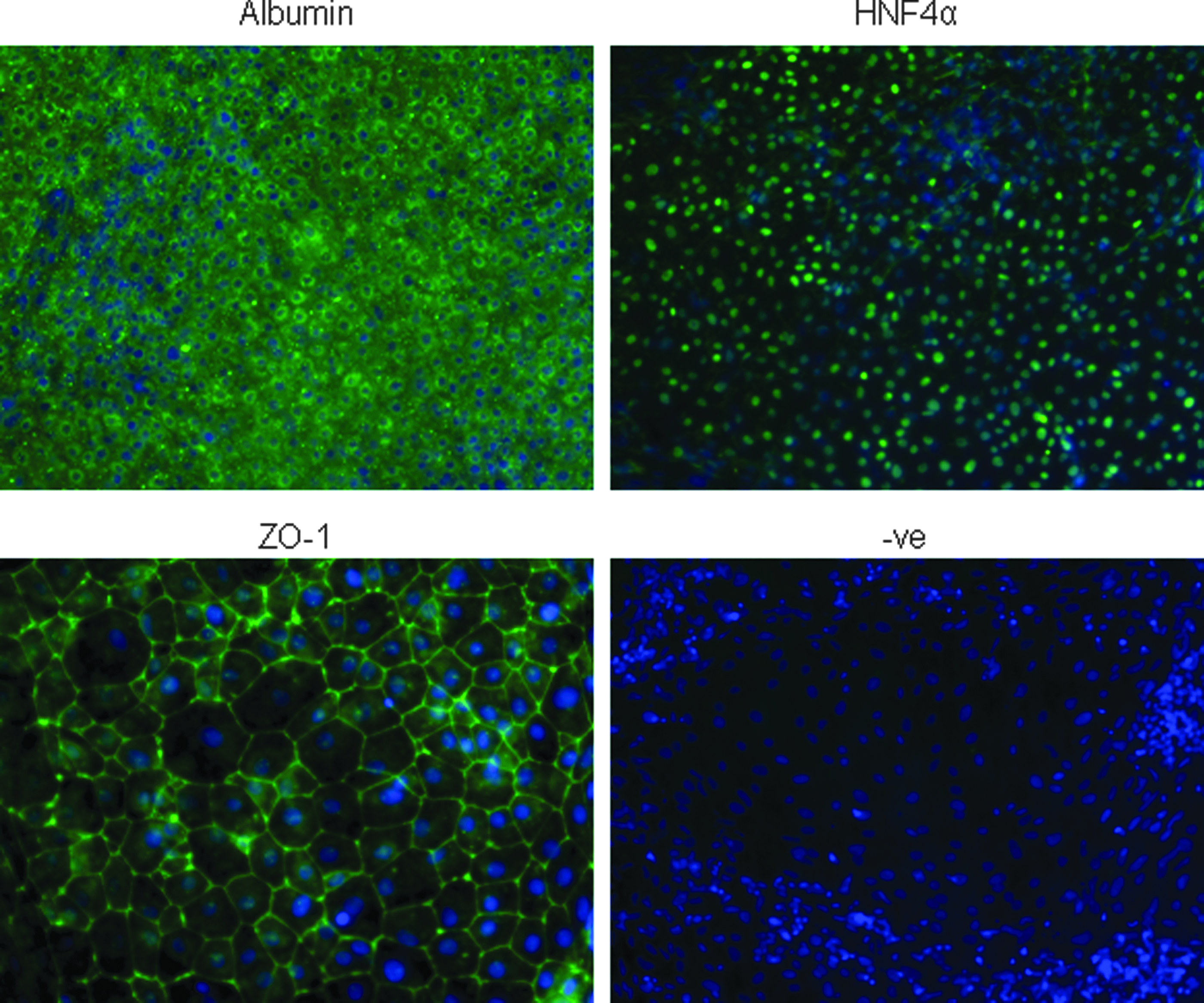
Immunofluorescence demonstrates expression of albumin, HNF4α and ZO-1. Markers of interest are shown in green, while nuclear staining with DAPI is shown in blue. Photographs of albumin and HNF4α were taken at 10× magnification. ZO-1 was photographed at 20× to highlight morphological detail. −ve, Negative. Color images available online at www.liebertpub.com/cell
(
Seeding density is critical to the functional profile of RC-10–derived HLCs
To examine the effect of cell density on stem cell–derived HLC function, we assessed three different seeding densities (1×10⁵, 2×10⁵, and 3×10⁵ cells per well). The results demonstrate that a 50% variance in seeding density led to significant alterations in CYP3A activity, highlighting this as an important variable in our process (Fig. 6B). Results are given as the mean of three experimental replicates±SD.
Large-scale production of somatic cells from pluripotent stem cell populations will require tissue culture formats suited to automation. Mechanical, colony-based passaging has been important in establishing growth conditions that support long-term culture and maintenance of stable karyotype, but it also introduces variability in the starting populations of cells undergoing differentiation. In reporting the production of HLCs from single-cell suspensions of hESCs seeded at defined densities, we have demonstrated a necessary technical advance required for the translation of pluripotent stem cell technology. We have also highlighted seeding as a critical stage in culture definition, which will require close attention if functional somatic cells are to be produced by automation.
We believe that the hESC screening protocol described could also be employed to select iPSC lines for hepatocyte differentiation. Moreover, our approach could be extended to the differentiation of other cell types from pluripotent stem cells. Exploration into other facets of cell biology, such as epigenetic modification, microRNA expression (Heinrich and Dimmeler, 2012), large intergenic noncoding RNA function (Guttman et al., 2011), and posttranslational modification (Zhou et al., 2012), may offer further insights into cellular pluripotency and differentiation. Meanwhile, improvements in stem cell scale-up are reported regularly, and two recent publications have described the maintenance of pluripotent cells in suspension culture (Olmer et al., 2010; Zweigerdt et al., 2011). These are exciting developments and offer significant advantages over the limitations imposed by adherent cell culture. In combination with defined and scalable directed differentiation, these advances are likely to improve the quality and cost-effectiveness of cell production for basic research and its application.
We believe our study has provided a major step forward to producing hepatocyte like cells in large quantities. However, the optimization of hepatic function from single-cell preparations remains an important consideration, and this will require further attention if we are to generate comparable metabolic function to our current gold standard stem cell–derived HLCs (Szkolnicka et al., 2013).
Footnotes
Acknowledgments
Mr. Sebastian Greenhough was supported by a Knowledge Transfer Partnership award between Roslin Cellab Limited and the University of Edinburgh. Dr. David Hay was supported by a Research Councils UK (RCUK) fellowship.
Author Disclosure Statement
Dr. David Hay is the chief scientific officer, director and shareholder of FibromEd Limited. John Gardner and Helen Bradburn are employees of Roslin Cellab Limited. Sebastian Greenhough was an employee of Roslin Cellab Limited during this project.