
Abstract
Abstract
Differentiating stem cells using gene delivery is a key strategy in tissue engineering and regenerative medicine applications. Nonviral gene delivery bypasses several safety concerns associated with viral gene delivery; however, leading nonviral techniques, such as electroporation, subject cells to high stress and can result in poor cell viabilities. Inhibition of Rho-associated coiled-coil kinase (ROCK) has been shown to mitigate apoptotic mechanisms associated with detachment and freezing of induced pluripotent stem cells and embryonic stem cells; however, inhibiting ROCK in mesenchymal stromal cells (MSCs) for improving gene delivery applications has not been reported previously. In this study, we hypothesized that ROCK Inhibitor (RI) would improve cell viability and gene expression in primary human umbilical cord mesenchymal stromal cells (hUCMSCs) when transfected via Nucleofection™. As hypothesized, the pre-treatment and post-treatment of hUCMSCs transfected via nucleofection with Y-27632-RI significantly improved survival rates of hUCMSCs and gene expression as measured by green fluorescent protein intensity. This study provides the first comparative look at the effect of Y-27632-RI on hUCMSCs that underwent transfection via nucleofection and shows that using Y-27632-RI in concert with nucleofection could greatly enhance the utility of differentiating and reprogramming hUCMSCs for tissue engineering applications.
Introduction
M
Over the past 5 years, several observations have been made regarding stem cell apoptosis related to detachment and freezing protocols as related to RhoA guanosine triphosphate (GTP) signaling pathways (Ohgushi and Sasai 2011; Xu et al., 2012; Zhang et al., 2011; Zhang et al., 2013). In particular, several research groups have noted that inhibition of the Rho-associated coiled-coil kinase (ROCK) appeared to increase cell survival by mitigating negative effects associated with cell dissociation and thawing (Claassen et al., 2009; Emre et al., 2010; Gauthaman et al., 2010a; Gauthaman et al., 2010b; Shi et al., 2013). The Y-27632 ROCK Inhibitor (Y-27632-RI) appeared to be especially useful for improving stem cell viability in human induced pluripotent cells (iPSCs) and embryonic stem cells (ESCs) (Joo et al., 2012; Narumiya et al., 2000). Furthermore, the use of Y-27632-RI was shown not to affect the pluripotency of ESCs (Watanabe et al., 2007). Chatterjee et al. (2011) were the first group to use RI to aid in the transfection of human iPSCs via Nucleofection™, an electroporative technique developed by Lonza Group Ltd. (Basel, Switzerland).
Human umbilical cord mesenchymal stromal cells (hUCMSCs) have a number of advantages over other cell sources and hold great potential for clinical translation, as we have reviewed extensively (Bailey et al., 2007a; Wang et al., 2009). Unfortunately, hUCMSCs are difficult to transfect, and few studies are available on the transfection of hUCMSCs. On the basis of its aforementioned success in other applications, we hypothesized that Y-27632-RI would improve cell viability and transfection efficiency for hUCMSCs that are transfected via nucleofection. In this study, transfection efficiency, gene expression, and cell viability were evaluated for hUCMSCs transfected via nucleofection with green fluorescent protein (GFP), with or without Y-27632-RI.
Materials and Methods
Procurement and expansion of hUCMSCs
hUCMSCs were isolated from Wharton's jelly of human umbilical cords obtained from the Kansas University Medical Center Hospital (IRB# 10951), Lawrence Memorial Hospital (IRB# LMH 08-2), and Stormont-Vail Hospital (IRB approved, no reference number) for a total of five cords used in the study (n=5). Four cords were from males that were born at full term and one cord was from a female born at 38.3 weeks, all under normal delivery conditions. Maternal age was not available. We isolated hUCMSCs according to our previous published protocol (Devarajan et al., 2013). hUCMSCs were cultured in traditional hUCMSC medium [10% fetal bovine serum (FBS-MSC Qualified) and 1% penicillin-streptomycin in low glucose Dulbecco's Modified Eagle Medium (DMEM); Life Technologies, Grand Island, NY, USA)]. hUCMSC medium was changed three times per week, and hUCMSCs were maintained at 37°C with 5% CO2 in a cell culture–grade incubator. hUCMSCs were trypsinized with 0.05% trypsin-EDTA (1×) (Life Technologies) at 80–90% confluency. All hUCMSCs were expanded to passage 2 (P2) for the experiments. Five umbilical cords (n=5) in total were used for this study. All experiments were performed in triplicate for each cord.
Cell characterization
At P2, a subculture of cells from each cord was characterized through cell-surface marker identification via flow cytometry on a MoFlo XDP fluorescence-activated cell sorter (FACS) (Beckman Coulter, Brea, CA, USA). hUCMSCs were characterized using the following antibodies and secondary antibodies: STRO-1 mouse immunoglobulin M (IgM) (2.5:200; 1 mg/mL; R&D Systems, Minneapolis, MN, USA); Alexa Fluor 568® rabbit anti-mouse IgG (2:200; 2 mg/ mL; Life Technologies); CD105 mouse IgG (2.5:200; 1 mg/mL; R&D Systems); Qdot® 525 donkey anti-mouse IgG (2:200; 1 μM; Life Technologies); human CD45 preconjugated to Qdot® 800 (2:200; Life Technologies); human CD73 preconjugated to fluorescein isothiocyanate (FITC) (5:200; BD Biosciences, San Jose, CA, USA); human CD34 preconjugated to Brilliant Violet (5:200; BD Biosciences); human CD90 preconjugated to allophycocyanin (APC) (5:200; BD Biosciences). A total of 20,000 events were recorded for each sample. Positive identification of cell markers was defined as fluorescent emission that exceeded the fluorescent threshold of cells stained with corresponding isotype (negative) controls. The isotype controls used in these studies were rabbit IgG Alexa Fluor 568, donkey IgG Qdot 525, IgG2 Qdot 800 (all from Life Technologies), and IgG1 FITC, IgG1 Brilliant Violet, and IgG1 APC (all from BD Biosciences). The cell characterization experiments were repeated three times for each cord.
RI treatments and transfection
On the day of transfection, medium from all wells was removed, and cells were washed twice with phosphate-buffered saline (PBS). Afterward, cells were incubated for 1 h at 37°C in traditional hUCMSC medium (10% FBS-MSC Qualified/1% penicillin-streptomycin/low-glucose DMEM) or traditional hUCMSC medium with 10 μM of Y-27632-RI (Reagents Direct, Encinitas, CA, USA). After 1 h, hUCMSCs were washed twice with PBS, trypsinized, and then resuspended in 95 μL of 4D Nucleofector™ P1 Primary Solution (P1PS) (Lonza) at a density of 500,000 cells per 95 μL in a 50-mL conical tube (Phenix Research Products, Candler, NC, USA). 5 μL of P1PS or 5 μL of pmaxGFP (1 mg/mL; Lonza) was added to each sample, depending on the group, to bring the final cell suspension volume to 100 μL. hUCMSCs were separated into five groups consisting of three replicates per group. Groups 1 and 3 received no pmaxGFP. Groups 2, 4, and 5 each received 5 μL of pmaxGFP (1 mg/mL; Lonza). Groups 1, 2, 3, and 4 were cultured in traditional hUCMSC medium before and after transfection, while Group 5 was cultured in traditional hUCMSC medium with 10 μM of Y-27632-RI (Reagents Direct) before and after transfection. Our preliminary experiments revealed that a concentration of 10 μM of Y-27632-RI used both before and after transfection were preferred for effective transfection. hUCMSC suspensions from Groups 1 and 2 were immediately transferred to six-well plates (BD Biosciences) containing 1.5 mL of traditional hUCMSC medium and incubated at 37°C. hUCMSC suspensions from Groups 3, 4, and 5 were transferred to 100-μL 4D Nucleofection™ cuvettes. Each cuvette was gently tapped twice and then placed in a 4D Nucleofector™ (Lonza) and nucleofected with the program FF-104. hUCMSCs were incubated at room temperature (∼22°C) for 10 min and then transferred to a six-well plate (BD Biosciences) containing 1.5 mL of traditional hUCMSC medium (Groups 3 and 4) or traditional hUCMSC medium with 10 μM of Y-27632-RI (Reagents Direct) (Group 5) and incubated at 37°C.
Fluorescent microscopy
At 24 h and 48 h after transfection, hUCMSCs were collected for analysis. A 0.5-μL amount of Hoechst 33342 dye (Life Technologies) was added to each well, and hUCMSCs were incubated for 10 min at 37°C. Afterward, hUCMSCs were imaged using an Olympus IX81 inverted epifluorescence microscope (Olympus America, Center Valley, PA, USA) with an Olympus LUCPlanFL 20X 0.4 NA objective (Olympus). Images were captured using the software, SlideBook (Intelligent Imaging Innovations (3i), Denver, CO, USA). A mercury arc lamp was used with the following excitation filters (Excitation/Emission) for image collection: Hoechst (387±11 nm/447±60 nm) and GFP (494±20 nm/531±22 nm). For each sample that was imaged, a montage was generated from 25 (five by five arrangement) neighboring fields of view that were aligned together to generate one comprehensive composite image of the sample. All experiments were repeated three times for each umbilical cord at 24 h and 48 h.
FACS analysis
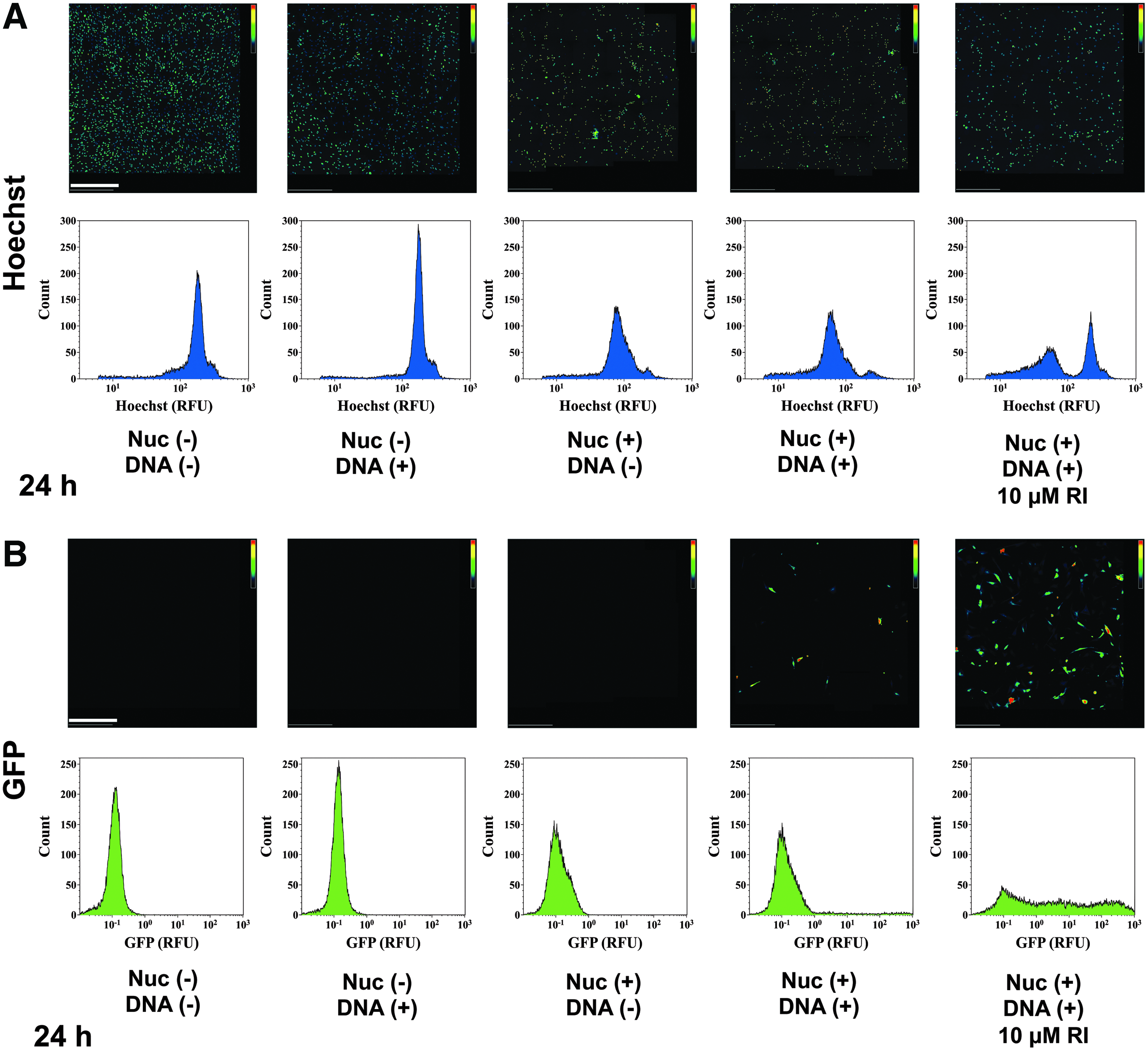
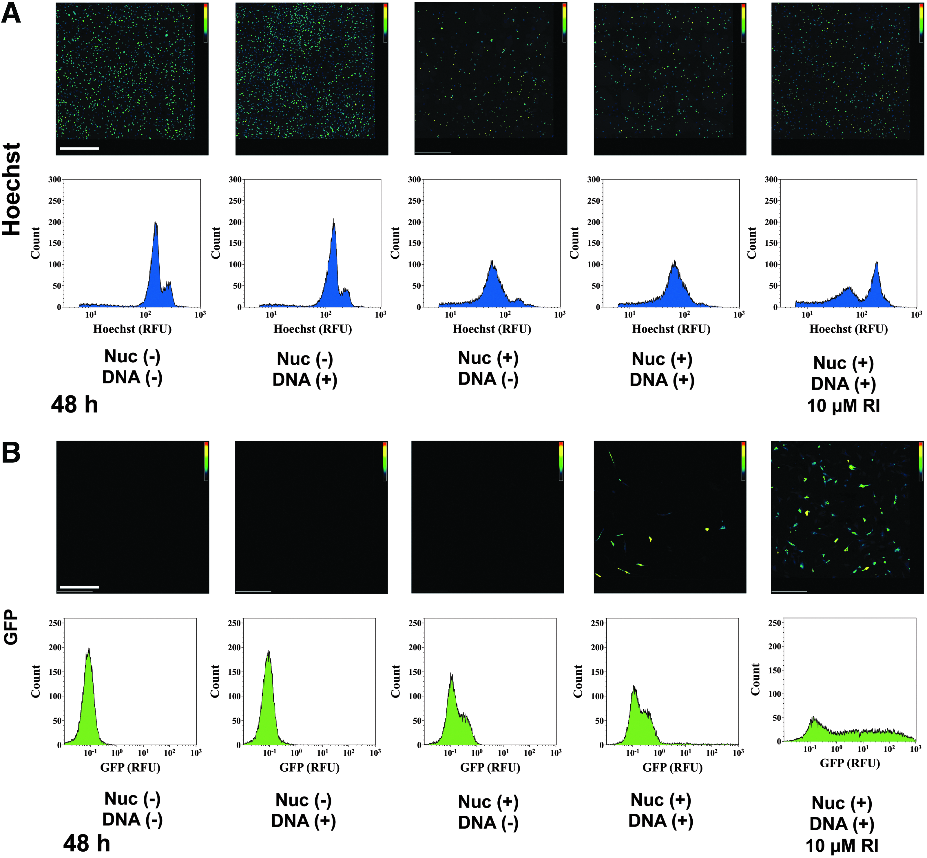
Immediately after imaging, cells were washed twice, trypsinized, and transferred into 5-mL polypropylene round-bottomed tubes (BD Biosciences). A 0.5-μL amount of propidium iodide (PI) (1 mg/mL; Life Technologies) was added to each sample just before analysis. hUCMSCs were analyzed via flow cytometry on the Beckman Coulter MoFlo XDP FACS. A total of 20,000 events were recorded for each sample analyzed. Flow cytometry was used to analyze both cell viability and transfection efficiency. Live hUCMSCs were characterized as hUCMSCs expressing Hoechst at an intensity of 102 relative fluorescence units (RFU) or above, with expression of PI at an intensity below 100 RFU. Dead hUCMSCs were characterized as hUCMSCs that expressed Hoechst at an intensity below 102 RFU and expressed PI at an intensity of above 100 RFU. GFP-positive hUCMSCs were characterized as live hUCMSCs that expressed GFP at an intensity of 100 RFU or greater. Transfection efficiency was determined by dividing the number of live GFP-positive cells in a sample by the total population of the sample. All experiments were repeated three times for each cord at 24 h and 48 h post-transfection. An example of how cell populations were gated is provided in Figure 1 and the statistics for all samples from an entire umbilical cord are displayed in Table 1.

Flow cytometry gating parameters used to quantify cell numbers. The set of histograms displayed are an arbitrary selection of a single replicate for each treatment from one umbilical cord out of five tested. Nuc (+/−) designates whether cells were Nucleofected™ or not. DNA (+/−) designates whether cells received 5 μg of pmaxGFP or not. RFU, relative fluorescence units; RI, ROCK Inhibitor. Color images available online at www.liebertpub.com/cell
RI, Rock Inhibitor.
Statistics
All values are reported as means±standard deviations. A one-way analysis of variance (ANOVA) was performed with a Tukey's post hoc test to assess statistical significance with n=5 cords. Statistical significance was set at p<0.05.
Results and Discussion
As hypothesized, hUCMSCs treated with RI displayed significantly increased survival rates and transfection efficiencies after nucleofection than hUCMSCs that were not treated with RI. Gene delivery is a powerful tool for reprogramming hUCMSCs as demonstrated by Baksh et al. (2007) and Devarajan et al. (2013). Until now, nonviral delivery methods have suffered from poor transfection efficiency with high cell viability or high transfection efficiency with poor cell viability. RhoA GTP signaling pathways are critical for inducing several apoptotic mechanisms in response to unfavorable environmental changes (Ichikawa et al., 2013; Ohgushi et al., 2010). Inhibiting ROCK can reduce apoptosis associated with detachment and freezing protocols (Harb et al., 2008; Ichikawa et al., 2012; Koyanagi et al., 2008; Kurosawa, 2012; Olson, 2008). One of the key disadvantages of using nonviral physical delivery methods such as electroporation is the need to dissociate cells from adherent surfaces. However, by using Y-27632-RI to mitigate some of the apoptotic mechanisms induced by cell detachment and electric shock, it might be possible to rescue positively transfected cells from cell death, which was the primary goal of this study.
Flow cytometry analysis revealed that the cell populations were mostly nonhematopoietic because the hUCMSCs were 98.1±0.04% negative for CD34 expression and 90.4±1.1% negative for CD45 expression. The expression of CD90, a key mesenchymal stem cell marker, was detected in 98.5±0.54% of cells. The expression of remaining key mesenchymal stem cell markers, CD73 (12.0±7.8%), CD105 (10.2±0.15%), and STRO-1 (3.4±0.33%), were low and relatively variable, suggesting subpopulations may exist within each cell population that displayed surface epitopes consistent with mesenchymal stem cell markers, as reviewed by (Wang et al., 2009; Wang et al., 2011).
As was seen in the microscopy images at 24 h (Fig. 2A) and 48 h (Fig. 3A) after transfection, hUCMSCs that were not subjected to nucleofection displayed high cell densities compared to hUCMSCs that were subject to nucleofection. However, hUCMSCs that were treated with 10 μM of Y-27632-RI before and after transfection displayed a greater cell density than hUCMSCs that were subject to nucleofection and not treated with Y-27632-RI, as shown in microscopy images and corroborated by flow cytometry data.
(
(
There was a clear increase in both cell viability and transfection efficiency between the experimental group of hUCMSCs that was nucleofected and treated with Y-27632-RI and the group of hUCMSCs that was nucleofected and not treated with Y-27632-RI at both 24 h and 48 h after transfection (Fig. 4). Cell viability was 3.3 times greater in hUCMSCs treated with Y-27632-RI than hUCMSCs that were not treated with Y-27632-RI 24 h after transfection (p<0.05), whereas cell viability was 3.2 times greater in hUCMSCs treated with Y-27632-RI than hUCMSCs not treated with Y-27632-RI 48 h after transfection (p<0.01). Transfection efficiency was 4.6 times greater in hUCMSCs treated with Y-27632-RI than hUCMSCs not treated with Y-27632-RI 24 h after transfection (p<0.01). At 48 h after transfection, transfection efficiency was 4.8 times greater in hUCMSCs treated with Y-27632-RI than hUCMSCs not treated with Y-27632-RI (p<0.05).
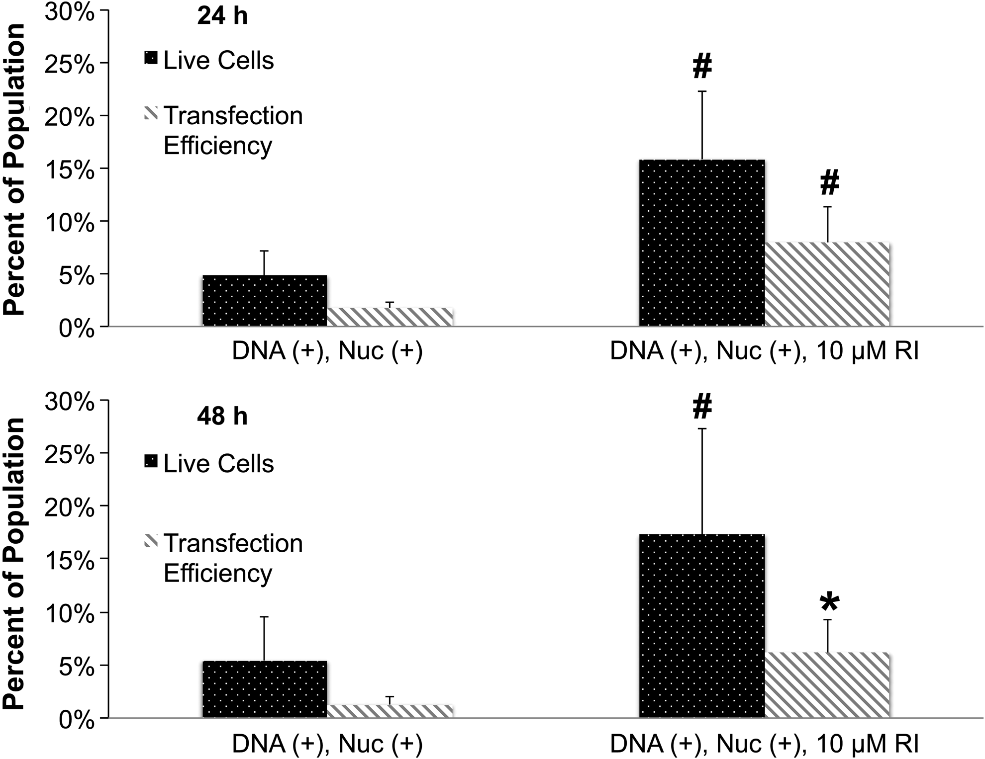
Live/dead and transfection efficiency collected via flow cytometry 24 h and 48 h after transfection. Groups that did not express GFP were not plotted in the bar chart. (*) Statistically significant difference (p<0.05) from hUCMSCs that underwent Nucleofection™ without RI supplement; (#) statistically significant difference (p<0.01) from hUCMSCs that underwent Nucleofection™ without RI supplement. The results are representative of cells collected from five different umbilical cords (n=5) and are reported as statistical means. All experiments were repeated three times. Error bars represent standard deviations. Nuc (+/−) designates whether cells were Nucleofected™ or not. DNA (+/−) designates whether cells received 5 μg of pmaxGFP or not. RI, ROCK Inhibitor.
The difference in GFP intensity may have been a result of an increased number of live cells present and able to readhere to a surface when treated with Y-27632-RI as opposed to hUCMSCs that were not treated with Y-27632-RI. The flow cytometry histograms were consistent with microscopy images in showing that a greater number of hUCMSCs treated with Y-27632-RI survived and expressed GFP at varying intensities at both 24 h (Fig. 2B) and 48 h (Fig. 3B) after transfection than hUCMSCs that were not treated with Y-27632-RI. Furthermore, the data from the histograms suggested that there might be a relationship between GFP expression and cell density. Further formal studies are needed to verify if an actual relationship exists.
hUCMSCs treated with Y-27632-RI at both 24 h and 48 h post-transfection displayed an increase in cell viability and a far more substantial increase in transfection efficiency compared to hUCMSCs that were not treated with Y-27632-RI. Thus, future studies are needed to explore whether a synergistic phenomenon is occurring in which Y-27632-RI is not only rescuing dying cells, but also improving cell health to facilitate expression of GFP in cells that may not have been previously able to express GFP. Further long-term studies are needed to determine whether Y-27632-RI can prolong and sustain gene expression in hUCMSCs. Additionally, follow-up studies are needed to determine whether Y-27632-RI can negatively affect multipotency character and downstream differentiation of hUCMSCs for tissue engineering applications (Watanabe et al., 2007; Pakzad et al., 2010).
For the first time, it was demonstrated that Y-27632-RI enhanced survival and gene expression in mesenchymal stromal cells for an electroporative gene delivery strategy. Transfection efficiency significantly increased four-fold and cell viability increased three-fold in hUCMSC populations that were treated with 10 μM of Y-27632-RI before and after transfection compared to hUCMSC populations not treated with Y-27632-RI. Although this study focused on hUCMSCs and provides an example for evaluating the effect of Y-27632-RI, other cell types undergoing electroporation may benefit from Y-27632-RI treatment, although dosing levels, application of treatment, and timing of treatment should be tailored for each cell type and application. The use of Y-27632-RI provides an opportunity to benefit strategies that combine both stem cell therapy and gene therapy for regenerative medicine applications.
Footnotes
Acknowledgments
We would like to acknowledge the efforts of the nursing staffs at the University of Kansas Hospital (Kansas City, KS), Lawrence Memorial Hospital (Lawrence, KS), and Stormont-Vail (Topeka, KS) for assisting us in obtaining human umbilical cords. Furthermore, we would like to acknowledge the efforts of Peggy Keefe and Austin Smith for their assistance on this project. This project was funded by the National Institutes of Health (NIH) and the state of Kansas.
Author Disclosure Statement
The authors declare that there are no conflicts of interest.