
Abstract
Abstract
Mesenchymal stem cells (MSCs) exhibited self-renewal and less differentiation, making the MSCs promising candidates for adult somatic cell nuclear transfer (SCNT). In this article, we tried to produce genome identical pigs through hand-made cloning (HMC), with MSCs and adult skin fibroblasts as donor cells. MSCs were derived from either adipose tissue or peripheral blood (aMSCs and bMSCs, respectively). MSCs usually showed the expression pattern of CD29, CD73, CD90, and CD105 together with lack of expression of the hematopoietic markers CD34and CD45. Flow cytometry results demonstrated high expression of CD29 and CD90 in both MSC lines, while CD73, CD34, and CD45 expression were not detected. In contrary, in reverse transcription-polymerase chain reaction (RT-PCR) analysis, CD73 and CD34 were detected indicating that human antibodies CD73 and CD34 were not suitable to identify porcine cell surface markers and porcine MSC cellular surface markers of CD34 might be different from other species. MSCs also had potential to differentiate successfully into chondrocytes, osteoblasts, and adipocytes. After HMC, embryos reconstructed with aMSCs had higher blastocyst rate on day 5 and 6 than those reconstructed with bMSCs and fibroblasts (29.6% ± 1.3% and 41.1% ± 1.4% for aMSCs vs. 23.9% ± 1.2% and 35.5% ± 1.6% for bMSCs and 22.1% ± 0.9% and 33.3% ± 1.1% for fibroblasts, respectively). Live birth rate per transferred blastocyst achieved with bMSCs (1.59%) was the highest among the three groups. This article was the first report to compare the efficiency among bMSCs, aMSCs, and fibroblasts for boar cloning, which offered a realistic perspective to use the HMC technology for commercial breeding.
Introduction
T
Pigs are abundant with fat that could be removed by biopsy of the adipose tissue through a small incision. Adipose MSC cell lines could be established by using a standard procedure. On the other hand, PB is considered the most appropriate source of MSCs because of convenient sampling and noninvasive procedure. Accordingly, we have used selected 1-year-old elite boars from different breeds, including Large Whites (LW-), Landraces (L-), and Durocs (D-) as somatic cell donors. All donor animals were purebred with documented lineage. MSCs were established from both PBs and adipose tissues and characterized with the above-mentioned markers. After SCNT, both in vivo and in vitro developmental competence of reconstructed embryos were observed and compared between different cell lines, including aMSCs, bMSCs, and subcutaneous fibroblasts derived from ear samples.
Materials and Methods
Except where otherwise indicated, all chemicals were obtained from Sigma-Aldrich (St Louis, MO). All animal care-related procedures described in this study were reviewed and approved by The Institutional Review Board on Bioethics and Biosafety of BGI.
Isolation of donor bMSCs, aMSCs, and adult skin fibroblast
Somatic cell donors were from purebred adult boars. For bMSCs, 5–10 mL blood was collected into a plastic tube containing heparin from precaval vein. Samples were transported at 4°C within 18 hours. Mononuclear cells were separated as described earlier (Bosch et al., 2006). Briefly, whole blood was added over Histopaque® 1077 gradient (density, 1.007; Sigma-Aldrich) in 15 mL conical centrifuge tube (BD, Becton Dickinson, Lincoln Park, NJ) and then centrifuged at 400 g for 30 minutes. Nucleated cells were collected from the opaque interface and washed thrice with phosphate-buffered saline (PBS; Invitrogen, Grand Island, NY). The cell pellet was suspended in low-glucose Dulbecco's modified Eagle's medium (L-DMEM; Invitrogen) supplemented with 15% (v/v) fetal bovine serum (FBS; Invitrogen), MEM nonessential amino acids solution (NEAA; Invitrogen), 10 ng/μL recombinant human fibroblast growth factor basic (bFGF; Invitrogen), and 30 IU/mL heparin in the gelatin-coated dish (BD). The bMSC culture medium was replaced with the minimum essential medium α (MEM-α; Invitrogen) supplemented with 15% (v/v) FBS (Invitrogen), NEAA (Invitrogen), and 10 ng/μL bFGF (Invitrogen) 48 hours after the first seeding. Adherent fibroblast-like cells were allowed to grow for 7–14 days, with the secondary medium replacement every third day.
Adipose tissue samples (∼30 mm × 30 mm × 30 mm each) were obtained from the anterior abdominal wall of pigs by surgical blade under anesthesia. The tissue was finely minced into 2–4 mm pieces with sterilized scissors and digested with 200 IU/mL collagenase type IV (Invitrogen) for 1 hour at 37°C. The digested cell solution was pelleted by centrifugation at 300 g for 10 minutes, washed with PBS, and resuspended in the L-DMEM supplemented with 10% (v/v) FBS and cultured until confluence.
For fibroblasts, porcine ear tissue sample was cut into 1 mm3 pieces with a sterile scissor and washed with PBS several times. Pieces of tissue were placed on the bottom of culture dish (BD). About 24 hours later, DMEM supplemented with 10% FBS was gently added. Fibroblasts grew out from explants within 5 days, and the medium was replaced every third day.
All types of cells were cultured in 5% CO2, at 38.5°C with maximum humidity. Once 90%–95% confluent, adherent cells were harvested by treatment with 0.25% (w/v) trypsin-EDTA solution (Invitrogen) and passaged with the split ratio 1:3. All experiments were performed using MSCs or fibroblasts after 3–10 passages.
Flow cytometry analysis
Cells were harvested by trypsinization and washed with PBS. After resuspension in PBS to achieve 1 × 10⁵ concentration, cells were labeled with PE-conjugated anti-CD29, anti-CD90, anti-CD34, and anti-CD73, and FITC-conjugated anti-CD45, respectively. All antibodies were from human species resource due to the lack of corresponding antibodies in porcine, obtained from eBioscience, San Diego, CA. Cells were stained with FITC- and PE- isotype mouse IgG1 (eBioscience) as control. Samples were individually run through flow cytometry (BD FACSCalibur, Lincoln Park, NJ). Typically for each tube, more than 10,000 cell populations were collected and data were analyzed by FlowJo software (Treestar, Inc., San Carlos, CA).
Reverse transcription-polymerase chain reaction (RT-PCR) analysis
Total RNA of cultured cells was extracted with TRIzol® Reagent (Invitrogen). Briefly, 5 × 10⁵ cells were lysed with 500 μL TRIzol, the lysate was added with 100 μL chloroform, and centrifuged in 8000 g for 15 minutes at 4°C. The upper layer was collected and added with same volume isopropanol. The RNA pellet was formed after centrifugation with 8000 g for 15 minutes at 4°C, and then washed with 75% ethanol and dissolved in RNase-free H2O. cDNA was transcribed with the PrimeScript™ II First-Strand cDNA Synthesis Kit (Takara, Dalian, Liaoning, China) and polymerase chain reaction (PCR) was performed with rTaq (Takara). PCR steps included denaturation at 95°C for 5 minutes, followed by 35 cycles at 95°C for 30 seconds, 60/65°C for 30 seconds, and 72°C for 30 seconds, and final extension at 72°C for 5 minutes. Primer sequences are listed in Table 1. We used umbilical cord and umbilical cord blood cDNA mix extracted from the umbilical cord tissue of the newborn piglet as positive control. Electrophoresis was carried out with 1.5% agarose gel for 30 minutes, and the gel was stained with ethidium bromide for 10 minutes.
MSC, mesenchymal stem cells.
Induction of multilineage differentiation
bMSCs and aMSCs were cultured in their traditional medium until an ∼90% confluence was obtained. Culture media were then replaced with the osteogenic medium (STEMPRO® Osteogenesis Differentiation Kit; Invitrogen) and chondrogenic medium (STEMPRO Chondrogenesis Differentiation Kit; Invitrogen), following the supplier's instructions. For inducing adipocytes, MSCs were cultured in L-DMEM containing 10% (v/v) FBS, 0.5 mM 1-methyl-3-isobutylxanthine, 1 μM dexamethasone, 10 mg/L insulin, and 0.1 mM indomethacin as described previously (Fu et al., 2012). All three types of differentiation media were refreshed every 2–3 days and the conditions were maintained for about 20 days. After differentiation, cells were fixed with 4% (w/v) formaldehyde solution and stained with 0.1% (w/v) Alizarin Red S, 1% (w/v) Alcian Blue, and 0.3% (w/v) Oil Red O for analyzing osteogenic, chondrogenic, and adipogenic lineage, respectively.
Hand-made cloning
As described earlier (Du et al., 2007), cumulus-oocyte complexes were collected from slaughterhouse-derived sow ovaries by slicing, then matured in bicarbonate-buffered TCM-199 supplemented with 10% (v/v) cattle serum (CS), 10% (v/v) pig follicular fluid, 1 IU/mL pregnant mare serum gonadotropin (PMSG; PROSPEC, Rehovot, Israel), and 5 IU/mL human chorionic gonadotropin (hCG; PROSPEC) at 38.5°C, 5% CO2 at maximum humidity for 42 hours. Cumulus cells were then removed by repeated pipetting in 1 mg/mL hyaluronidase in Hepes-buffered TCM-199. Zona removal and oriented enucleation were performed with a procedure based on partial digestion of the zona pellucida followed by oriented enucleation by hand using a sharp blade (Du et al., 2005; Kragh et al., 2004). Cytoplasts were fused with three types of donor cells using a fusion chamber (BTX microslide 0.5 mm fusion chamber, model 450; BTX, San Diego, CA). Fused pairs were incubated for 1 hour, and then a second fusion with another cytoplast was performed. Reconstructed embryos were incubated in the PZM-3 medium (Yoshioka et al., 2002) supplemented with 5 mg/mL CB and 10 mg/mL cycloheximide for 4 hours at 38.5°C in 5% CO2, 5% O2, and 90% N2 with maximum humidity, and then washed thoroughly with the in vitro culture (IVO) medium before culture. The embryos were cultured for 5 days and ready for transfer.
Embryo transfer
Sows that showed a standing estrous by applying back pressure reflex were considered to be in estrus and were used in the experiments. After 5 days, developed blastocysts were surgically injected into the joint of the oviduct and uterine horn with a flexible transfer catheter (Cat. 273466; Guangzhou Yongran Trading Co., Ltd., Guangdong, China). The average of embryos per transfer was 89, with the minimum of 51 and maximum of 141. Anesthesia was achieved by intramuscular injection of Compound ketamine (Shenyang veterinary pharmaceutical factory, Shenyang, China), followed by a gas mixture of 5% isoflurane (Hebei Jiupai Pharmaceutical Co., Ltd., Hebei, China), oxygen (0.8 L/min), and nitrous oxide (0.5 L/min). B-ultrasound (WED-2000AV; Shenzhen Well.D Medical Electronics Co., Ltd., Shenzhen, China) was performed once, 2 weeks from 30 days after transfer. Piglets were naturally delivered.
Cell number determination of D5 and D6 blastocysts
Cell numbers were determined when the reconstructed embryo developed to D5 and D6 blastocysts. Embryos were loaded on a microscope slide into a 2 μL glycerol drop containing 20 μg/mL Hoechst 33342, and covered carefully with a cover glass. The cell number was counted under Olympus IX51 inverted microscope (Olympus, Shinjuku, Tokyo, Japan) with U-MWU2 filter unit.
Statistical analysis
All statistical analyses were performed using the one-way ANOVA test with a 5% significance level. Results were expressed as mean ± standard error of the mean (SEM).
Results
Isolation of MSCs and fibroblasts
Three bMSC, two aMSC, and eight fibroblast primary cell lines were derived from individual blood or tissues (Table 2). The morphology of MSCs was similar to skin fibroblasts, with a rapid adherence to plastic and spindle-like morphology. In the beginning of culture of bMSCs, lots of anuclear red blood cells were attached on the substrate, but their number has decreased rapidly during the medium change. A few sparse bMSC groups appeared after 5 days in culture, and started colony formation after 10 days. A lot of aMSCs were attached to the dish bottom 24 hours after first seeding, and formed a confluent layer within 7 days. Skin fibroblasts migrated from the explant in 5 days and grew to confluence within 2 weeks. All three types of cells were passaged after having reached confluence by trypsinization and reseeding in a fresh 24-well plate (BD). Monolayers formed from bMSCs, aMSCs (Fig. 1), and fibroblasts could passage stably in vitro with the obvious nucleus and typical spindle-shape morphology. Donor cells used for hand-made cloning (HMC) were passaged no more than 10 times.

Phase contrast micrograph of cell line bMSCs–D1711 derived from peripheral blood at P1
Large Whites (-LW), Landraces (-L), and Durocs (-D).
a, b: Values in the same column with different superscripts are significantly different (p < 0.05).
Characterization of mesenchymal stem cells
To confirm phenotypic characteristics of cells from different cellular source as MSCs, we chose two bMSC cell lines (bMSCs—D1711 and bMSCs—LW3515) and one aMSC cell line (aMSCs—LW17009) randomly to detect surface markers with FACs analysis (Fig. 2). All these cell lineages were positive for CD29 (range from 92.4% to 98.5%) and CD90 (range from 87.2% to 96.3%) expressed in MSCs, whereas they were negative for both CD34 (below 2.69%) and CD45 (below 0.787%) as hematopoietic cell markers. CD73, which was positive in human MSCs, was not expressed in our cell lines of MSCs (Image not showed).
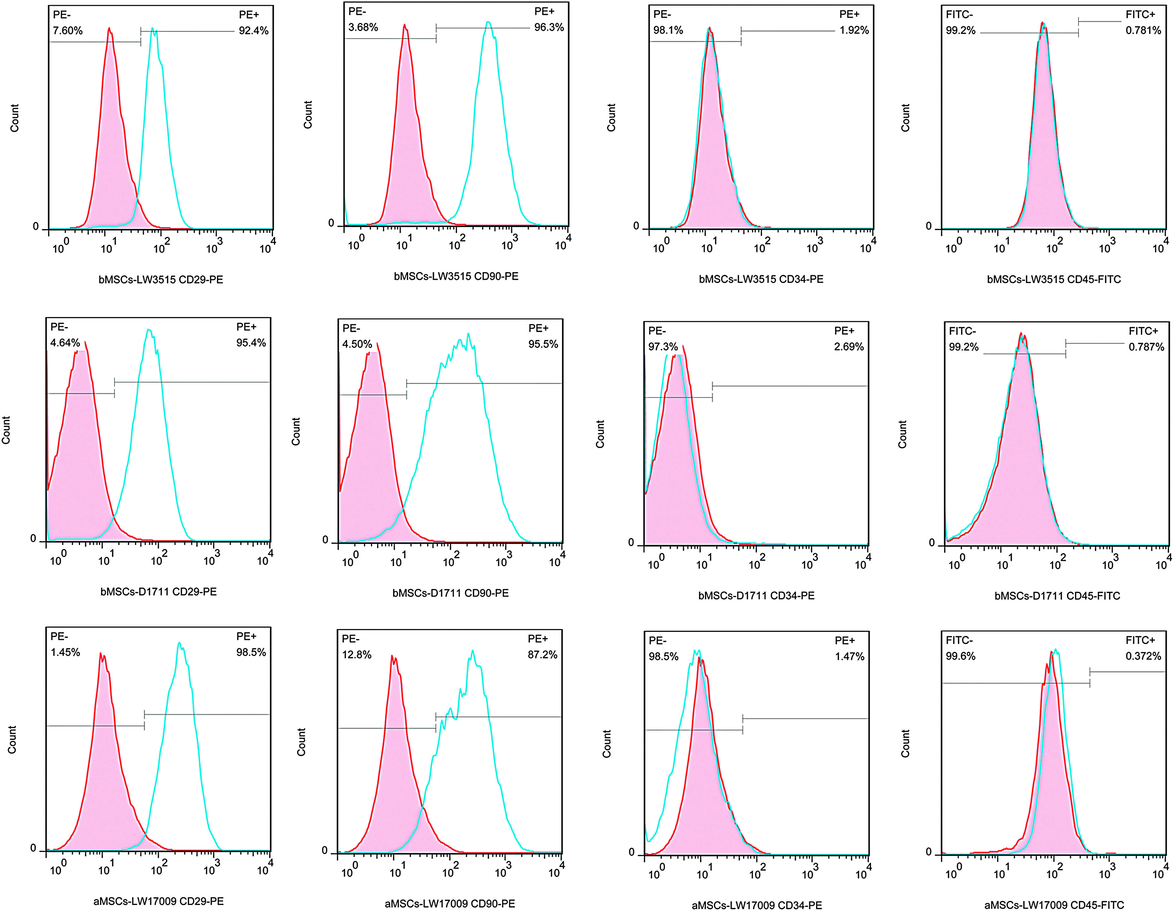
Flow cytometry analysis—Histogram plots of expression of cell surface markers. bMSCs—LW3515, bMSCs—D1711, and aMSCs—LW17009 cell lines were both positive for CD29 and CD90 and negative for CD34 and CD45. Histogram of red line tilted with pink was negative PE or FITC—isotype control, and blue line with none coloring filled was the tested sample.
We chose one cell line of each aMSCs and bMSCs (aMSCs—LW17009 and bMSCs—D1711) that had been tested by FACS analysis and another random sample (aMSCs—LW073504 and bMSCs—D7807) to test the RNA expression. Total four MSCs with one positive sample were detected with two hematopoietic cellular surface markers and five MSC markers that had been reported by previous reports (Dariolli et al., 2013; Li et al., 2014). All analyzed bMSCs and aMSCs retained similar PCR results with positive of mesenchymal markers of CD29, CD73, CD90, and CD105 and negative of the hematopoietic cell marker of CD45. However, they all expressed hematopoietic cell markers of CD34 (Fig. 3).

RT-PCR analysis of hematopoietic and endothelial cell markers of MSCs. aMSCs—LW073504, aMSCs—LW17009, bMSCs—D1711, and bMSCs—D7807 were positive for expression of CD29, CD34, CD73, CD90, and CD105, and negative for expression of CD45. Umbilical cord and umbilical cord blood cDNA mix as positive control. Positive and negative controls are indicated by P and N, respectively.
Multilineage differentiation
The differentiation potential of porcine MSCs was assessed by culturing under specific conditions. Twenty days after changing with the osteogenic-inducing medium, bMSC (bMSCs—D1711) and aMSC (aMSCs—LW17009) cell lines presented a strong staining with Alizarin Red, showed calcium deposition, and referred to successfully differentiate into osteoblasts (Fig. 4A, B). By inducing chondrocytes, monolayer MSCs trended to gather to clump after 3 days of culture with the differentiate medium. The clump could be stained with Alcian Blue, which suggested the presence of acidic mucosubstances and confirmed well the chondrogenic differentiation (Fig. 4C, D). For adipogenic inducing, the MSC morphology changed from spindle to more round shape under the conditional adipose-inducing medium, and cells were accumulated with intracellular lipid and stained positive with Oil Red O (Fig. 4E, F).

In vitro multilineage differentiation potential of MSCs derived from PB and adipose tissue. Twenty days after changing with osteogenic-inducing medium, bMSC (bMSCs—D1711) and aMSC (aMSCs—LW17009) cell lines presented a strong staining with Alizarin Red
In vitro development of reconstructed embryos
As shown in Table 2, three bMSCs, two aMSCs, and eight ear fibroblasts were used as donor for HMC, for more details see Table 2. When data were pooled together, embryos reconstructed with aMSCs had a higher blastocyst rate on day 5 and 6, compared to bMSCs and fibroblasts, (29.6% ± 1.3% and 41.1% ± 1.4% for aMSCs vs. 23.9% ± 1.2% and 35.5% ± 1.6% for bMSCs and 22.1% ± 0.9% and 33.3% ± 1.1% for fibroblasts, respectively). There was no difference in total cell number of bMSC-, aMSC-, and fibroblast-derived blastocysts on day 5 (54.0 ± 0.5 for bMSCs vs. 55.4 ± 0.5 for aMSCs and 55.4 ± 0.5 for fibroblasts), Table 2.
In vivo development after HMC
Pregnancy rates of porcine were higher in both bMSCs and aMSCs than fibroblasts (60.0% for bMSCs vs. 64.3% for aMSCs and 46.7% for fibroblasts). However, the farrowing and average live birth (No. of delivered live cloned piglets/transferred blastocysts) rates achieved with bMSCs were higher than that obtained with aMSCs and fibroblasts (50.0% and 1.59% for bMSCs vs. 35.7% and 0.81% for aMSCs, and 23.3% and 0.88% for fibroblasts; for, more details see Table 2). We obtained total 36 live cloned piglets, 31 from bMSCs, 12 from aMSCs, and 20 from fibroblasts, respectively; see Table 3 for more details. In addition, 10 mummies were delivered from four recipients: two from cell line bMSCs—D7807, delivered by one recipient and 8 cloned from cell line F—D5607 and delivered by three recipients.
Discussion
Fibroblasts are usually used as donor cells for porcine SCNT and separated from ear tissue. MSCs obtained from adipose tissue (Bosch et al., 2006) or PB (Faast et al., 2006) exhibited self-renewal and less differentiation, making the MSCs promising candidates for adult somatic cell nuclear transfer (NT). The most widely used culture medium for MSCs is low-glucose DMEM with 10% to 15% FBS. This culture medium was suitable for our aMSCs, however, not bMSCs. For bMSC culture, we used two-step procedure, which resulted in low red blood cell attachment and provided more space for MSCs to grow.
It was found that the blood contained a type of adherent fibroblast-like cells and appeared to be mesenchymal stem cells (Ab Kadir et al., 2012). It was also reported that surface markers of MSCs derived from human PB were similar to those derived from cord blood and bone marrow (Kassis et al., 2006; Kim et al., 2012). PB MSCs were an easy and convenient source for providing cells for SCNT, for cell therapy, or biotechnical research in other scientific fields.
In our study, phenotypic characteristics of porcine MSCs were not clearly defined, most probably due to the lack of cross reaction with human antibodies. Accordingly, we used both flow cytometry and RT-PCR for identification. Porcine MSCs have expressed CD29, CD90, and CD105 and negatively of CD45, in accordance with a previous study (Comite et al., 2010). Hemopoietic cell marker CD34 and matrix receptor CD73 could be detected by RT-PCR, but not by flow cytometry in our study, which indicated that human antibodies were not suitable to detect porcine cellular surface antigens CD34 and CD73. However, it was unexpected that porcine CD34 was expressed in our MSCs. CD34 is considered a kind of hematopoietic progenitor cell antigen, it is not expressed in human MSCs, but there were few reports about detection of CD34 expression in porcine MSCs using both FACs and RT-PCR, and it was also reported that ovine MSCs expressed CD34 (Lyahyai et al., 2012). We considered that cellular surface markers of porcine MSCs, especially CD34, may be different from those in other species. As the result of multilineage differentiation, our separated MSCs had the capability of differentiating into osteoblasts, chondrocytes, and adipocytes, which indicated that the adipose tissue and PB contain a population of multipotent cells. The surface markers of porcine MSCs, even human MSCs, were not definite until recently. It was sure that MSCs exhibited less differentiation, making the MSCs promising candidates for adult somatic cell NT, which might have a good effect on the cloned embryo development both in vitro or vivo.
Three PB MSCs, two adipose MSCs, and eight ear-derived fibroblasts were used in this study for HMC. D5 and D6 blastocyst rates were higher in aMSCs than bMSCs and fibroblasts, indicating a better preimplantation development of SCNT embryos from aMSCs. Similar results were also reported in previous studies (Lee et al., 2010; Li et al., 2013). This may explain that the expression profiling of aMSC-NT embryos is more similar to that of the in vivo-derived embryos compared to fetal fibroblast NT embryos (Kumar et al., 2007). MSCs with a relatively undifferentiated genome expression pattern might serve as suitable donors and could be more efficiently reprogrammed to reactivate expression of early embryonic genes in porcine NT (Kumar et al., 2007).
The pregnancy and farrowing rates achieved with bMSCs were higher than those obtained with aMSCs and fibroblasts. The donor cell lines of F-L6707 and F-D4803 that transferred into six recipients did not have any sign of pregnancy during the whole pregnancy cycle. In our experiment, fetuses of 13 recipients (including 2 fetuses of bMSCs, 4 of aMSCs, and 7 of fibroblasts) were absorbed in the uterine horns during the process of pregnancy. This phenomenon often happened in cloned fetuses in our laboratory (data not published). In total, we obtained 63 live cloned piglets, 31 from bMSCs, 12 from aMSCs, and 20 from fibroblasts, respectively. The live birth rate obtained with bMSCs also was higher than those with aMSCs and fibroblasts, indicating that embryos cloned with bMSC cells survived better in the late stage of fetal development.
In our study, adult boar cells were used as donors for SCNT, considering the future application in elite animal breeding. Adipose tissue and PB MSC cell lines could be obtained easily and embryos derived from these cells had good developmental potential in both preimplantation and gestation stages. MSCs were considered appropriate sources for donors in SCNT, especially for future application in agriculture.
Footnotes
Acknowledgments
We appreciate the contribution of members of our porcine cloning group, and thank the staff of our experiential station for good care of pregnant sows and cloned piglets. The study was supported by National Cultivate New Varieties of Genetically Modified Organisms Major Project (2014ZX0801007B-003), Shenzhen Municipal Basic Research Program (JCYJ20140717153620436), and Shenzhen Strategic Emerging Industry Development Special Fund (NYSW20130326010019).
Author Disclosure Statement
The authors declare that no conflicting financial interests exist.