
Abstract
Abstract
A major obstacle of widespread commercial application of bovine somatic cell nuclear transfer is the low overall efficiency, that is, healthy calf—late pregnancy per transferred embryo rate. In this study, we report a series of experiments with a limited number of embryos created with handmade cloning (HMC) and transferred without or after open pulled straw vitrification. Embryo reconstruction was performed by using in vitro matured oocytes and adult ear skin fibroblasts. In two experiments, a total of 53 D7 blastocysts were developed from 188 reconstructed embryos. Fresh transfer of seven blastocysts into six recipients has resulted in three early pregnancies, two of them developed over 90 days and eventually resulted in healthy calves (33% offspring/transfer rate). In another two experiments, a total of 11 D7 blastocysts were obtained from 36 reconstructed embryos. Out of these, eight have reexpanded 18 hours after vitrification and warming. Transfer of these blastocysts into eight recipients has resulted in four early pregnancies and two live births; 25% offspring/transfer rate. These results indicate that low overall efficiency may not be an intrinsic feature of cattle cloning, and selection of the right procedures may help to overcome the actual limitations.
Introduction
T
However, the principal problem was and still is the low overall efficiency of the process. For most practical purposes—especially for agricultural applications—the decisive parameter is the healthy offspring per embryo transfer rate. A considerable variation exists between species, but this rate is generally low, around 1% to 10%, much lower than that achieved with in vivo or in vitro fertilized counterparts. It means a little advancement since the initial success 20 years ago (Wilmut et al., 1997), with very modest improvements during the past decade.
Various efforts have been made to overcome this problem, including treatment of somatic cells with chemical or biological agents to facilitate reprogramming or to increase the ability of enucleated oocytes to induce the process; to find the optimal time and cell cycle stage for enucleation, fusion, and activation; and to optimize activation and the in vitro culture period afterward (Akagi et al., 2014; Kwon et al., 2017; Wilmut et al., 2015).
Many of these efforts resulted in improvement in the laboratory phase, but none of them had a confirmed and generally accepted positive effect on the in vivo development, especially on advancement to the late pregnancy period and birth of healthy offspring. It should be noted that—due to financial and technical reasons—very few research group had the possibility to proceed from the laboratory phase to transfers and in vivo follow-up of development. Accordingly, in most laboratories, somatic cell nuclear transfer is still done as described 20 years ago, with small modifications related to species-specific requirements.
Meanwhile, the rapid advancement in genomics and epigenomics has allowed to reveal considerable differences between the gene expression profiles of cloned embryos and their in vitro fertilized or in vivo produced counterparts (Niemann et al., 2008, 2011). As early abortions are usually associated with abnormalities in placental development (Ogura et al., 2013), intervention to correct the related abnormal gene expression profiles seems to be a feasible possibility (Loi et al., 2016). However, it may require manipulation of the genome function with possible additional biological, legal, and bioethical consequences.
Recent data indicate that in some species, an alternative nuclear transfer technique may also be efficient. A representative work in pigs based on more than 200,000 reconstructed embryos has reported around 75% pregnancy rates after transfers, and ∼75% of pregnancies resulted in healthy piglets, although the offspring per reconstructed embryo rate remained low (1.5%) (Liu et al., 2015).
Another group, using the same approach reported that transfer of more than 5000 porcine cloned embryos resulted in around 90% pregnancy rates, and 90% of these pregnancies resulted in healthy piglets. The number of piglets per embryo transferred was an impressing 15% (Callesen et al., 2014). The common feature of these experiments was that they used a simplified method for reconstruction called zona free or handmade cloning (HMC) (Peura et al., 1998; Vajta, 2007) and in vitro culture until the late morula/blastocyst stage. A recent comparative experiment has demonstrated that porcine embryos produced by HMC had better in vitro developmental rates and higher cell numbers than their traditionally cloned counterparts (Liu et al., 2017).
HMC was originally introduced in cattle and applied later successfully in other species, including sheep, pigs, goat, and water buffalo (Du et al., 2007; Khan et al., 2014; Peura and Vajta, 2003; Shah et al., 2009; Vajta et al., 2006). However, in spite of the promising efficiency in vitro (around and above 50% blastocyst per reconstructed embryo rates in cattle) (Vajta et al., 2003), during the past 17 years there was no opportunity to make a large-scale representative embryo transfer experiment. Accordingly, reported pregnancy and calving rates were sporadic and often obtained under suboptimal circumstances, including lack of appropriate incubators or ad hoc laboratory arrangements.
The aim of our study was to investigate the achievable healthy calf per transferred cloned embryo rate with a limited number of embryos under optimized laboratory conditions and with an experienced embryo transfer team. HMC was performed as described earlier with slight modification in activation protocol and maturation/embryo culture media. Both fresh and cryopreserved embryo transfers were performed to test the feasibility of the latter approach, as well.
Materials and Methods
Unless otherwise specified, all chemicals and disposables were obtained from Sigma Chemical Co. (St. Louis, MO), and Nunc (Roskilde, Denmark), respectively.
In vitro maturation of oocytes
Ovaries were obtained from a local slaughterhouse and transported to the laboratory in physiological saline at 33–35°C. Follicles between 3 and 6 mm diameter were punctured with a syringe and 18-gauge needle. After sedimentation, oocytes were collected in holding medium (TCM199 with 4 mM bicarbonate, 18 mM Hepes, and 50 μg/mL gentamicin) supplemented with 10% fetal bovine serum (FBS). Grade I and II oocytes with a homogeneous, dark cytoplasm and compact cumulus investment were matured in four-well dishes (25–30 oocytes per well) in bicarbonate-buffered TCM199 medium supplemented with 0.6 mM glutamine, 0.2 mM pyruvate, 0.01 U/mL follicle-stimulating hormone (FSH) and lutenizing hormone (LH), 1 μg/mL estradiol, 50 μg/mL gentamicin, 10 ng/mL epidermal growth factor (EGF), and 10% FBS at 38.5°C in 5% CO2 atmosphere for 21 hours.
Cytoplast preparation
Removal of cumulus cells was performed with vortexing in 0.3 mg/mL hyaluronidase solution for 6 minutes. Oocytes with visible polar body and intact cytoplasm were incubated in Demecolcine (2.5 μg/mL) for 2 hours, then zonae pellucidae were digested in 2 mg/mL pronase dissolved in holding medium supplemented with 2% FBS. Zone-free nude oocytes were transferred to drops of 20 μL of holding medium with 30% FBS in groups of 10 or 12 oocytes per drop. By using the extrusion cone as the orientation point, part of the cytoplasm containing the metaphase plate was removed with a hand-held microblade (AB technology, Pullman, WA). Enucleated halves greater than or equal to 50% of the original cytoplasmic volume were selected for embryo reconstruction. The manipulation was performed under a stereomicroscope (SZX16 Olympus, Japan) with a 40 × magnification.
Somatic cell preparation
Adult skin fibroblast monolayers were prepared according to Tovar et al. (2008). Briefly, ear skin biopsied, obtained under sterile conditions, were digested in 1 mg/mL collagenase with orbital agitation at 37°C for 18 hours, then the supernatant was carefully recovered and seeded in DMEM-F12 supplemented with 1 mM glutamine, 0.2 mM pyruvate, 10 ng/mL EGF, and 30% FBS. After passage on 18 days, subcultures were grown to 100% confluency in the same medium supplemented with 10% FBS, then cryopreserved by using a standard procedure. Thawed cells with a total two to four passage numbers, without serum deprivation, but at least 24 hours after total confluence were used for embryo reconstruction. After digestion with 0.025% trypsin, suspended cells were kept at RT for maximum 3 hours.
Pairing and electrofusion
One somatic cell was paired with two cytoplasts by using the sandwich method as described earlier (Tecirlioglu et al., 2005). To facilitate attachment, one cytoplast was briefly incubated in 1 m/mL phytohemagglutinin in holding medium without FBS, and then dropped over a somatic cell. After attachment, the pair was transferred to a fusion chamber (Model 450, 01-000209-01; BTX Corp, San Francisco, CA) fusion chamber covered with 0.3 M mannitol, 0.05 mM CaCl2, 0.1 mM MgCl2, and 1 mg/mL polyvinyl alcohol (PVA). With an alternate current generated by an electrofusion machine (BLS, Budapest, Hungary), the pair was floated to one of the wires with the somatic cell far from the wire, then a second cytoplast was added to touch both the somatic cell and the first cytoplast.
Fusion was induced by a single direct current pulse of 2 kV/cm for 9 μs. Triplets were then carefully removed from the chamber and incubated in the holding medium with 20% FBS for 30 minutes to complete the fusion.
Activation and in vitro culture
After a 2-hour incubation in tissue culture medium (TCM) bicarbonate with 10% FBS in 5% CO2 and 38.5°C, reconstructed embryos were activated by incubation for 5 minutes at RT in 7% ethanol in holding medium supplemented with 20% FBS, followed by a 5-hour incubation with 5 μg/mL cytochalasin B and 10 μg/mL cycloheximide at 38.5°C in 5% CO2 atmosphere.
Subsequently, embryos were individually cultured in well of the wells (Vajta et al., 2000) in SOFaaci medium (Holm et al., 1999) supplemented with 0.3% bovine serum albumin (BSA) in 5% CO2, 5% O2, and 90% N2 at 38.5°C for 7 days. To avoid any inconsistency in gas content and temperature, incubation was performed in sealed foil bags (Vajta et al., 1997) cleavage rate was not determined, and medium was not refreshed during this period.
Vitrification and warming
Open pulled straw (OPS) vitrification and warming of D7 HMC blastocysts was performed as described earlier (Vajta et al., 1998) with two modifications: instead of pulled plastic straws, pulled glass Pasteur pipettes (Sigma-Aldrich) were used as carrier tools; and instead of adult cattle serum, FBS was used for protein supplementation of media. After warming, blastocysts were cultured for 18 hours under conditions described above, and reexpansion rates were assessed.
Embryo transfer and pregnancy monitoring
Synchronized recipient cows with corpus luteum above 16 mm diameter were selected for recipients. Embryo transfers to the ipsilateral uterine horn were made by using a 21" embryo transfer gun with steel-tipped sleeves (Agtech, Manhattan, KS). Implantation and pregnancy rates were assessed on D35, 45, and 90 by ultrasonography.
Results and Discussion
Results of four consecutive embryo transfer experiments—including two with fresh and two with vitrified embryos—are summarized in Table 1.
The cumulative in vitro developmental rate of reconstructed embryos (64/224; 29%) (Fig. 1a) was lower than reported earlier, most probably due to the supplementation of embryo culture medium with BSA instead of cattle serum (Vajta et al., 2003). However, after transfer of seven embryos into six recipients, cumulative pregnancy/embryo transfer rates achieved with fresh embryos were high (3/6; 50%) resulting in two live births (33% offspring/embryo transfer rate), the second one with cesarean section. The two cloned heifers are healthy, 15 and 11 months old at the time of submission, respectively (Fig. 1c).
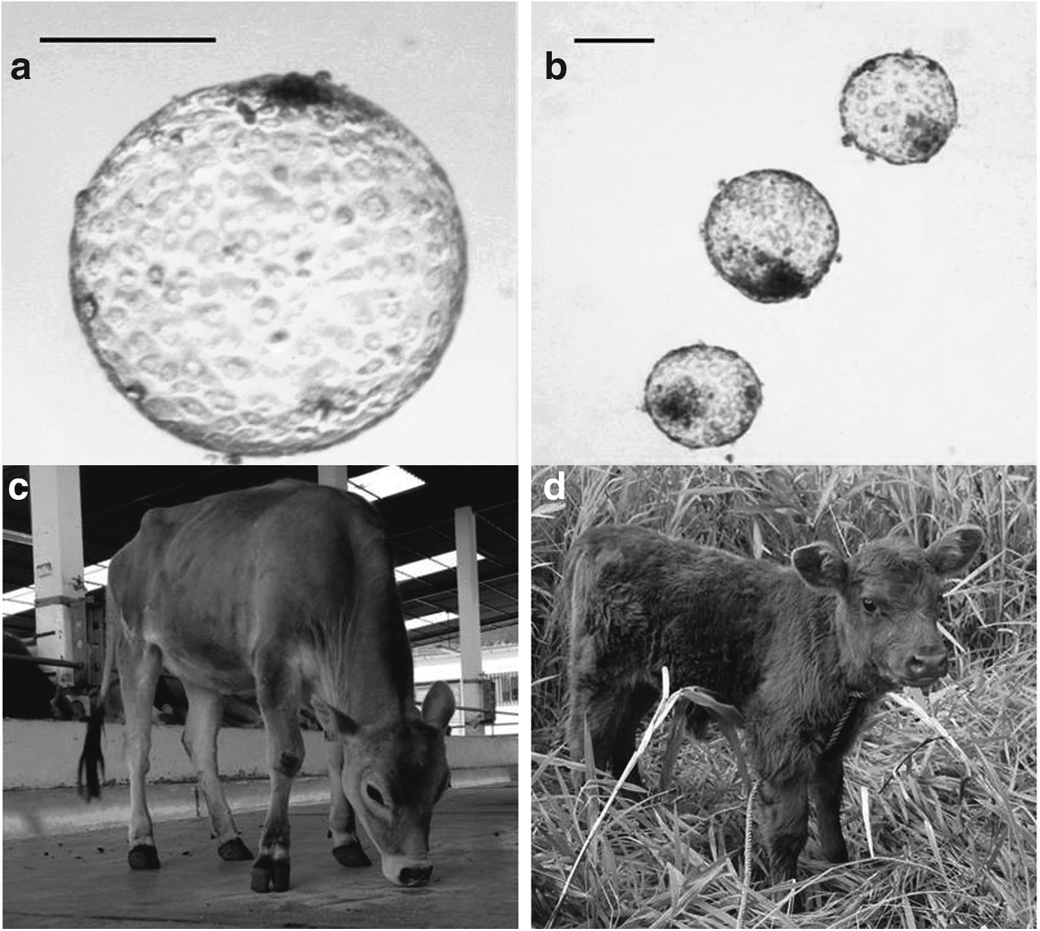
In the second series, 8 of the 11 vitrified D7 blastocysts have reexpanded after 18 hours of culture in vitro (73%) (Fig. 1b). Transfer of eight of these blastocysts in eight recipients resulted in four pregnancies (4/8, 50%) and two live births (25% offspring/embryo transfer rate), both of them with natural birth. The two female calves are healthy, 15 and 5 days old at the time of submission, respectively (Fig. 1d).
These results—together with those achieved previously in pigs—indicate that HMC may be competitive with or even superior than traditional cloning in terms of overall efficiency. Distinct features may contribute in this improvement, including (1) a high oocyte cytoplasm volume per somatic cell ratio as two cytoplasts contribute in the construction of one embryo (Tecirlioglu et al., 2005); (2) rapid and efficient manipulations decreasing the environmental stress of oocytes and embryos (Gerger et al., 2017; Vajta et al., 2006); (3) high fusion, activation, and in vitro embryo development rates due to the specifically adjusted parameters and environment (Vajta et al., 2003); (4) the zona-free situation that is commonly regarded as a handicap, but can be even beneficial both in vitro and after transfer (Vajta et al., 2010).
Additionally, HMC embryos seem to be tolerant to cryopreservation with vitrification. Advanced pregnancies and birth of clones after embryo vitrification are still regarded as exceptional achievements, with less than a dozen births reported in four species by seven groups worldwide. All of these successes were achieved with the use of minimum-volume vitrification techniques.
The OPS method with its high cooling and warming rates achieved in a protective narrow tube structure seem to be uniquely suitable for cryopreservation of zona-damaged (biopsied and traditionally cloned) and zona-free (HMC) embryos (Vajta et al., 1998). OPS or a slightly modified version were applied by four of the abovementioned groups and also used in our experiments. The achieved 25% offspring per transferred embryo rate allows a cautious optimism to overcome the logistic problems at on-farm application of cattle cloning.
Obviously the modest number of transfers does not allow for drawing clear conclusions. However, the 33% and 25% live offspring/transfer rate achieved with fresh and vitrified HMC embryos may indicate the need of a thorough investigation based on appropriate number of transfers without and with cryopreservation at the blastocysts stage.
Footnotes
Disclosure Statement
G.V. is director of a consulting company dealing with handmade cloning and OPS vitrification.