
Abstract
Abstract
Somatic cell nuclear transfer (SCNT) is required for the generation of transgenic animals as disease models. During the in vitro development of SCNT embryos, the quality of matured oocytes is one of the major factors regulating the developmental potential of embryos. Time-lapse monitoring systems are new tools that assess the developmental capacity of embryos for use in embryo transfer. In this study, we investigated the effect of fibroblast growth factor 10 (FGF 10) on the developmental potential of SCNT embryos. After the in vitro maturation (IVM) of oocytes in IVM medium containing 10 ng/mL FGF 10 (10 F), the polar body extrusion rate was significantly higher than in the control. However, there was no difference in the percentage of fused embryos between the groups. The cleavage and blastocyst formation rates of embryos were significantly increased in the 10 F compared with the control. In addition, the total cell number was higher and the apoptotic index was lower in the 10 F than control at day 7. The messenger RNA (mRNA) expression of genes involved in apoptosis (baculoviral inhibitor of apoptosis repeat containing 5 [BIRC5] and caspase 3 [CASP3]) and development (octamer-binding transcription factor 4 [POU5F1] and sex determining region Y box 2 [SOX2]) increased after 10 F treatment. Furthermore, the kinetics of the first cleavage was faster and the percentage of embryos at cell block was significantly lower in the 10 F group than in the control. These results demonstrate that exposure of oocytes to FGF 10 during IVM promotes developmental competence.
Introduction
A
The addition of follicular fluid to maturation medium can positively affect the rates of in vitro fertilization and development (Guler et al., 2000). To improve the efficiency of SCNT, donor cell synchronization (Hyun et al., 2016), endoplasmic reticulum stress (Lin et al., 2016), and the interval between fusion and activation have been studied (Aston et al., 2006). Among them, the quality of oocytes derived from in vitro maturation (IVM) is a key factor influencing successful embryonic development (Lee et al., 2015). During oocyte maturation, the growth factors are generally used to improve the cumulus cell expansion, oocyte maturation (Conti et al., 2006; Shinada et al., 2006; Sugiura et al., 2010). The fibroblast growth factor 10 (FGF 10) is known as the effect of promotion of the oocyte maturation (McCarroll and Nechiporuk, 2013) and embryo developmental quality (Machado et al., 2015).
We previously reported that FGF 10-supplemented IVM medium improved the maturation and quality of porcine parthenotes (Son et al., 2017). Following the results of the previous study, treatment of FGF 10 on IVM medium efficiently used to improve the SCNT embryo quality. To improve the overall efficiency of SCNT, we treated porcine embryos with FGF 10 during IVM.
A previous study reported that the first two or three cell divisions are regulated by maternal factors after the activation of the embryo's genome (Camous et al., 1984), and that early cleaving embryos are more successful than late-cleaving embryos at developing to the blastocyst stage in vitro (Isom et al., 2012). Embryo quality related to its developmental potential and the timing of the first zygotic cleavage is a valuable indicator of embryonic competence (Lechniak et al., 2008). Time-lapse monitoring systems are generally used to confirm the embryo developmental quality and select the optimal cleavage-stage embryos by real-time monitoring (Shimoda et al., 2016) and the selection of a critical time point is essential to minimize the difference between embryos (Shoukir et al., 1997).
In this study, we investigated the effect of 10 ng/mL FGF 10 treatment on the developmental potential of SCNT embryos using a time-lapse monitoring system. To investigate the results by adding the FGF 10 during IVM, the polar body (PB) extrusion, fusion rate, and development rate were identified. By real-time monitoring system, the embryo cleavage time and the morphology were classified during in vitro development of SCNT embryo. The blastocyst quality was compared by total cell number, apoptotic cell number, and development-related gene expression in blastocysts. In summary, the addition of FGF 10 to the medium accelerated the kinetics of the first cleavage time and improved the quality of blastocysts compared with the control.
Materials and Methods
Oocyte collection and IVM
Prepubertal porcine ovaries were collected from a local slaughterhouse and transported to the laboratory in saline supplemented with 75 mg/mL penicillin G and 50 mg/mL streptomycin sulfate within 2 hours at 32°C–35.0°C. Cumulus–oocyte complexes (COCs) were aspirated from follicles with a diameter of 2–8 mm using an 18-gauge needle and a disposable 10-mL syringe. COCs were washed three times in 4-(2-hydroxyethyl)-1-piperazineethanesulfonic acid (HEPES)-buffered tissue culture medium (TCM)-199 containing 0.1% (w/v) bovine serum albumin (BSA).
Groups of 50 COCs were matured in 500 μL of TCM-199 (Gibco, Grand Island, NY) containing Earle's salts, 0.57 mM cysteine, 10 ng/mL epidermal growth factor, 0.5 μg/mL follicle-stimulating hormone, 0.5 μg/mL luteinizing hormone, and 10% (v/v) porcine follicular fluid under mineral oil for 44 hours at 38.8°C in a humidified atmosphere of 5% (v/v) CO2 and 95% (v/v) air. TCM-199 was supplemented with 0 or 10 ng/mL FGF 10 (Con and 10 F, respectively). The concentration of FGF 10 was selected based on a study previously published by our laboratory (Son et al., 2017).
SCNT and in vitro culture
Following maturation, cumulus cells were removed by gently pipetting in the presence of 1 mg/mL hyaluronidase for 2–3 minutes. After a 30-minute recovery, the first PB and nucleosome were enucleated in HEPES-buffered TCM-199 containing 0.4% (w/v) BSA and 7.5 μg/mL cytochalasin B (CB) using a 20-μm glass pipette under an Oosight imaging system (Cambridge Research & Instrumentation, Inc.). The donor cell was inserted in the perivitelline space surrounding the cytoplasm. The karyoplast–cytoplast complexes were fused in a Fusion medium containing 0.3 M
Inserted donor cells were aligned to the negative electrode in a fusion chamber (Nepa Gene) and exposed to a direct current of 110 V/cm for 60 μseconds. After the fusion, the reconstructed embryos were activated in 7.5 μg/mL CB for 3 hours and then transferred to porcine zygote medium (PZM)-5 supplemented with 0.4% (w/v) FAF-BSA. On days 2 and 7, the cleavage and blastocyst formation rates were recorded as the number of cleaved embryos.
Embryo culture in a Primo Vision culture dish
The Primo Vision culture dish is composed of microwells forming a matrix of four rows with four wells per row. The culture dish was loaded with early embryo culture medium (PZM-5 containing 4 mg/mL BSA) on the day before starting the time-lapse imaging experiment to allow the media to equilibrate before adding the embryos. Each microwell was filled with 40–60 μL of culture medium. After activation or reconstruction, embryos were cultured in PZM-5 containing 4 mg/mL BSA at 38.5°C in 5% (v/v) O2, 5% (v/v) CO2, and 90% (v/v) air with maximum humidity for 7 days.
Time-lapse cinematography
In vitro embryo development was monitored using a real-time monitoring system with Imaging Capture Software. During the 168-hour culture period, 2016 images of embryos were taken at 5-minute intervals. Image stacks were analyzed using Primo Vision Analyzer Software. The timing of the first appearance of embryos at two-cell, four- to eight-cell, morula, early blastocyst, and expanded blastocyst stages was recorded.
Assessment of blastocyst quality
To detect fragmented DNA, terminal deoxynucleotidyl transferase dUTP nick end labeling (TUNEL) assay was performed. The blastocysts were fixed in 4.0% (w/v) paraformaldehyde (PFA) in phosphate-buffered saline (PBS) overnight at 4°C and then incubated in 0.1% (v/v) Triton X-100 at 38.8°C for 30 minutes. Blastocyst transferred on fluorescein-conjugated dUTP and terminal deoxynucleotidyl transferase (the in situ Cell Death Detection Kit; Roche, Manheim, Germany) in the dark at 38.8°C for 1 hour. The total number of mitotic and apoptotic cells was scored. Nuclei were stained with 1 μg/mL Hoechst 33342 for 30 minutes, and embryos were washed in 0.1% (w/v) BSA-PBS. Blastocysts were mounted onto glass slides and examined under an inverted Olympus IX-71 (Tokyo, Japan) microscope. At least 10 blastocysts from each group were examined.
Quantitative real-time polymerase chain reaction
To confirm the gene expression, the messenger RNA (mRNA) was isolated from 20 blastocysts using the Dynabeads mRNA Direct Kit (DynalAsa, Oslo, Norway). First-strand complementary DNA (cDNA) synthesis was achieved by reverse transcription of mRNA using an oligo(dT)12–18 primer and SuperScript III reverse transcriptase (Invitrogen, Carlsbad, CA). Real-time polymerase chain reaction (PCR) was performed in a DNA Engine OPTICON 2 System (MJ Research, Waltham, MA) in a final reaction volume of 20 μL that contained SYBR Green Master Mix and a double-stranded DNA-binding fluorophore (qPCR Kit; FINNZYMES, Espoo, Finland). The primers used for PCR are listed in Table 1.
BIRC5, baculoviral inhibitor of apoptosis repeat containing 5; CASP3, caspase 3; F, forward; POU5F1, octamer-binding transcription factor 4; R, reverse; SOX2, sex-determining region Y box 2.
The PCR conditions were as follows: 10 minutes at 94°C, followed by 39 cycles of 30 seconds at 94°C, 30 seconds at 60°C, and 55 seconds at 72°C, and a final extension of 5 minutes at 72°C. The relative gene expression was analyzed by the 2−ΔΔCt method (Livak and Schmittgen) after normalization against the β-actin mRNA level.
Statistical analysis
The general linear model procedure within Statistical Analysis System Software (Cary, NC) was used to analyze data from all experiments. Significant differences were determined by Tukey's multiple range tests. A paired Student's t-test was used to compare the relative gene expression between groups. p-Values of ≤0.05 were considered statistically significant.
Results
Effect of FGF 10 on porcine embryo development in vitro
The effect of FGF 10 on the in vitro development of porcine SCNT embryos was investigated. The PB extrusion rate was significantly higher in 10 F than in the Con (Con, 43.7% ± 3.4% vs. 10 F, 53.1% ± 3.2%, p ≤ 0.05, Table 2). The matured oocytes were subjected to SCNT. There was no difference in the donor cell fusion rate between both groups (Con, 47.6% ± 2.6% vs. 10 F, 48.4% ± 1.6%, p ≤ 0.05, Table 2). The number of two- to four-cell-stage embryos was significantly increased at day 2 in the 10 F compared with Con (Con, 48.1% ± 4.3% vs. 10 F, 62.0% ± 4.7%, p ≤ 0.05, Table 2). Furthermore, the blastocyst formation rate at day 7 was higher in the FGF 10-treated group than in the untreated group (Con, 11.6% ± 2.9% vs. 10 F, 20.2% ± 2.7%, p ≤ 0.05, Table 2).
p ≤ 0.05.
PB, polar body.
Time-lapse image analysis of the in vitro development of porcine SCNT embryos
We examined the development of SCNT embryos at 24-hour intervals (Fig. 1). At 12 hours, the number of embryos at the two-cell stage was higher and the kinetics of development to the next cell stage were faster in the 10 F-treated group than in the control. Likewise, after 24 hours, the embryo development to the next stage appeared faster in 10 F than the Con in every step. On day 2, 25% and 38% of Con embryos reached two- and four-cell stages, respectively, whereas 25% and 75% of 10 F embryos reached these stages, respectively. The morula formation at 72 hours was 17% in 10 F, however, it did not appear at that time in Con.

The ratios of the different developmental stages of SCNT porcine embryos cultured in vitro up to 120 hours. Twenty-four and 48 hours was ratio of embryos at each stage per fused one and other times was ratio of embryos at each stage per cleaved one. Data were derived from four independent data points for each treatment and developmental stages. SCNT, somatic cell nuclear transfer.
At 96 hours, all embryos in both groups reached the four-cell stage and the percentage of morula formation was higher in 10 F than Con (respectively, four-cell and morula; Con, 12% and 88%; 10 F, 8% and 92%). On day 5, the number of blastocysts was higher in the 10 F than in the Con (Con, 38% and 10 F, 67%), and on day 6, all embryos developed to the blastocyst stage (Fig. 1).
Time-lapse image analysis of porcine SCNT embryo cleavage in vitro
Using time-lapse monitoring system we found that the first cleavage time of SCNT embryos to be shorter in 10 F than in the Con (Con, 22.9 ± 2.8 hours vs. 10 F, 18.8 ± 0.9 hours, p ≤ 0.05, Fig. 2A). Also, the cleavage time of two-cell stage embryos to four-cell was significantly faster in 10 F group than control group (Con, 48.8 ± 5.1 hours vs. 10 F, 36.0 ± 3.0 hours, p ≤ 0.05, Fig. 2A). However, there were no differences in the cleavage time of embryos at morula (Con, 89.5 ± 3.3 hours vs. 10 F, 84.7 ± 3.2 hours) and blastocyst (Con, 114.4 ± 5.2 hours vs. 10 F, 119.3 ± 3.5 hours) stages between both groups (Fig. 2A). The morphology of embryos at the average cleavage time between Con and 10 F groups are shown in Figure 2B (two-cell, 21 hours; four-cell, 43 hours; morula, 87; and blastocyst, 116 hours).
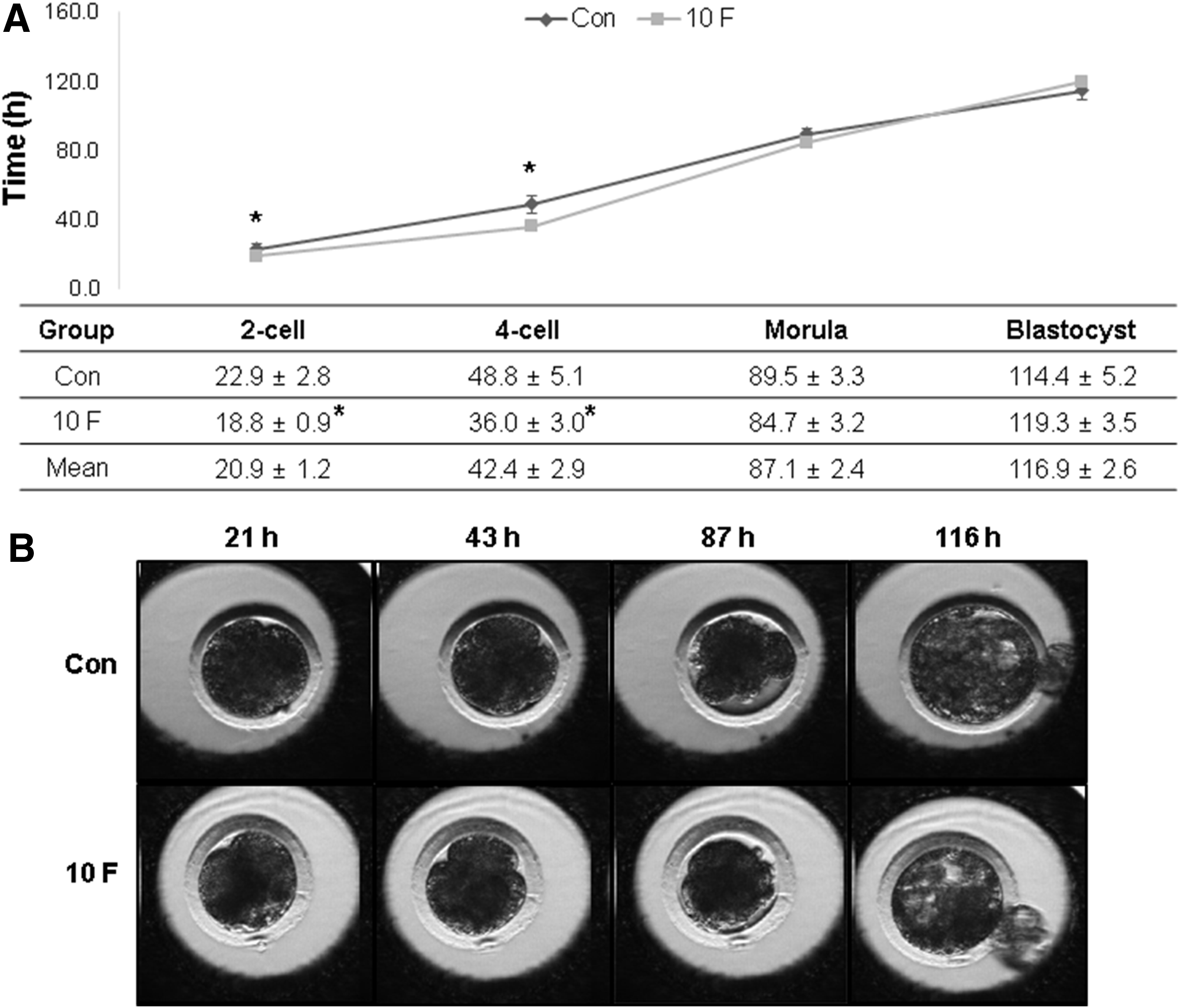
The timing of the formation of embryos at two-cell, four-cell, morula, and blastocyst stages
Relationship with cleavage time and kinetics on embryo development
During embryo development, we classified the percentage of blastocyst formation according to first cleavage time. Depending on whether cleavage occurred before or after 21 hours, we classified the embryos into early or late (Fig. 3A). The percentage of early cleaving embryos was higher in the 10 F than in the Con (Con, 19.3%; 10 F, 32.3%), whereas the percentage of late-cleaving embryos was lower in the former group than the latter one (Con, 80.7%; 10 F, 67.6%, Fig. 3B). Furthermore, the percentage of early cleaving embryos was higher than that of late-cleaving embryos in both groups (Con, 10.5% [early], 7.0% [late]; 10 F, 20.6% [early], 5.9% [late], Fig. 3C).

The morphological classification of early cleaving (before 21 hours) or late-cleaving (after 21 hours) SCNT embryos
Total cell number and gene expression of SCNT embryos treated with FGF 10
To confirm the effect of FGF 10 during porcine IVM on the developmental quality of embryos, day 7 blastocysts were subjected to the TUNEL assay. The total number of cells in blastocysts was significantly increased in the 10 F than in the Con (Con, 57.5 ± 4.0; 10 F, 70.0 ± 3.2, p ≤ 0.1, Fig. 4A). By contrast, the percentage of apoptotic cells were significantly decreased in the 10 F-treated group than in the untreated group (Con, 8.1 ± 0.1; 10 F, 5.8 ± 0.7, p ≤ 0.05, Fig. 4B).

The total number of cells
We evaluated the mRNA expression level of apoptosis (caspase 3 [CASP3] and baculoviral inhibitor of apoptosis repeat containing 5 [BIRC5]), reprogramming-related genes (octamer-binding transcription factor 4 [POU5F1], and sex-determining region Y box 2 [SOX2]). The expression of POU5F1, SOX2 (p ≤ 0.05), and BIRC5 (p ≤ 0.01) genes was higher in the 10 F group than in the Con (Fig. 4). By contrast, the expression of the proapoptotic gene CASP3 was significantly decreased in FGF 10-treated group compared with the untreated group (Fig. 4, p ≤ 0.01).
Discussion
In this study, we investigated the effect of FGF 10 during oocyte IVM on the developmental kinetics of porcine SCNT embryos. Major findings in the present study include: (1) treatment of 10 F result in a significantly higher rate of PB extrusion, cleavage, and blastocyst formation compared with control (p ≤ 0.05); (2) accelerated kinetics of embryo development of embryos in 10 F as determined by a time-lapse monitoring system; (3) the early cleavage embryo were much more in 10 F than Con and the early cleaved embryo developed more efficiently to blastocyst compared with late cleaved embryo; (4) an increased number of total cells, a decreased number of apoptotic cells, and significant changes in the expression of genes involved in development and cell apoptosis in 10 F.
Oocyte quality is reflected in an oocyte's intrinsic developmental potential (Gilchrist et al., 2008), which is critical for successful and reproducible SCNT, IVM, oocyte activation, and in vitro embryo culture (Wani et al., 2010). The maturation of oocytes is the most important step for successful SCNT (Jeong et al., 2008). FGF 10 is known as acts at the cumulus–oocyte complex by upregulating the expression of genes involved in cumulus cell expansion and maturation (Pomini Pinto et al., 2015; Son et al., 2017).
Similar results were reported in cattle, where FGF 10 stimulates cumulus expansion and improves oocyte developmental competence (Machado et al., 2015). Oocytes supplemented with FGF 10 during IVM also show improved developmental rates (Zhang et al., 2010). In this study, we investigated the effects of FGF 10 on the developmental potential of SCNT embryos on days 0, 2, and 7 and found the rates of PB exclusion, cleavage, and blastocyst formation to be higher in treated embryos than in the control.
These results are consistent with those in COCs that showed FGF 10 to improve oocyte maturation, cumulus expansion, and embryo development by improving the ability of cumulus cells to regulate meiosis and providing sufficient ooplasm for early embryonic survival. Based on these results, the addition of FGF 10 to the medium can enhance the developmental potential and quality of porcine oocytes.
We used a time-lapse monitoring system to track the SCNT embryo development and confirmed the positive effects of FGF 10 on the kinetics of embryo cleavage. The kinetics of embryo cleavage are useful in the selection of embryos with higher developmental potentials, which are more suitable for SCNT (Canto et al., 2012). Although a previous study reported no association between the timing of blastocyst formation and the completion of the first cleavage (Soom et al., 1997), recent other studies suggested that when comparing the development, the time point of the first, and especially the second, significantly higher numbers of early cleaving embryos became good quality blastocyst, as well as giving significantly higher pregnancy, implantation, and birth rate (Canto et al., 2012; Lee et al., 2012; Lundin et al., 2001; Pribenszky et al., 2010; Vandaele et al., 2006).
The difference in development to the morula or blastocyst stage of embryos could first be detected as a difference in the time at which the zygote cleaved to the two-cell stage (Hillery-Weinhold, 1991). The length of zygotic S-phase correlated with embryo development in vitro and the zygotes sired by low-fertility bulls detected with a delayed entry into S-phase (Eid et al., 1994). The time-lapse monitoring system is practical for evaluating the kinetics of embryo development, and we used this system to confirm the positive effects of FGF 10 in embryos at 24-hour intervals. We found that the timing of embryo development was faster in the 10 F group than Con (Fig. 1), and that the kinetics of development to the two- and four-cell stages was significantly faster in treated embryos than in untreated embryos (Fig. 2).
In addition, the number of blastocysts formed from early cleaved embryos was higher than that of blastocysts formed from late-cleaved embryos (Fig. 3). Taken collectively, these results indicate that the addition of FGF 10 to IVM medium can improve the kinetics of embryo development, thus bypassing the developmental cell block caused by late cleavage, and increase the ratio of blastocyst formation.
To assess the quality of blastocysts formed from SCNT embryos, we investigated the number of total and apoptotic cells as well as the expression of genes involved in apoptosis and development. Of all the morphological characteristics typically assessed in cleavage-stage embryos, the cell number is the single most important indicator of embryo viability (Machtinger and Racowsky, 2013). We found that the blastocyst cell number was higher in the 10 F than Con, whereas the apoptotic index was significantly low in the 10 F. It reveals that FGF 10 can improve the quality of blastocysts derived from SCNT embryos. We also investigated the expression of genes involved in apoptosis and development in blastocysts.
Compared with the control, the expression of POU5F1, SOX2, and BIRC5 significantly increased 10 F, whereas the expression of the proapoptotic gene CASP3 was decreased. Apoptosis affects scattered single cells and involves cell shrinkage, chromatin condensation, and the formation of small spherical bits of membrane, referred to as apoptotic bodies, which contain the nuclear fragmentation (Otoi et al., 1999).
Apart from epigenetic status of embryos, successful development depends on the proper expression of specific genes, such as OCT3/4, NANOG, and CDX2, which control pluripotency and early embryonic development (Saini et al., 2015). For example, Oct4 establishes and maintains the pluripotency of the inner cell mass, and its absence causes the inner cell mass to differentiate into the trophectoderm (Huang et al., 2011). Sox2 acts to maintain or preserve developmental potential that, like Oct4, it is expressed in the ICM, epiblast, and germ cells that are required in the lineage leading to epiblast formation, and in their absence trophectoderm is formed (Avilion et al., 2003). Cell proliferation is also important for embryo development, and BIRC5 plays a role in this process by inhibiting apoptosis (Jeon et al., 2011). Caspase, a member of the Bcl2 family, promotes apoptosis.
At least 11 caspase genes have been identified to mediate protein cleavage and induce apoptosis (Liang et al., 2015). Taken collectively, these results indicate that the addition of FGF 10 to the IVM medium improved the quality of SCNT embryos by improving the cell number, decreasing the ratio of cell death, and increasing target gene expression. We believe that these changes were due to the relative ease in which embryos bypassed the developmental cell block during cleavage, although further studies are needed to confirm this conclusion. Nevertheless, FGF 10 is likely to improve the efficiency of embryo implantation, a requirement for the successful production of nontransgenic and transgenic animals.
Many investigators have tested different culture media in an effort to improve the quality of embryos produced in vitro and to mimic the physiological conditions that embryos are exposed to in vivo (Arias et al., 2013). In this study, we found that FGF 10 promoted the development of good-quality blastocysts from approximately matured oocyte by FGF 10 treatment during IVM. By supplement of FGF 10 during IVM, we confirmed the beneficial effects of FGF 10 on SCNT embryos. A significantly higher fusion and rapid development of oocytes compared with the control.
In concept of embryo kinetics, the first and second cleavage formation were faster in FGF 10-treated group, and from the early cleaved embryo, the blastocyst formation and capacity were significantly improved, which may have been due to the relative ease with which embryos bypassed the developmental cell block. In summary, we regarded that the percentage of implantation will be increased when we use the 10 ng/mL FGF 10 during oocyte IVM for producing SCNT embryo. However, we need to study more and investigate the effect of FGF 10 on the implantation of SCNT embryos and the production of nontransgenic and transgenic animals.
Footnotes
Acknowledgment
Grant sponsor: Research Center for Production Management and Technical Development for High Quality Livestock Products through the Agriculture, Food and Rural Affairs Research Center Support Program, Ministry of Agriculture, Food and Rural Affairs, Republic of Korea (Grant No.: 715003-07).
Author Disclosure Statement
The authors declare they have no conflicting financial interests.