
Abstract
Abstract
Bone marrow mesenchymal stem cells (BMSCs) can transdifferentiate into different types of cells and may serve as a cell source for tissue engineering. Resveratrol has been shown to possess many benefits, including activation of osteogenesis. Furthermore, Wnt/β-catenin signaling has been known to promote osteogenic differentiation in many cells. In this study, we investigated the role of resveratrol in osteoblastic differentiation of canine BMSCs. Resveratrol treatment of canine BMSCs remarkably increased alkaline phosphatase activity and calcium nodules, inhibited the function of glycogen synthase kinase 3β, led to an increase in stabilization and nuclear accumulation of β-catenin, and upregulated expression of osteoblast-related marker gene expression. In addition, resveratrol caused rapid activation of ERK1/2. Collectively, our results indicate that resveratrol promotes osteoblastic differentiation of canine BMSCs by activating the Wnt/β-catenin; ERK/MAPK signaling pathways are also involved in osteogenic differentiation of canine BMSCs.
Introduction
M
Therefore, increasing the efficiency of BMSC differentiation is a key aim of this study. To improve the treatment of bone diseases, new methods to induce canine BMSCs to differentiate into osteoblasts are needed. Therefore, for the treatment of pet diseases, it is important to understand the mechanisms underlying the differentiation of BMSCs into osteoblasts and to improve the ability of BMSCs to differentiate into osteoblast. However, the extremely complex set of signaling molecules and pathways such as Wnt/β-catenin, transforming growth factor beta, and bone morphogenetic protein (BMP) signaling strictly control and regulate the differentiation time and direction of BMSCs in vivo (Augello and De, 2010; Long et al., 2017; Müller-Deubert et al., 2017).
A previous study showed that Wnt/β-catenin signaling plays an important role in osteogenic differentiation by directly stimulating gene expression of runt-related transcription factor 2 (RUNX2) (Gaur et al., 2005). Wnts activate the canonical pathway through their interaction with receptors of the frizzled (fz) family and co-receptors of the low-density lipoprotein receptor-related 5/6 (LRP5/6) family (Logan and Nusse, 2004; Tamai et al., 2000); result in destruction of a complex that consists of axin, adenomatous polyposis coli (APC), and glycogen synthase kinase 3β (GSK-3β) (He et al., 2004); and then stabilizes β-catenin levels and affects its subcellular localization, in which it activates the T cell factor (TCF)/lymphoid enhancer factor-1 (LEF) transcription system involved in the regulation of cell cycle progression and differentiation (Gordon and Nusse, 2006; Peifer and Polakis, 2000).
Resveratrol (trans-3,5,4′-trihydroxystibene) is a natural polyphenol found in red grapes, peanuts, berries, and pomegranates (Nagaoka et al., 2007; Rimando et al., 2004; Soleas et al., 1997) and has been shown to have many health benefits including neural protection and anti-inflammatory and life span extending activities in various organisms (Bastianetto et al., 2000; de la Lastra and Villegas, 2005; Valenzano et al., 2006). Studies have demonstrated that resveratrol could enhance osteogenic differentiation (Boissy et al., 2005; Mizutani et al., 1998.; Peltz et al., 2012), and inhibit adipogenesis (Rayalam et al., 2008; Yang et al., 2008). However, to our knowledge, the effects of resveratrol on the canine BMSCs have not been reported. In this study, we examined the effects of resveratrol on the proliferation and osteogenic differentiation of canine BMSCs in vitro.
We found that the Wnt/β-catenin signaling pathway is involved in resveratrol-stimulated osteogenic differentiation of canine BMSCs. In addition, resveratrol promoted the transcription of genes involved in osteogenes and led to rapid activation of ERK1/2.
Materials and Methods
Ethics statement
All procedures involving animals were conducted in accordance with the Guide for the Care and Use of Laboratory Animals (Ministry of Science and Technology of China, 2006), and the study design was approved by the animal ethics committee of Northwest A&F University (Yangling, China).
Materials
Chemicals were obtained from Sigma-Aldrich Chemical (St. Louis, MO) unless otherwise specified.
Canine BMSCs isolation and culture
Canine BMSCs were obtained from beagle dogs (2 months old, 1.5 kg, from Wugong County). In brief, dogs were injected with 10 mg/kg ketamine. Then, 10 mL of bone marrow was obtained from the iliac crest under sterile conditions according to previously reported method (Sun et al., 2010). Then, the samples were incubated in the minimum essential medium α (α-MEM; Gibco) supplemented with 10% fetal bovine serum (FBS; Gibco) and 100 U/mL penicillin/streptomycin (North China Pharmaceutical Co., Ltd., Shijiazhuang, China), and maintained in a humidified incubator with 5% CO2 at 37°C. The nonadherent cells were removed 24 hours after seeding, and the medium was replaced with fresh α-MEM containing 10% FBS. The medium was replaced every 3 days. In this study, cells within five passages were used.
Osteogenic differentiation of canine BMSCs
Canine BMSCs were initially cultured in α-MEM, after the cells adhered to the dish, and the cells were grown in osteogenesis-inducing medium (OM; α-MEM with 10% FBS, 10 mM β-glycerophosphate, 0.3 mM ascorbic acid and 1 × 10⁻⁵ mM dexamethasone). The medium was replaced every 4 days.
Proliferation of canine BMSCs
To assess the effect of resveratrol on canine BMSCs proliferation, we used the previously reported method (De Boer et al., 2004). In brief, the cells were seeded at a density of 3 × 10⁴ cells per well in a 24-well plate. After the cells were attached, the medium was changed to OM containing 0, 10, 20, 40, and 100 μM resveratrol. Control untreated cells were cultured in α-MEM with 10% FBS. Cell numbers were determined after 4 days, using a red blood cell count plate (Qiujing), and proliferation was expressed as the number of population doublings per day.
Western blot analysis
Canine BMSCs cultured in osteogenic medium with or without resveratrol for different time intervals were harvested and lysed using radioimmunoprecipitation assay buffer (RIPA). The protein concentrations were determined by the BCA Protein Assay Kit (Heart). Cell lysates were mixed with sodium dodecyl sulfate–polyacrylamide gel electrophoresis (SDS-PAGE) loading buffer (CW Bio), boiled for 10 minutes, and then resolved on 10% SDS-PAGE. After electrophoresis, the gel was transferred to polyvinylidene difluoride (PVDF) membrane and blocked with 3% bovine serum albumin (MP Biomedicals) in TBST (0.1% (v/v) Tween-20 in Tris-buffered saline) for at least 2 hours at 25°C.
The primary antibodies used were as follows: anti-β-actin (No. 4970, 1:4000; Cell Signaling Technology), anti-ERK1/2 (No. 4695, 1:2000; Cell Signaling Technology), anti-P-ERK1/2 (No. 4370, 1:2000; Cell Signaling Technology), anti-β-catenin (ab32572, 1:2000; Abcam), anti-GSK-3β (ab32391, 1:2000; Abcam), and anti-P-GSK-3β (ab75814, 1:2000; Abcam). Blots were incubated in primary antibody overnight at 4°C. Membranes were then rinsed and incubated at 25°C with horseradish peroxidase-conjugated secondary antibody for at least 2 hours. The membranes were washed three times with 1 × TBST, and immunoblots were visualized using ECL (Bio-Rad) kit according to manufacturer's instructions.
Immunofluorescence staining
Canine BMSCs were cultured in differentiation medium for 4 days, washed twice with phosphate-buffered solution (PBS), and then fixed for 30 minutes in 4% paraformaldehyde. The cells were washed thrice with PBS again and permeabilized with 0.2% Triton X-100 for 15 minutes. Cells were then rinsed thrice with PBS and incubated for 1 hour in a blocking buffer (1% FBS in PBS) followed by incubation at 4°C overnight with the β-catenin primary antibody (1:100; Abcam). Cells were then rinsed thrice with PBS, incubated with secondary donkey anti-rabbit antibody (1:100; Santa Cruz Biotechnology) at 37°C for 1 hour, and washed three times with PBS. The nuclei were stained with 4′,6-diamidino-2-phenylindole (DAPI) (1:1000; Beyotime) for 5 minutes at 25°C. After washing with PBS, the images were captured using a fluorescence microscope (Olympus, Tokyo, Japan).
Reverse transcription quantitative polymerase chain reaction
Canine BMSCs were cultured in differentiation medium for 3, 7, and 14 days. The cells were washed twice with ice-cold PBS, and total RNA was extracted using RNAiso Plus (Takara, Beijing, China) following the manufacturer's instructions. All potential DNA contamination was removed by RNase-free DNase treatment. Then complementary DNA (cDNA) was synthesized using PrimeScript™ RT Reagent Kit (Takara). The primers used in this study are given in Table 1. Real-time polymerase chain reaction (PCR) was performed using SYBR green PCR Mix (Takara) and was carried out on Quant Studio™ 6 Flex System (Applied Biosystems).
Primers Used in the Reverse Transcription–Quantitative Polymerase Chain Reaction Analysis
Marker genes of osteogenic cell: RUNX2, runt-related transcription factor 2; ONN, osteonectin; OCN, osteocalcin; GAPDH, glyceraldehyde-3-phosphate dehydrogenase.
F, forward; R, reverse.
Inner mitochondrial membrane potential detection
Canine BMSCs were cultured in different medium for 4 days, and then the mitochondrial membrane potential (MMP) was measured by JC-1, a cationic fluorescent dye. In brief, cells were seeded in a 24-well plate. After 96 hours of treatment, the JC-1 stain was added to each well, followed by incubation at 37°C for 20 minutes. Images were then captured using a fluorescence microscope (Olympus).
Reactive oxygen species detection
Generation of reactive oxygen species (ROS) was assessed after 4 days in treated and untreated canine BMSCs. Intracellular ROS levels were measured by 2′-,7′-dichlorofluorescin diacetate (DCFH-DA) according to standard protocol. Canine BMSCs were seeded in a 24-well plate and cultured in different medium for 96 hours, and then they were incubated with 10 μg/mL DCFH-DA at 37°C for 30 minutes. The DCFH-DA fluorescence intensities were detected by a fluorescence microscope (Olympus).
Alkaline phosphatase staining and alkaline phosphatase activity detection
Canine BMSCs were cultured for 7 days, alkaline phosphatase (ALP) staining was performed using an ALP staining kit (Nanjing Jiancheng Bioengineering Institute). The cells were fixed with 4% formaldehyde (Sinopharm Chemical Reagent Co., Ltd.) for 30 minutes and stained with the ALP reagent for 1 hour at 25°C. Then images were captured using an inverted microscope (Olympus), and the number of positive cells was obtained by randomly counting the number of positive cells in three fields of vision, repeating three times, and averaging.
ALP activity was quantitated using an ALP assay kit (Beyotime). Canine BMSCs were cultured in different medium for 7 days; these cells were lysed in 150 μL lysis buffer. After supernatants were collected, enzymatic reactions were performed according to manufacturer instructions. Absorbance was read at 405 nm with a spectrophotometer (BioTek Epoch). ALP activity was normalized to protein concentration in parallel experimental plates. A minimum of three independent experiments were performed for quantitation of ALP activity.
Alizarin red staining
Canine BMSCs were cultured for 14 days, and mineralization levels in treated and untreated cells were determined by alizarin red staining as previously described (Li et al., 2015). In brief, cells were induced for differentiation, fixed with 4% paraformaldehyde for 30 minutes, and stained with 0.1% alizarin red (pH 8.3) for 1 hour. They were then washed thrice with PBS, and images were captured using an inverted microscope (Olympus). Alizarin red dye was then eluted with 10% cetylpyridinium chloride (Dalian Meilun Biotechnology Co., Ltd.) overnight at 4C, and the optical densities were measured at 540 nm with a spectrofluorometer (BioTek Epoch).
Statistical analysis
All experiments were repeated independently at least three times, and data were analyzed by one-way analysis of variance using SPSS 19.0 software (SPSS, Inc.). Data were expressed as mean ± standard deviation, and significance at p < 0.05 was considered.
Results
Effects of resveratrol on cell proliferation, ROS levels, and MMP in canine BMSCs
To understand the effects of resveratrol on cell proliferation, ROS levels, and MMP in canine BMSCs, the cells were exposed to various concentrations of resveratrol in OM, and cell proliferation, MMP, and ROS levels were determined. Low dose of resveratrol did not cause any significant effect on proliferation of canine BMSCs (Fig. 1A, B), whereas high doses (40 and 100 μM) reduced proliferation. Proliferation of BMSCs was completely inhibited at 100 μM resveratrol (Fig. 1A, B).
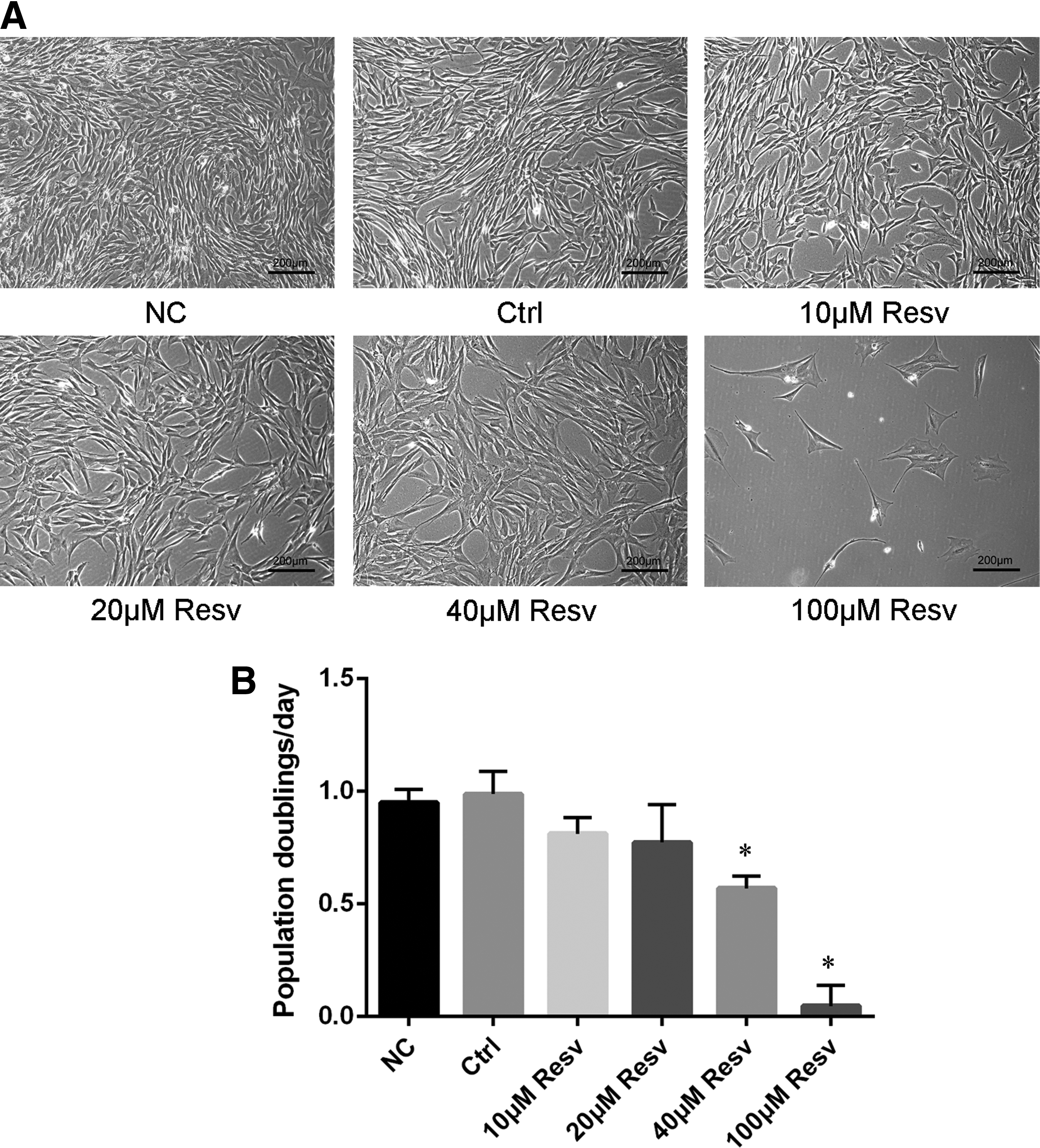
Effect of resveratrol on morphology and proliferation of canine BMSCs.
Resveratrol is known to either protect cells from apoptosis or induce apoptosis (Li et al., 2017; Sha et al., 2008). Decline in MMP and mitochondrial damage are early signs of apoptosis (Ly et al., 2003). Therefore, we detected the MMP of the cells by JC-1 staining. Lower red/green fluorescence intensities represent lower MMP and higher mitochondrial damage. As given in Figure 2A, strong red and weak green fluorescent signals were observed on treatment with different concentrations of resveratrol (0, 10, 20, 40, and 100 μM), indicating no significant effect of resveratrol on the MMP in canine BMSCs.
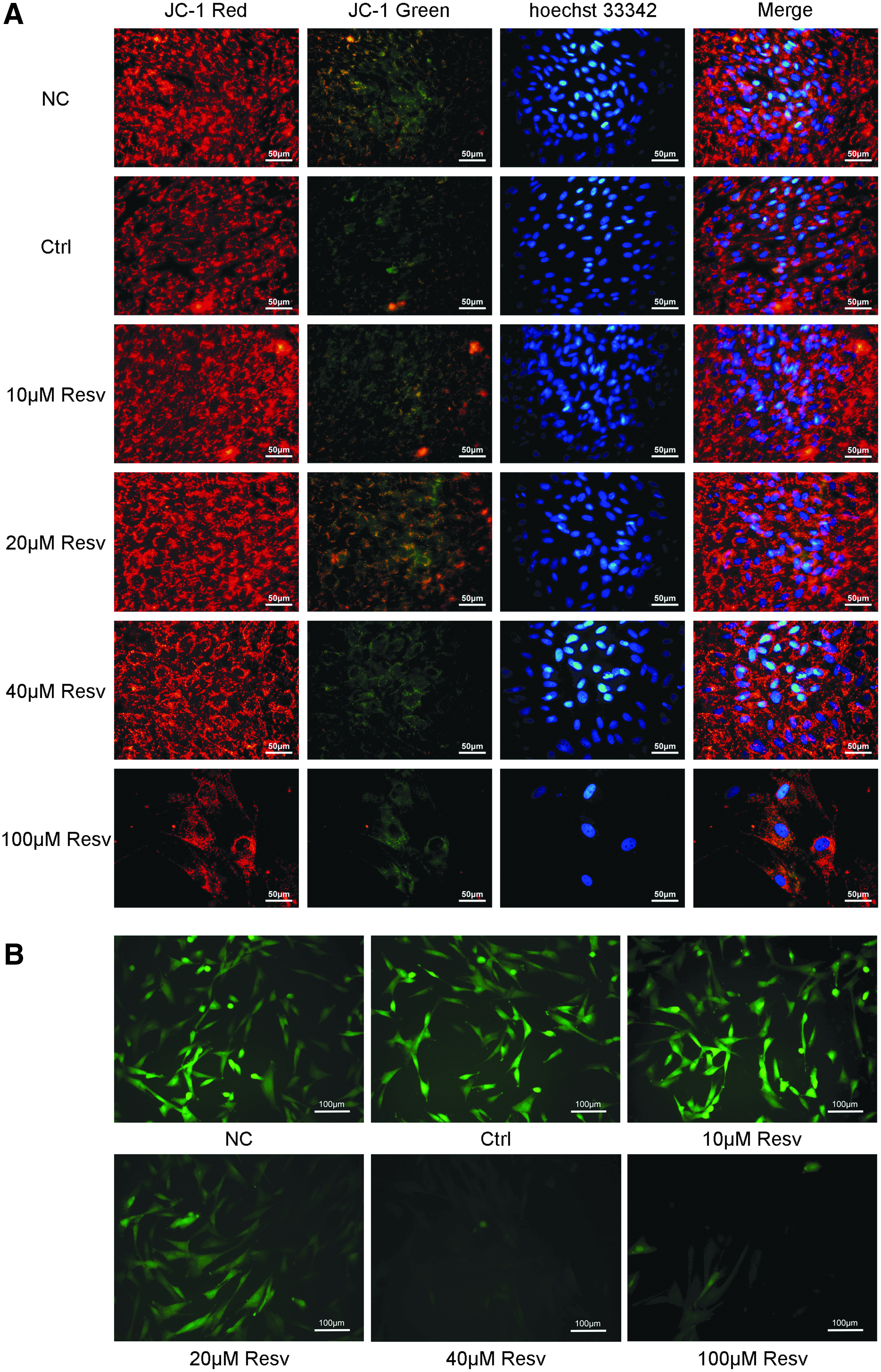
Changes in the MMP and ROS of canine BMSCs grown for 4 days in different medium as indicated. JC-1 Red: normally, mitochondria JC-1 use its potential as a red fluorescent emitter to form a polymer. JC-1 Green: in the destruction of mitochondrial function, JC-1 as a monomer dispersed and distributed in the cytoplasm was detected as green fluorescence. Hoechst33342: Hoechst33342 staining of the nucleus. Merge: the Superposition of red fluorescence (polymer), green fluorescence (monomer), and Hoechst33342 in canine BMSCs
Redox imbalance because of excessive or insufficient ROS production is known to induce apoptosis in cells (Lin et al., 2014; Qiu et al., 2015). Resveratrol possesses the ability to scavenge oxidatively generated free radicals and can either protect or induce apoptosis in cells (Kao et al., 2010). Thus, we detected ROS levels in resveratrol-treated canine BMSCs. As given in Figure 2B, the ROS levels in canine BMSCs were significantly reduced on treatment with high concentrations of resveratrol (40 100 μM). Treatment with 40 and 100 μM of resveratrol significantly reduced ROS levels and led to redox imbalance in canine BMSCs.
Resveratrol enhances ALP activity of canine BMSCs
As ALP is a well-recognized biochemical maker for osteoblasts, we examined the ALP activity in canine BMSCs in response to the various concentrations of resveratrol for 7 days. As given in Figure 3, the resveratrol (10, 20, and 40 μM) groups showed increase in ALP activity compared with the control group. This promotion was visualized by ALP enzyme histochemistry (Fig. 3A), the number of ALP-positive cells (Fig. 3B), and enzyme activity assays (Fig. 3C), where 20 μM resveratrol generated the highest increase in ALP activity and the number of ALP-positive cells. Based on the results of cell proliferation, ROS and MMP levels, and ALP activity of canine BMSCs, 20 μM resveratrol was used for further experiments.
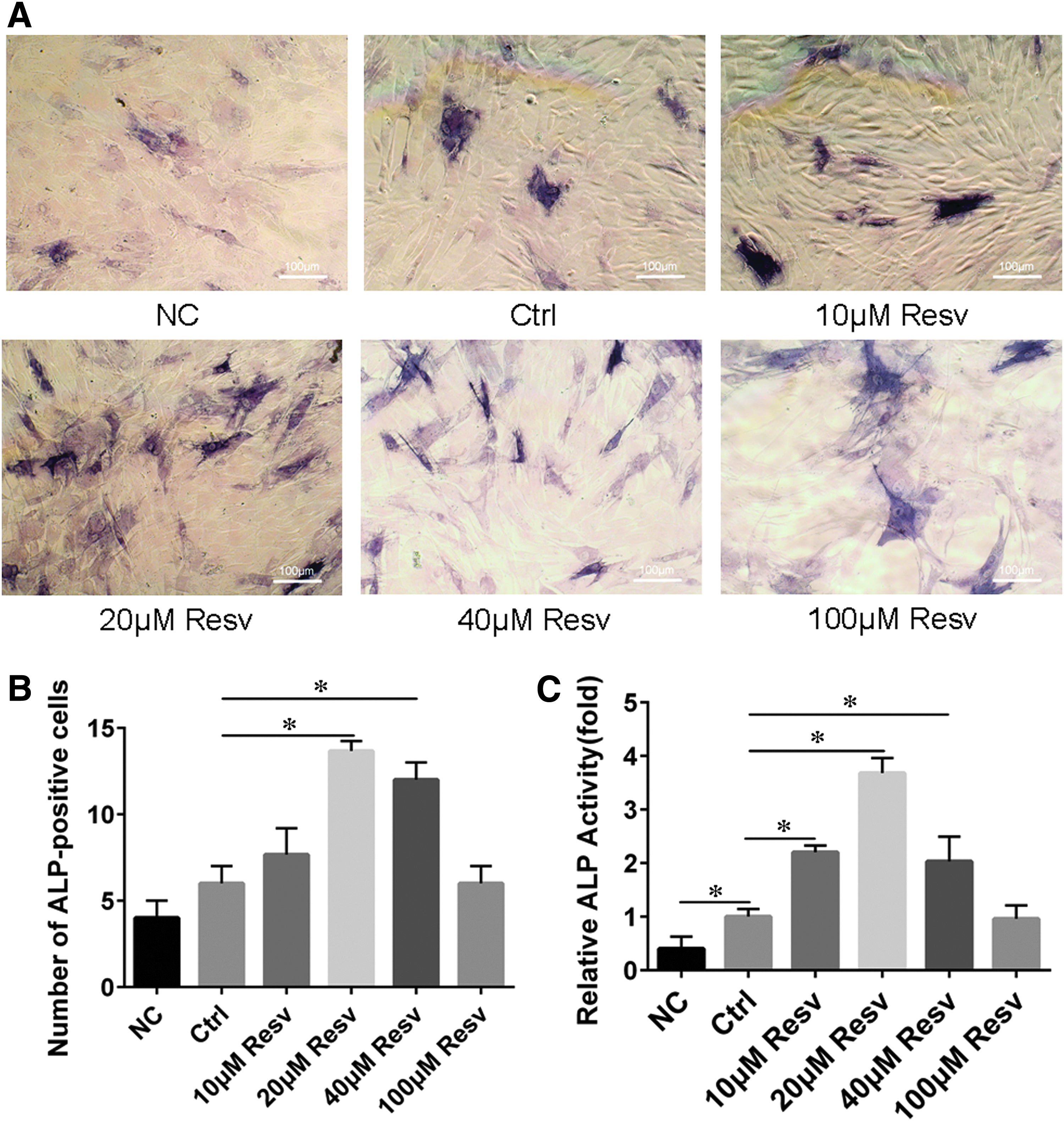
Resveratrol enhances ALP activity of canine BMSCs.
Resveratrol increases calcium deposits of canine BMSCs
Exposure of 20 μM resveratrol to canine BMSCs for 14 days led to an increase in alizarin red staining, indicating increase in mineralization and calcium deposits. As given in Figure 4, when the cells in the noninduced group were cultured for 14 days, the alizarin red staining assay was negative, which showed that the cells did not form mineralized nodules. However, for the normal induction group, cells formed calcified nodules. Compared with the normal induction group, the resveratrol group formed more and bigger calcified nodules (Fig. 4A). Absorbance value indicated that resveratrol-treated samples deposited 2.0 times more calcified matrix than the normal induction samples (Fig. 4B). These results suggest that resveratrol promotes the canine BMSCs to differentiate into osteoblasts.
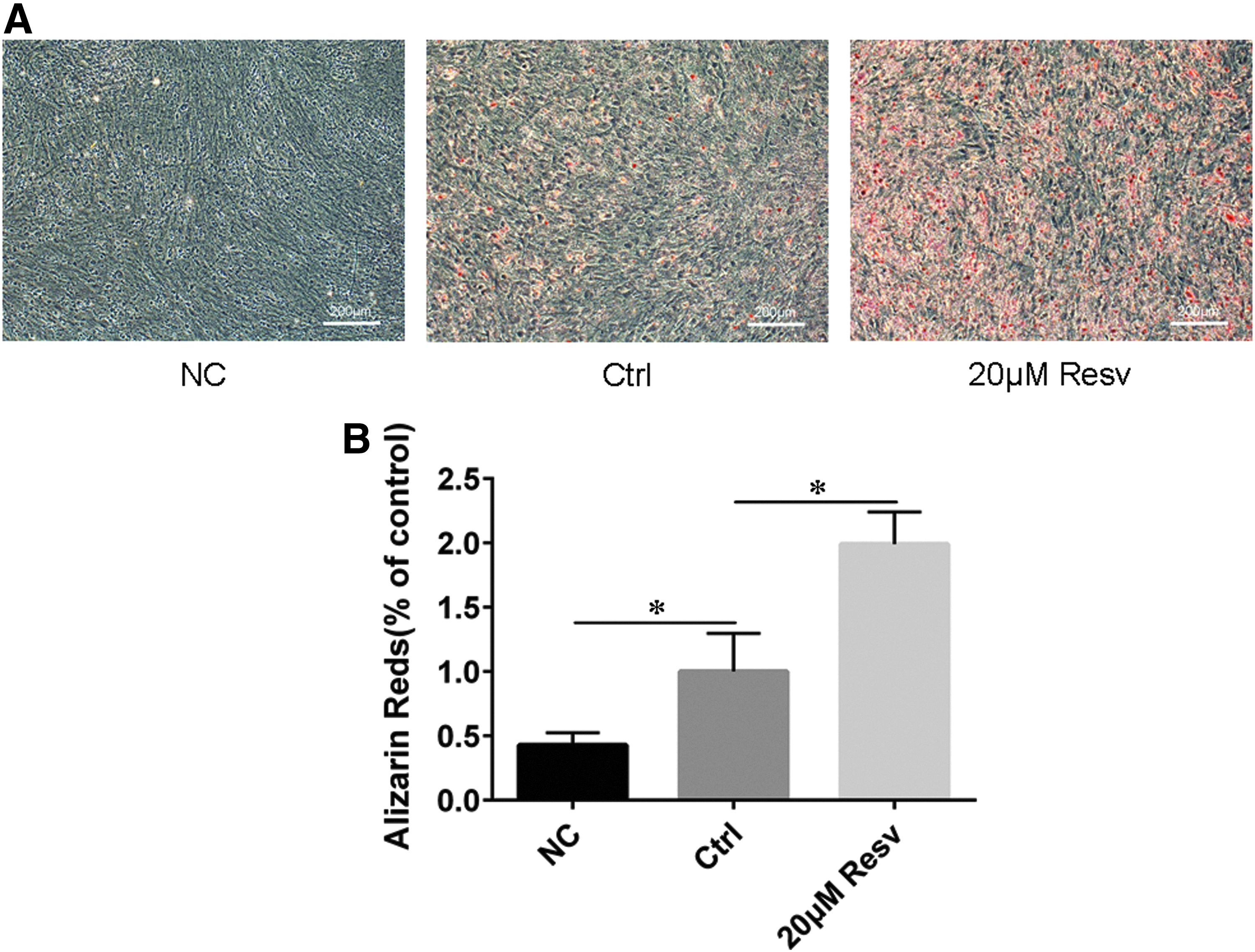
Resveratrol enhances calcified matrix of canine BMSCs.
Resveratrol promotes osteogenesis-related marker gene expression
The effect of resveratrol on the expression of osteogenic-related genes during osteoblast differentiation was studied using real-time PCR. Canine BMSCs were incubated with 0 and 20 μM resveratrol during differentiation for 3, 7, and 14 days. Changes in the RUNX2 (a bone-specific transcription factor), osteocalcin (OCN), and osteonectin (ONN) messenger RNA (mRNA) expression levels were measured. As given in Figure 5, 20 μM resveratrol significantly increased the expression of ONN and RUNX2 mRNA in cells at all three time intervals, whereas OCN mRNA expression was evidently increased in cells treated with 20 μM resveratrol for 14 days. These results are consistent with previous studies stating that resveratrol promotes osteogenic differentiation (Liu et al., 2016).
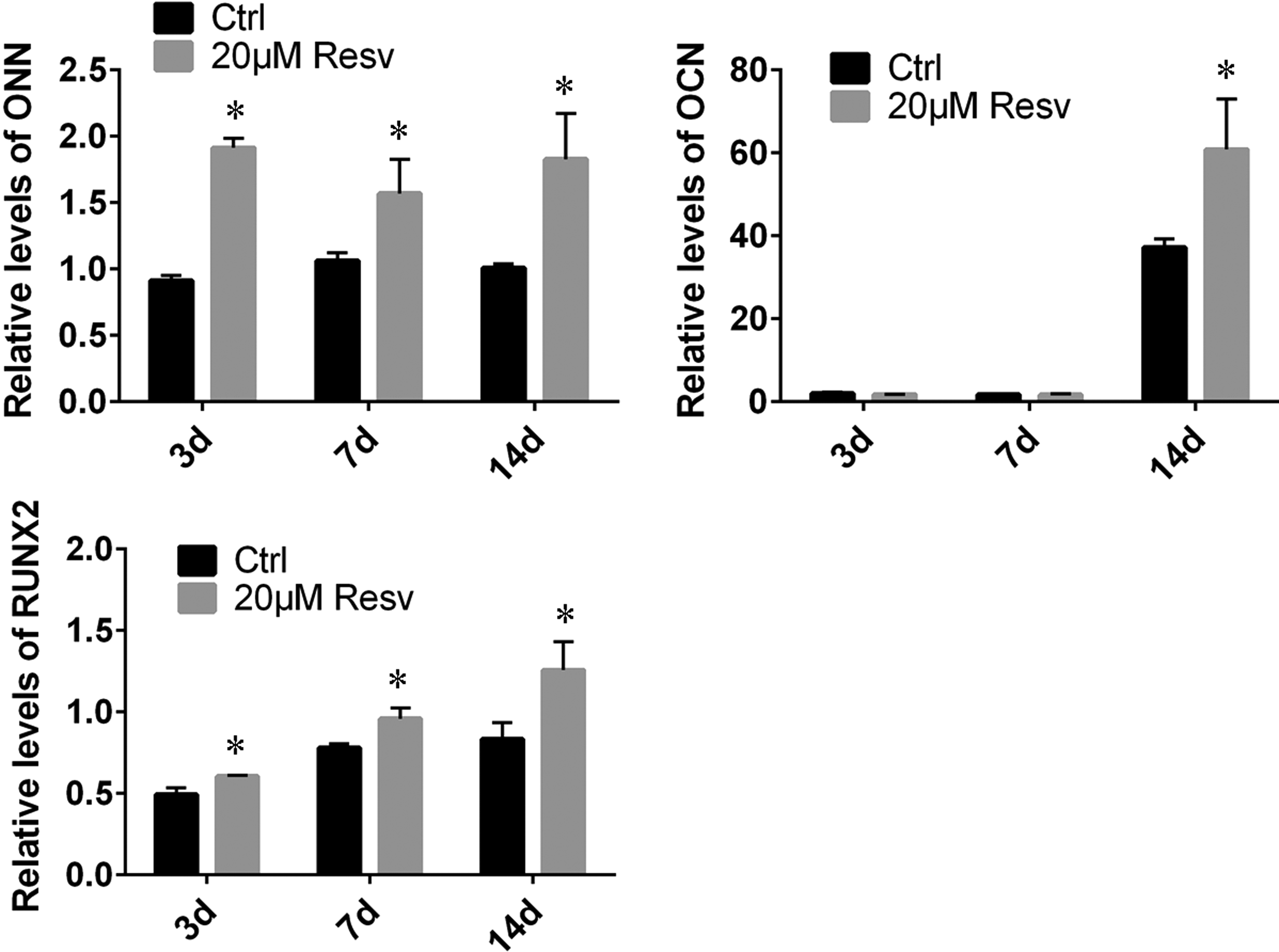
Resveratrol enhanced the mRNA expression of osteoblast differentiation markers in canine BMSCs. Canine BMSCs were incubated in α-MEM with 10% FBS, osteogenic medium containing 0 and 20 μM resveratrol for 3, 7 and 14 days, collected the cells and detected expression of RUNX2, ONN and OCN mRNA by qRT-PCR (n = 3). Error bars denote mean ± SD. *p < 0.05. mRNA, messenger RNA; OCN, osteocalcin; ONN, osteonectin; PCR, polymerase chain reaction; RUNX2, runt-related transcription factor 2.
Resveratrol promotes osteogenic differentiation through the Wnt/GSK-3β/β-catenin pathway
The Wnt/Gsk-3β/β-catenin signaling pathways are reported to be involved in osteogenic differentiation (Gaur et al., 2005; Gordon and Nusse, 2006). Inactivation of the GSK-3β by phosphorylation can stabilize β-catenin protein and lead to an increased transcription of its target genes (Zhou et al., 2009). Therefore, we examined the level of GSK-3β phosphorylation in 20 μM resveratrol-treated canine BMSCs. As given in Figure 6A, the level of p-GSK-3β was significantly increased in the presence of 20 μM resveratrol, indicating that GSK-3β activity was inhibited by resveratrol.
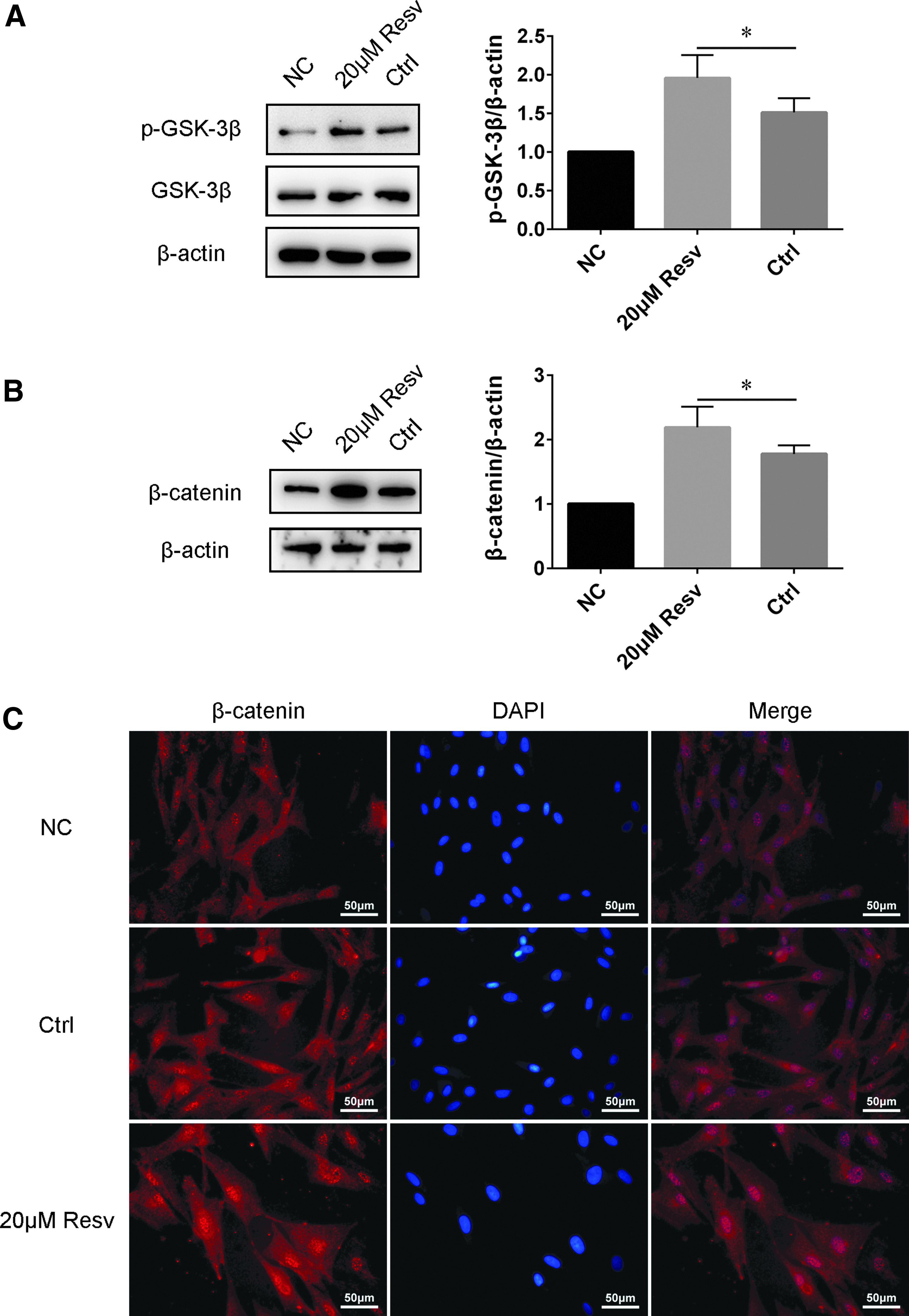
Resveratrol activated Wnt/β-catenin signaling pathways in canine BMSCs.
Then we examined the level of β-catenin and its subcellular distribution. As given in Figure 6B, treatment of canine BMSCs with 20 μM resveratrol for 96 hours resulted in an increase in the levels of total β-catenin. Immunofluorescence results showed that the nuclear accumulation of β-catenin was upregulated in resveratrol-treated canine BMSCs (Fig. 6C). These results indicate that resveratrol is involved in activation of the Wnt signaling pathway to promote osteogenic differentiation.
Resveratrol causes a rapid activation of ERK1/2
A previous study showed that the ERK signaling pathway plays an important role in osteoblastic differentiation (Dai et al., 2007). Resveratrol has been reported to promote human BMSCs osteoblastic differentiation through ER-dependent ERK1/2 activation (Klinge et al., 2005). Therefore, we examined the levels of phosphorylation of ERK1/2 in 20 μM resveratrol-treated canine BMSCs. As given in Figure 7, resveratrol-mediated activation of ERK1/2 in canine BMSCs was rapid (in 15 minutes) with the activation lasting for 6 hours.
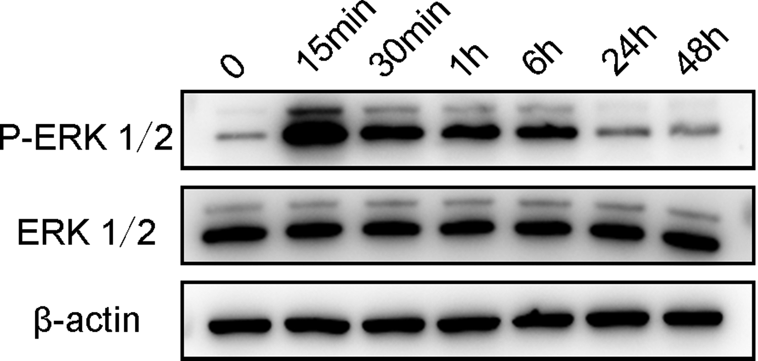
Resveratrol caused a rapid activation of ERK in canine BMSCs. Canine BMSCs were incubated in osteogenic medium with 20 μM resveratrol for 15 and 30 minutes and for 1, 6, 24, and 48 hours, collected the cells and detected expression of ERK1/2 by Western blot. Resveratrol quickly activated ERK1/2 in 15 minutes in canine BMSCs, and lasted 6 hours.
Discussion
As an antioxidant, resveratrol is used for the prevention and treatment of atherosclerosis, coronary heart disease, ischemic heart disease, and hyperlipidemia (Tamaki et al., 2014; Tomayko et al., 2014). Studies have shown that resveratrol inhibits proliferation and promotes apoptosis in many types of tumor cells. This effect may be related to resveratrol-mediated inhibition of Wnt/β-catenin signaling, but the specific mechanism is still not very clear (Chen et al., 2012). Resveratrol can promote osteogenic differentiation in mouse and human mesenchymal stem cells by activating Wnt/β-catenin signaling (Dai et al., 2007; Peltz et al., 2012; Zhou et al., 2009). This effect of resveratrol is related to the concentration of resveratrol and the type of cells involved.
In this study, to investigate the role of resveratrol in differentiation of canine BMSCs, the cells were exposed to various concentrations of resveratrol in OM. High resveratrol concentrations (40 and 100 μM) inhibited proliferation and reduced ROS levels in canine BMSCs. Lower concentration of resveratrol (20 μM) did not cause significant effects on canine BMSC proliferation but resulted in an increase in the activity of ALP. Alizarin red staining of cells in the resveratrol-treated group revealed formation of osteoblasts and matrix mineralization; absorbance value indicated that the resveratrol-treated samples deposited more calcified matrix than the normal induction samples. These results suggested that 20 μM resveratrol facilitated the differentiation of canine BMSCs into osteoblasts.
The effect of resveratrol on osteogenesis depends on the cellular environment and the expression of osteogenic markers such as RUNX2, ONN, and OCN. RUNX2 is a crucial transcription factor regulating osteogenesis and differentiation of mesenchymal cells (Deng et al., 2008). It has been reported that an increase in the expression of β-catenin by Wnt signaling is enough to promote RUNX2 expression that in turn leads to osteogenic differentiation (Day et al., 2005). OCN is a mature osteoblast marker of osteogenic differentiation (Maroni et al., 2012). In our experiments, resveratrol evidently increased relative expression of RUNX2, ONN, and OCN mRNA. Thus, resveratrol accelerated the process of osteogenic differentiation of canine BMSCs.
The Wnt/Gsk-3β/β-catenin signaling pathway plays an important role in the process of osteogenic differentiation. The crucial step of this pathway is the signal through LRP5/6 that leads to inhibition of GSK-3β, followed by stabilization of β-catenin. β-catenin then translocates to the nucleus and interacts with coactivators of lymphoid-enhancer binding factor (LEF)/T cell-specific transcription factors (TCFs) to play a role in differentiation (Bodine and Komm, 2006). Therefore, we investigated whether resveratrol promoted osteogenic differentiation in canine BMSCs through the canonical Wnt/β-catenin signaling.
Our results showed that β-catenin was upregulated in canine BMSCs treated with 20 μM resveratrol and was translocated into the nucleus, indicating activation of the Wnt/β-catenin signaling pathway by resveratrol. In addition, we tested the inhibitory activity to GSK-3β by resveratrol. Western blot analysis showed that resveratrol upregulated GSK-3β phosphorylation, indicating its inhibition and activation of Wnt/β-catenin signaling in canine BMSCs. Resveratrol treatment led to increased β-catenin expression and inactivation of GSK-3β. These results prove that resveratrol could increase the ability of BMSCs to differentiate into osteoblasts through regulation of the target genes of β-catenin signaling.
The differentiation of BMSCs is regulated by a set of signaling molecules and pathways. ERK/MAPK signaling is thought to be closely related to cell development, proliferation, and differentiation (Draganova et al., 2015; Rhee et al., 2016). Growing evidence suggests that ERK/MAPK signaling is involved in osteogenic differentiation (Dai et al., 2007; Klinge et al., 2005). Resveratrol treatment caused rapid activation of ERK1/2 showing that this signaling pathway is also involved in osteogenic differentiation of canine BMSCs. Thus, resveratrol can activate the ERK/MAPK signaling pathway to regulate the osteogenic differentiation of canine BMSCs.
Conclusion
Resveratrol activated Wnt/β-catenin signaling during osteogenic differentiation of canine BMSCs. This resulted in nuclear β-catenin accumulation and translocation followed by activation of downstream target genes essential for osteoblast differentiation, thus promoting osteogenesis in canine BMSCs. The ERK/MAPK signaling pathway was also involved and activated by resveratrol for osteogenic differentiation of canine BMSCs.
Footnotes
Acknowledgment
This study was supported by funds from the Natural Science Foundation of China, grant number: 31772818.
Author Disclosure Statement
The authors declare that there are no financial conflicts of interest.