
Abstract
Abstract
Accumulating evidence suggests that a low pluripotency of donor nuclei might lead to abnormal development of cloned embryos and underlie the inefficiency of mammalian somatic cell nuclear transfer (SCNT). To improve the pluripotency of SCNT embryo, RepSox, a defined small-molecule compound that functions in inhibiting the transforming growth factor β signaling pathway, was used to treat nuclear-transferred porcine oocytes after activation. We found that the developmental ability of porcine SCNT embryos (defined as the blastocyst rate) was significantly increased (13.5% vs. 21.8%) when 25 μm RepSox was added to the porcine embryo culturing system for 14–16 hours after activation. Of note, RepSox treatment significantly increased the transcriptional expression of the pluripotency gene NANOG at the four- and eight-cell stages. Furthermore, according to the TUNEL (TdT-mediated dUTP nick end labeling) staining and expression levels of the apoptosis-regulated gene Caspase 3 and proapoptotic gene Bax, the percentage of apoptotic cells in blastocyst cells was not affected after RepSox treatment. These results indicated that treatment with RepSox enhanced the developmental competence of porcine SCNT embryos through improvements in nuclear pluripotency.
Introduction
In biomedical applications, pigs have been widely used as animal models for human diseases and genetically defined models for surgery and xenotransplantation, according to their anatomical, physiological, and genomic characteristics (Wang et al., 2016). The pig genome was recently sequenced (Groenen et al., 2012), and analyses of pig genomes have provided insights into porcine demography and evolution. Furthermore, genetic modification (GM) of pig cells and somatic cell nuclear transfer (SCNT) can be performed in pigs, which have promoted GM pigs as the most sought after large animal models for biomedical applications.
However, despite decades of intense trials, the establishment of porcine embryonic stem cells (ESCs) has proven elusive, and a similar lack of success has been observed with other ungulate species (Esteban et al., 2010). Nevertheless, GM techniques to create human disease porcine models or improve porcine agricultural production traits are still facing a major technological bottleneck caused by incomplete reprogramming of SCNT and abnormalities of cloned animals.
SCNT represents a promising technique for the reprogramming of terminally differentiated somatic cells into totipotent states by transplanting a donor nucleus into an enucleated oocyte (Wilmut et al., 1997). However, some technical bottlenecks persist in SCNT, hindering its application. Many studies have shown that incomplete reprogramming and abnormal gene expression profiles are likely associated with abnormal development of cloned embryos and the low success rates observed in all mammals produced through SCNT (Humpherys et al., 2002).
Moreover, the generation of induced pluripotent stem cells (iPSCs) can reprogram terminally differentiated somatic cells into pluripotency, demonstrating their great potential use in generating SCNT animals (Takahashi and Yamanaka, 2006). Therefore, the factors that are beneficial for the generation of iPSCs may provide another strategy to improve SCNT.
The transforming growth factor β (TGF-β) family plays important roles in embryonic development, tissue homeostasis, adult immunity, and wound repair. RepSox, a small-molecular compound, acts as an inhibitor of the Tgfbr1 kinase. It can inhibit the TGF-β signaling pathway. Previous studies have suggested that RepSox can dramatically improve the induction of mouse fibroblasts into iPSCs through inhibiting the TGF-β signaling pathway in the generation of iPSCs, and the addition of TGF-β1 or TGF-β2 to the culture medium can suppress this effect (Ichida et al., 2009). However, the developmental capacity of nuclear transfer embryos by regulating the expression of TGF-β has not been reported.
Hence, in the present study, we investigated the role of RepSox in pig somatic reprogramming by using RepSox to treat nuclear-transferred oocytes after activation. The results showed that RepSox treatment enhanced the developmental competence of porcine SCNT embryos in vitro. RepSox treatment improved the somatic cell-inherited nuclear pluripotency of cloned embryos and improved the transcriptional expression of the pluripotency gene NANOG.
Materials and Methods
Chemicals and reagents
All chemicals for embryo culture and manipulation were purchased from Sigma–Aldrich Corp. (St. Louis, MO), unless specified otherwise.
Preparation of somatic cells for SCNT
The nuclear donor cells in our experiment were porcine embryonic fibroblasts (PEFs), obtained from a Landrace fetus on day 32 of pregnancy. After removal of the head, limbs, and internal organs, the remaining tissues were cut into small pieces. After 3 hours of digestion in 0.25% trypsin-EDTA at 37°C the resultant cell suspension was filtered through several layers of gauze. Isolated cells were washed several times in phosphate-buffered saline (PBS) and cultured in Dulbecco's modified Eagle's medium supplemented with 10% fetal bovine serum at 37°C in a humidify atmosphere containing 5% CO2 in air. When reaching 80% confluence, PEFs at passages 3–10 were prepared by standard trypsinization immediately before SCNT.
Collection and in vitro maturation of porcine oocytes
Ovaries were collected from prepubertal gilts at a local abattoir and transported to the laboratory in 0.9% (wt/vol) NaCl at 30°C–35°C within 2 hours. Ovarian follicles 2–6 mm in diameter were incised with a scalpel. Cumulus–oocyte complexes were picked out with Tyrode lactate-HEPES containing 0.1% polyvinyl alcohol (PVA) (Silva et al., 2008); those with uniform cytoplasm and at least three layers of cumulus cells were selected and washed three times in Tyrode lactate-HEPES/PVA.
Approximately 200 oocytes were transferred into 2 cm dishes containing 2 mL pre-equilibrated maturation medium (TCM-199) (Lin et al., 2009), supplemented with 0.1% PVA, 3.05 mM
SCNT and embryo culture
In vitro-matured oocytes were denuded of cumulus cells in HEPES-buffered TCM-199 (30 mM NaCl, 0.595 mM NaHCO3, 0.3% bovine serum albumin, 0.1% HEPES, 50 mg/mL penicillin G, and 60 mg/mL streptomycin) supplemented with 0.1% hyaluronidase. Those with an intact zona pellucida were enucleated by aspirating the first polar body and ∼20% of the adjacent cytoplasm, using a micropipette with an inner diameter of 20 mm in HEPES-buffered TCM-199 containing 7.5 mg/mL cytochalasin B. Thereafter, a single-round PEF cell was injected into the perivitelline space through the same hole to form a couplet.
After 2 hours in HEPES-buffered TCM-199 at 38.5°C, the reconstructed couplets were fused and activated simultaneously with a single DC pulse of 120 kV/cm for 30 ms using a BTX Electro Cell Manipulator 2001 (BTX, San Diego, CA). The fusion and activation medium was composed of 0.3 M mannitol, 1.0 mM calcium chloride dehydrate, 0.1 mM magnesium chloride hexahydrate, and 0.5 mM HEPES. After activation, ∼50 reconstructed embryos were transferred into each well of four-well Nunc dishes containing 500 μL porcine embryo culture medium [PZM-3 (Wang et al., 2011) with a double dose of
For RepSox treatment, reconstructed embryos were divided into four groups and incubated with 0, 5, 10, and 25 μM RepSox for 14–16 hours. The 0 μM group (nontreated) served as a control. Percentages of cleavage and blastocyst formation were evaluated separately on day 2 and 6 after activation. Cleavage embryos were evaluated under a stereomicroscope. Blastocysts were fixed with 4% formaldehyde for 40 minutes at room temperature, washed three times in PBS, permeabilized in PBS containing 0.1% Triton X-100 for 40 minutes, and stained with Hochest 33342 for 15 minutes. Numbers of cells per blastocyst were determined during examination with an epifluorescence microscope.
Real-time polymerase chain reaction
To investigate the abundance of mRNA in porcine embryos, 50–100 embryos were collected for each stage. Total RNA was extracted from the samples using the Qiagen AllPrep DNA/RNA Micro Kit (Qiagen) according to the manufacturer's instructions. After RNA isolation, reverse transcription was conducted using the TIANscript RT Kit (Tiagen, Beijing, China). The synthesized cDNA was used for quantitative real-time polymerase chain reaction (PCR). The housekeeping gene, H2AFZ, was used as an internal control (primer sequences are shown in Supplementary Table S1).
The PCR was conducted using TaKaRa SYBR Premix Ex Taq (TaKaRa, Tokyo, Japan). Primer validation tests were conducted for each designed primer to verify that the amplification efficiencies were similar for each cycle. The program used for the PCR included an initial temperature of 95°C for 30 seconds, followed by 40 cycles at 95°C for 5 seconds and at 60°C for 34 seconds.
Real-time fluorescence data were collected during the extension time. The relative quantification method based on the comparative values for the threshold cycles (Ct) was used to identify the abundance of the message. The transcript abundance of each gene was calculated relative to that of the internal control gene H2AFZ. ΔCt was calculated by subtracting the Ct values of each gene from the Ct values for H2AFZ. The control group Ct values served as calibrators and were subsequently used to obtain ΔΔCt values. Fold differences in transcript abundance were obtained using the Default 2−ΔΔCt.
At least three biological and three experimental replicates were used for each assay. The quantitative real-time PCR results were compared through one-way analysis of variance (ANOVA) using the Statistical Analysis System software SAS 6.12 (SAS Institute, Cary, NC). Differences with p < 0.05 were considered significantly different.
TdT-mediated dUTP nick end labeling assay
TUNEL (TdT-mediated dUTP nick end labeling) assays were carried out with In Situ Cell Death Detection Kit (Roche Diagnostics, Basel, Switzerland) according to the manufacturer's instructions.
Statistical analyses
All experiments were repeated with at least three replicates. Differences in cleavage, blastocyst rates, and apoptosis cells index were tested for significance using chi-square. The data were considered significant when the p-value was less than 0.05 (*) or 0.01 (**). Differences in blastocyst cell number and dead cell index were compared using ANOVA. Results of real-time PCR were analyzed using the 2−ΔΔCt method to compare the relative transcription levels of target genes in each sample. The SPSS statistical package 16.0 (SPSS, Inc., Chicago, IL) was used for all analyses.
Animal ethics statement
All experiments involving animals were conducted according to the Guidelines for the Care and Use of Laboratory Animals established by the Beijing Association for Laboratory Animal Science and approved by the Animal Ethics Committee of the Institute of Zoology, Chinese Academy of Sciences.
Results
The small-molecule compound RepSox could promote in vitro development of porcine SCNT embryos
To evaluate the effect of RepSox on the in vitro development of porcine SCNT embryos, the reconstructed SCNT embryos were treated with various concentrations of RepSox (Fig. 1A) at 0 (control), 5, 10, and 25 μM for 14–16 hours after activation. On day 2 and 6 after activation, cleavage and blastocyst rates were observed to quantify the efficiency of RepSox addition (Fig. 1B).
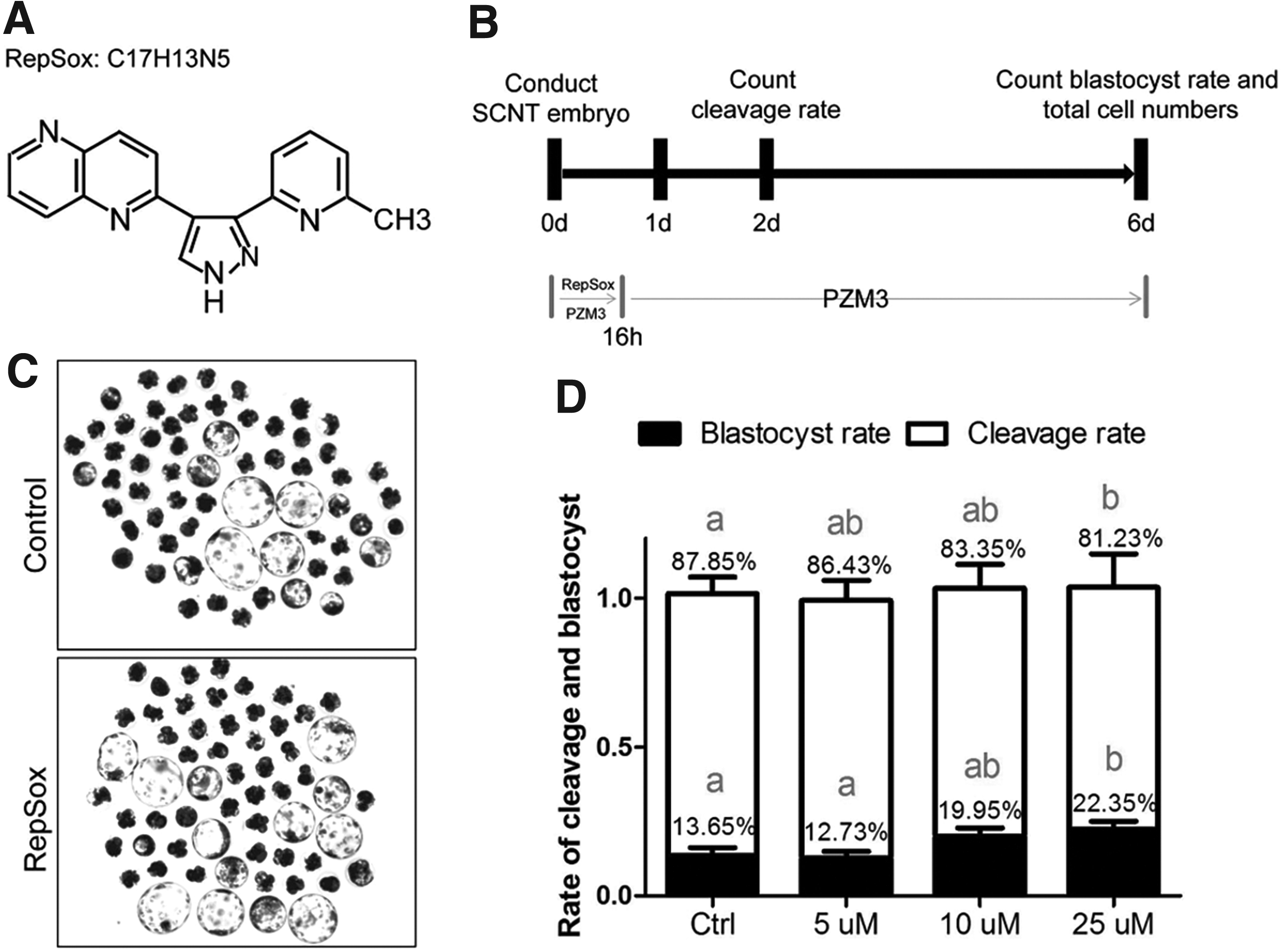
TGF-β inhibitor RepSox promotes SCNT embryos in vitro development.
The results showed that treatment with 25 μM RepSox significantly increased the development (defined as the rate of blastocysts) of SCNT embryos compared with untreated and other treatment groups (p < 0.05) (Fig. 1D). Therefore, 25 μM RepSox treatment was applied for all subsequent experiments. We observed that 25 μM RepSox treatment had no effect on the 48-hour cleavage rate. However, the 144-hour blastocyst rate was significantly improved compared with untreated SCNT embryos (Fig. 1D, 21.8% vs. 13.5%, p < 0.05). The blastocyst cell number was not significantly different between the 25 μM TGF-β inhibitor RepSox group and the untreated group (32.71 ± 2.008 vs. 34.00 ± 1.618 p > 0.05).
Potential mechanism: the clone efficiency was improved by increasing the pluripotency-related gene NANOG
To reveal the potential mechanism of RepSox-mediated improvement of porcine SCNT efficiency, the expression of pluripotency-related genes was examined. The real-time PCR (qPCR) results revealed that the expression levels of the pluripotency-related genes OCT4, NANOG, SOX2, and c-Myc were higher in the 25 μM RepSox treatment group compared with the control group in four- to eight-cell stage embryos. Of note, the expression level of NANOG was significantly increased (p < 0.05) (Fig. 2A), which indicated that RepSox enhanced the porcine SCNT reprogramming efficiency by increasing the expression of the pluripotency-related gene NANOG.
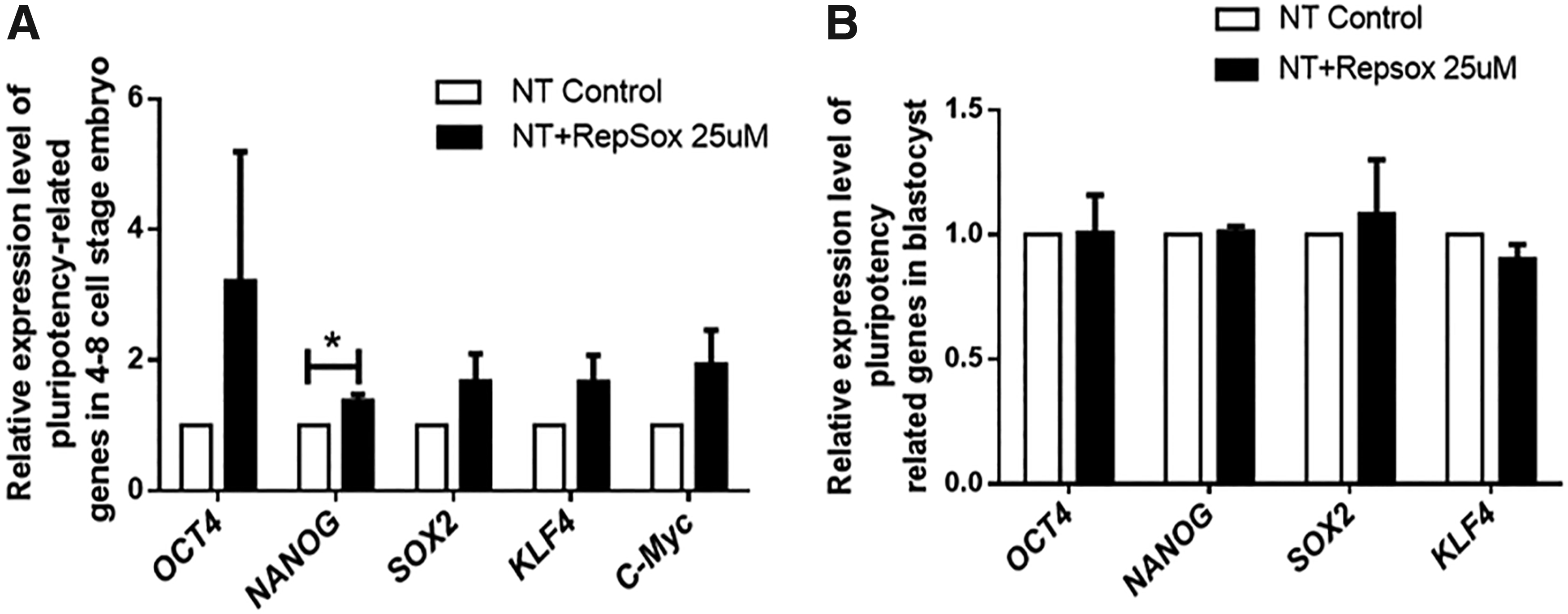
Relative expression of pluripotency-related genes in SCNT porcine embryos.
However, there was no significant difference in the expression level of NANOG in blastocysts derived from the RepSox treatment group compared with the control group (p > 0.05) (Fig. 2B), which suggested that zygotic pluripotency-related gene activation, especially NANOG activation, could directly promote SCNT embryo developmental potency. Additionally, real-time PCR (RT-PCR) analysis showed that TET1 expression was also upregulated in RepSox-treated porcine SCNT embryos, but this increase was not significant (Fig. 2A).
RepSox treatment had no significant effect on porcine SCNT blastocyst apoptosis
Based on a TUNEL assay, the approximate dead cell index values per embryo are shown (Fig. 3B). In our experiment, the apoptotic rates (TUNEL index) were higher in the 25 μM group than in the control group (Fig. 3C), but this difference was not significant (p > 0.05). These results indicated that the increasing developmental rate in the 25 μM RepSox group was not due to abnormal cell apoptosis.
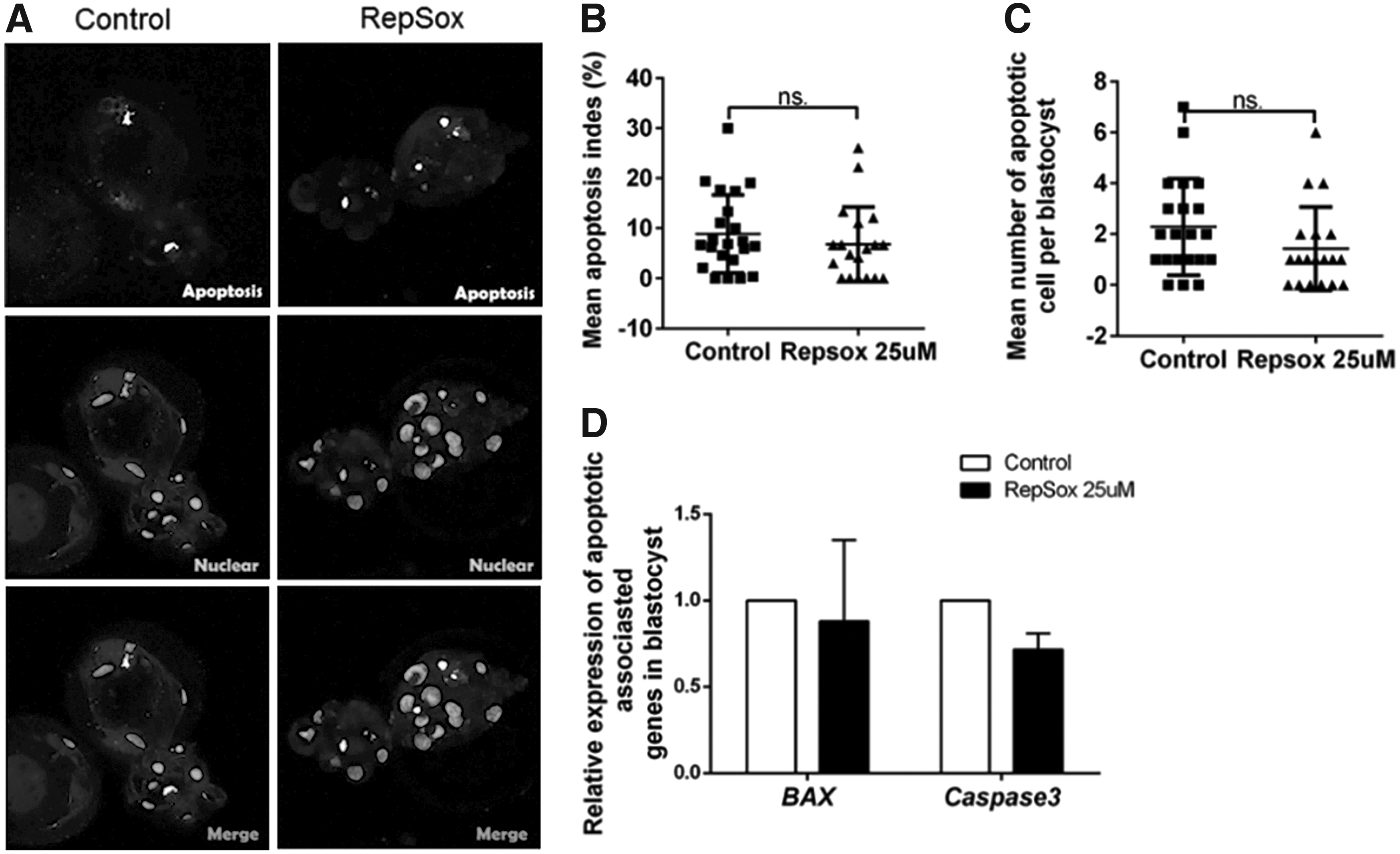
Cloning embryo's apoptosis levels, between the RepSox-treated group and the control group, did not change significantly.
The expression levels of the apoptotic-related genes Bax and Caspase 3 during the blastocyst stage were studied by real-time PCR. Despite the lower mRNA levels of Bax and Caspase 3 in the 25 μM group compared with the control group, there were no significant differences in the blastocyst stage (Fig. 3A).
Discussion
SCNT and the generation of iPSCs are two dominant strategies for reprogramming terminally differentiated somatic cells into a pluripotency status in mammals. SCNT is the main technology to create GM pigs, as there are no well-identified porcine iPSCs and ESCs (Telugu et al., 2010). Although newly developed GM technologies provide unlimited possibilities to produce GM pigs, the main obstacles for GM in livestock are a low cloning efficiency and abnormalities of the cloned animals.
Several molecules that can efficiently reprogram cells to generate iPSC lines have been successfully used to enhance nuclear reprogramming by regulating the expression of specific genes and to improve the developmental competence of SCNT embryos. In the generation of iPSCs, the small-molecule compound RepSox can dramatically improve the efficiency of mouse fibroblast differentiation into iPSCs via inducing the expression of NANOG (Ichida et al., 2009).
Our result revealed that RepSox could enhance the efficiency of reconstructed embryo reprogramming by inducing the expression of NANOG at the four- to eight-cell embryonic stage, but interestingly, the expression of NANOG in the blastocyst was not significantly different (p > 0.05) between the RepSox treatment group and the control group. It is well known that porcine zygotic genome activation occurs at the four- to eight-cell stage in the pig, which is the most critical period of embryonic reprogramming. Thus, RepSox might play a role in promoting pluripotency-related gene expression at the four- to eight-cell stage, which contributes directly to the improvement of the somatic cell reprogramming status and promotes embryonic development.
Zygote gene activation and embryonic reprogramming might have been completed at the blastocyst stage; therefore, it is easy to understand the absence of significant differences in NANOG expression between the RepSox-treated group and the control group.
Apoptosis, which occurs spontaneously in normal preimplantation embryos, can eliminate abnormal, detrimental, or superfluous cells and thus regulates embryo cell numbers (Fabian et al., 2005). However, apoptosis may be a normal feature in mammalian preimplantation development, even in vivo, and it may play an active role in the developing embryo through the removal of genetically abnormal cells. In contrast to these beneficial effects, apoptosis may have detrimental effects if either the number of apoptotic cells or the ratio of these cells to normal cells is elevated (Metcalfe et al., 2004). As mentioned, apoptosis occurs more frequently after SCNT than after in vitro fertilization in the control treatment in a bovine model (Park et al., 2004). It can be hypothesized that the inhibition of blastomere apoptosis improves preimplantation development after SCNT (Park et al., 2004).
However, we found that RepSox treatment had no significant effect on porcine SCNT blastocyst apoptosis in our research, and the expression of the apoptosis-related genes Bax and Caspase 3 was not significantly different in the RepSox treatment compared with the control group. Therefore, we concluded that the apoptotic potential of embryos was not enhanced after RepSox treatment, which suggested an improvement of the porcine clone efficiency via RepSox treatment.
Footnotes
References
Supplementary Material
Please find the following supplemental material available below.
For Open Access articles published under a Creative Commons License, all supplemental material carries the same license as the article it is associated with.
For non-Open Access articles published, all supplemental material carries a non-exclusive license, and permission requests for re-use of supplemental material or any part of supplemental material shall be sent directly to the copyright owner as specified in the copyright notice associated with the article.