
Abstract
Abstract
Periodontitis is a chronic inflammatory disease that can lead to the loss of periodontal bone tissue. The osteogenic potential of periodontal ligament stem cells (PDLSCs) is significantly decreased in periodontitis microenvironment. However, the mechanism is still unclear. We used Porphyromonas gingivalis lipopolysaccharide (LPS) as a stimulator of PDLSCs to mimic the periodontal inflammatory environment. The mineralization capability was restrained in LPS-stimulated PDLSCs, and the level of miR-148a increased, while the level of Neuropilin 1 (NRP1) decreased. Downregulation of miR-148a could reverse the osteogenesis deficiency of PDLSCs under LPS treatment. In addition, the expression of miR-148a in PDLSCs was negatively correlated with the expression of NRP1. Furthermore, overexpression of NRP1 upregulated the osteogenesis ability of LPS-stimulated PDLSCs, while inhibition of NRP1 eliminated the stimulative effect of miR-148a inhibitor on osteogenic differentiation. These data illustrated that the inflammatory environment mimicked by LPS inhibits osteogenesis by upregulation of miR-148a and subsequent downregulation of NRP1. We also found, compared to healthy periodontal tissues, miR-148a level increased, while NRP1 level decreased in periodontitis tissues. These two phenomena also exist in PDLSCs that come from the upper two types of tissues. To summarize, the decline of osteogenic potential of PDLSCs under inflammatory condition of periodontitis is related to miR-148a/NRP1 functional axis. This study may provide a novel strategy in the molecular aspect for the therapy of periodontitis.
Introduction
Periodontitis is a highly prevalent chronic inflammatory disease, accompanied by the main symptoms of periodontal bone destruction, tooth loosening, and eventually leading to tooth loss, which seriously weakens the quality of human life (Winning and Linden, 2017). In healthy periodontal microenvironment, the formation and absorption of alveolar bone are in a dynamic equilibrium state. When inflammation occurs, this homeostasis will be broken, resulting in bone loss (Henderson and Kaiser, 2018; Modinger et al., 2018).
Gram-negative bacteria, especially Porphyromonas gingivalis, are the initiators of periodontitis. Their production lipopolysaccharide (LPS) is the major stimulus factor for periodontal inflammation and bone resorption (Hienz et al., 2015; Shirasugi et al., 2018). One type of multipotent stem cells exists in periodontal ligament tissue, namely periodontal ligament stem cells (PDLSCs), which can differentiate into odontoblasts, osteoblasts, adipocytes, and chondrocytes in vitro (Xie and Liu, 2012), and form periodontal ligaments, cementum, and alveolar bone in vivo (Park et al., 2011; Yang et al., 2009). Previous research found that PDLSCs can be implanted into periodontal defects and efficaciously promote periodontal regeneration (Yang et al., 2009). Thus, PDLSCs possess great potential as a source for stem cell therapy and tissue engineering.
Remarkably, some studies indicated that the differentiation and regeneration abilities of PDLSCs derived from periodontitis microenvironment are evidently inhibited (Li et al., 2018; Liu et al., 2011). In addition, researchers proved that the inflammatory environment induced by LPS also has a negative impact on self-renewal and differentiation potential of PDLSCs (Li et al., 2014). However, the molecular mechanism involved in the differentiation of PDLSCs under an inflammatory microenvironment remains unclear.
MicroRNAs (miRNAs) are short and noncoding RNA molecules composed of 18–22 nucleotides, which can bring about the repression or degradation of genes through binding to the 3′-untranslated regions of target mRNAs. By targeting specific mRNAs, miRNAs play critical roles in regulating wide spectrum of cellular functions, including cell proliferation, apoptosis, differentiation, energy metabolism, tissue morphogenesis, and carcinogenesis (Carthew and Sontheimer, 2009; Friedman et al., 2009; Ha and Kim, 2014).
Various miRNAs are considered to be involved in different steps of bone remodeling (Gamez et al., 2014; Jing et al., 2015), and are hypothesized as “molecular switches” that control the osteogenic differentiation process of mesenchymal stem cells (MSCs) (Martin et al., 2016). For example, miR-130a and miR-27b can enhance osteogenic differentiation of bone marrow MSCs by decreasing the gene expression of peroxisome proliferator-activated receptor γ (Seenprachawong et al., 2018). Furthermore, miRNA-214 can suppress osteogenic differentiation of PDLSCs by targeting activating transcription factor 4 (ATF4) (Yao et al., 2017).
MicroRNA-148a (miR-148a) has been considered to play a key role in inflammation and carcinoma in numerous studies. The small RNA could mediate the downregulation of Neuropilin 1 (NRP1) to inhibit the invasion and tumorigenic potential of medulloblastoma cells (Yogi et al., 2015). Moreover, recent studies have found that miR-148a takes part in bone remodeling and osteogenic differentiation of stem cells.
One study discovered that silencing miR-148a in ovariectomized mice can successfully inhibit bone resorption and improve bone mass (Xiao et al., 2018). Another study validated that the level of miR-148a decreased during the induction of osteogenic differentiation of MSCs (Manochantr et al., 2017). Furthermore, Hongzhi Liu et al. (2018) noticed that miR-148a inhibits MSC-mediated fracture healing by lowering the level of insulin-like growth factor 1 (IGF1). Interestingly, it also has been shown that the expression of miR-148a in inflammatory periodontitis tissue is higher than in healthy periodontal tissue (Luan et al., 2018). However, no information is acquirable about whether miR-148a is involved in regulating osteogenic differentiation of PDLSCs in an inflammatory environment.
In this study, we find out the potential molecular mechanism about the decreasing mineralization capability of PDLSCs under local inflammatory environment caused by periodontitis. Abnormal expression of miR-148a will inhibit the osteogenesis of PDLSCs in a proinflammatory condition by directly targeting NRP1. This study may provide a novel therapeutic mechanism and a potential therapeutic target for bone defect caused by periodontitis.
Materials and Methods
Sample collection
Samples were collected in accordance with the guidelines of the Ethics Committee of the Affiliated Hospital of Nantong University and with the informed consent of donors. The healthy periodontal tissues were derived from healthy teeth samples collected from eight individuals 25–38 years of age. Teeth were being extracted for orthodontic reasons. The periodontal tissues with periodontal inflammation were collected from eight patients (35–45 years of age) diagnosed with chronic periodontitis. All the individuals in our study were examined to be free from systematic disease history, smoking, alcohol, or special medication.
Isolation and culture of human PDLSCs
All experimental protocols were approved by the Ethics Committee of the Affiliated Hospital of Nantong University and informed consent was obtained from each subject. Participants ranged in age from 25 to 45 years. The Periodontal ligament tissue was digested in 3 mg/mL collagenase type I for 1 hour at 37°C. Then we obtained single-cell suspensions by passing the digested tissues through a 70 μm cell strainer (BD Falcon). Cell suspensions were cultured in low-glucose Dulbecco Modified Eagle Medium (HyClone, Logan) with 10% fetal bovine serum (FBS; Gibco, Carlsbad), 100 U/mL penicillin, and 100 μg/mL streptomycin (Gibco) at 37°C in a 5% CO2 incubator. The medium was changed every 3 days.
Cells were passaged at the ratio of 1:3 when they reached 85%–90% confluence. The cells from P3 to P5 were used in this study. Inflammatory conditions were mimicked by adding Porphyromonas gingivalis LPS (Sigma) at concentrations of 0, 0.01, 0.1, 1, or 10 μg/mL to the culture medium.
Western blot analysis
Total protein lysates were extracted from harvested cells using RIPA buffer supplemented with 1 mM PMSF and loading buffer (Beyotime, Shanghai, China). Protein concentrations were determined using a BCA protein assay kit (Beyotime), according to the manufacturer's specifications. Equal amounts of protein (100 μg) were subjected to 10% sodium dodecyl sulfate–polyacrylamide gel electrophoresis. The separated proteins were transferred onto polyvinylidene fluoride (Invitrogen, Thermo Fisher Scientific, MA) membranes in a blotting apparatus (Bio-Rad, Richmond, CA) at 300 mA for 90 minutes.
Membranes were blocked with 5% non-fat milk for 2 hours at 37°C, and then incubated overnight at 4°C with primary antibodies: GADPH (1:1000, anti-rabbit; Santa Cruz), RUNX2 (1:800, anti-mouse; Sigma), NRP1 (1:200, anti-rabbit; Santa Cruz), alkaline phosphatase (ALP), and OCN (1:500, anti-rabbit; R&D Systems). Then the membranes were probed with HRP-conjugated anti-rabbit or anti-mouse IgG antibody (Beyotime) for 2 hours at room temperature. Finally, the protein bands were visualized using Pierce™ ECL Plus Western Blotting Substrate (Thermo Fisher Scientific).
RNA isolation and real-time quantitative-polymerase chain reaction analysis
Total RNA was collected using TRIzol Reagent (Invitrogen, CA) as per manufacturer's instructions. To quantify miR-21 expression, complementary DNA (cDNA) was synthesized using TaqMan™ MicroRNA Reverse Transcription Kit (Thermo Fisher Scientific), followed by quantitative polymerase chain reaction (PCR) with AceQ qPCR SYBR Green Master Mix (without ROX) (Vazyme, Shanghai, China). We used small nuclear RNA U6 as the normalizing control.
For GADPH, RUNX2, ALP, OCN, and NRP1 detection, cDNA was synthesized using RevertAid RT Reverse Transcription Kit (Thermo Fisher Scientific). AceQ qPCR SYBR Green Master Mix (without ROX) (Vazyme) was then used for quantitative PCR of these genes. GAPDH was used for normalization. We use a Light Cycler 480 Real-Time PCR System (Roche Diagnostic, Mannheim, Germany) to test these levels.
The primer sequences used in the experiment were as follows: GAPDH: 5′-GAAGGTGAAGGTCGGAGTC-3′, 5′-GAAGATGGTGATGGGATTTC-3′; RUNX2: 5′-TCAACGATCTGAGATTTGTGGG-3′, 5′-TCAACGATCTGAGATTTGTGGG-3′; ALP: 5′-GGA CCATTCCCACGTCTTCAC-3′, 5′-CCTTGTAGCCAGGCCCATTG-3′; OCN: 5′-CCCAGGCGCTAC CTGTATCAA-3′, 5′-GGTCAGCCAACTCGTCACAGTC-3′; and NRP1: 5′-TGAGCCCTGTGGTTTATTCC-3′, 5′-CGTACTCCTCTGGCTTCTGG-3′. The 2^-ΔΔCt method was used for relative quantification of the gene expression.
Small interfering RNA and transfection
Small interfering RNA (siRNA) was purchased from Santa. The siRNA duplexes were transfected into PDLSCs at final concentrations of 50 nM using Lipofectamine 2000 (Invitrogen) according to the instructions. Cells were harvested 24 hours later, and the knockdown efficiency was further determined by real-time quantitative-polymerase chain reaction (RT-qPCR). Oligo sequences used were as follows: siNRP1-RNA: 5′-AACGATAAATGTGGCGATA-3′ and negative control siRNA: 5′-TTCTCCGAACGTGTCACGT-3′.
Plasmid constructs and plasmid transfection
The full-length NRP1 (GenBank Accession Number NM_003873) was extracted from the human cDNA library and connected to p-CMV-flag. The primers used for NRP1 were as follows: 5′-TGAGCCCTGTGGTTTATTCC-3′ (sense) and 5′-CGTACTCCTCTGGCTTCTGG-3′ (anti-sense). P-CMV-flag and P-CMV-flag-NRP1 were transfected in PDLSCs by Lipofectamine™ 2000 reagent (Invitrogen) according to the manufacturer's instructions.
MiRNA transfection
MiRNA negative control, miR-148a inhibitor, and miR-148a mimic were designed and synthesized by Gene Pharma (Shanghai, China). The PDLSCs were plated in a 12-well culture plate (Corning) to 45% confluence and transfected with miRNA negative control and miR-148a inhibitor and miR-148a mimic at final concentrations of 50 nM using Lipofectamine 2000 (Invitrogen) in accordance with the transfection reagent instructions.
Osteogenic differentiation
PDLSCs were plated into a 12-well culture plate (Corning) at a density of 2 × 10⁵ cells/well. After cell adhesion, the culture medium was replaced with an osteogenic medium supplemented with 10% FBS, 50 μg/mL ascorbic acid, 100 nM dexamethasone, and 5 mM β-glycerophosphate for 7–14 days. The media were changed every 2 days. The collected cells were then used for Alizarin red staining and ALP assay. Total protein and RNA were used for Western blot and RT-PCR analysis.
Alizarin red staining and ALP assay
The mineralization potential of the cells was assessed by Alizarin red staining when cells were cultured with the osteogenic medium for 14 days. The cells were fixed with 4% paraformaldehyde for 1 hour and then incubated with 40 mM Alizarin red S (Sigma) for 15 minutes in the dark. ALP staining were carried out after osteogenic induction for 14 days using the ALP assay kit (Beyotime) according to the the manufacturer's instructions.
Statistical analysis
All the experiments were repeated at least three times independently and the data were presented as mean ± standard deviation. Two group comparisons were tested with two-tailed paired Student's t-test. All statistical evaluation was performed with GraphPad Prism 6.01. One-way analysis of variance was applied to analyze the difference among multiple groups. Value of p < 0.05 was considered statistically significant.
Results
Inflammation inhibits osteogenic differentiation and increases miR-148a expression in PDLSCs
According to the usual research of medical literature, we simulated the microenvironment of periodontitis by treating PDLSCs with Porphyromonas gingivalis LPS. After incubating with osteogenic differentiation media for 14 days, alizarin red staining indicated that the higher the LPS stimulation concentration, the lower the mineralization capability of PDLSCs. However, LPS at 1 and 10 μg/mL had similar mineralization inhibition (Fig. 1A). Besides, the osteogenic differentiation transcription factor RUNX2 protein expression level also proved the same results (Fig. 1B). Subsequently, we detected the expression of miR-148a in normal PDLSCs and LPS-stimulated PDLSCs after osteogenic induction with 7 days through RT-qPCR analysis. The result showed that the level of miR-148a in PDLSCs increased with increasing LPS stimulation concentration.

Osteogenic potential and miR-148a level of PDLSCs under inflammatory condition. PDLSCs were cultured in an osteogenic medium containing LPS at concentrations of 0, 0.01, 0.1, 1, or 10 μg/mL.
Furthermore, the expression of the miR-148a in PDLSCs stimulated by 1 and 10 μg/mL LPS was similar. Moreover, it has a tendency to be opposite to the degree of mineralization of PDLSCs (Fig. 1C). These data suggest that miR-148a may be involved in the mineralization process of PDLSCs exposed to LPS. Also, we selected 1 μg/mL LPS for subsequent experiments. In addition, we found that the level of miR-148a in periodontal tissue and PDLSCs (P-PDLSCs) of periodontitis individual was higher compared with periodontal tissues and PDLSCs (H-PDLSCs) of healthy individual (Fig. 1D, E). All these results implicate that the increase of miR-148a level may inhibit the osteogenesis in PDLSCs under inflammation condition, which is closely related to the decrease of bone mass in periodontitis.
Inhibition of miR-148a restores osteogenic differentiation of PDLSCs in chronic inflammatory environment
Next, we want to confirm the relationship between miR-148a and the decrease of osteogenic capacity of LPS-stimulated PDLSCs. To investigate whether inhibiting miR-148a could reverse inflammatory impaired osteogenic differentiation of PDLSCs, we transfected miRNA negative control and miR-148a inhibitor into LPS-stimulated PDLSCs. The specificity and efficacy of miR-148a inhibitor were verified by RT-qPCR after transfection (Fig. 2A), which effectively reduced the level of miR-148a in LPS-stimulated PDLSCs.
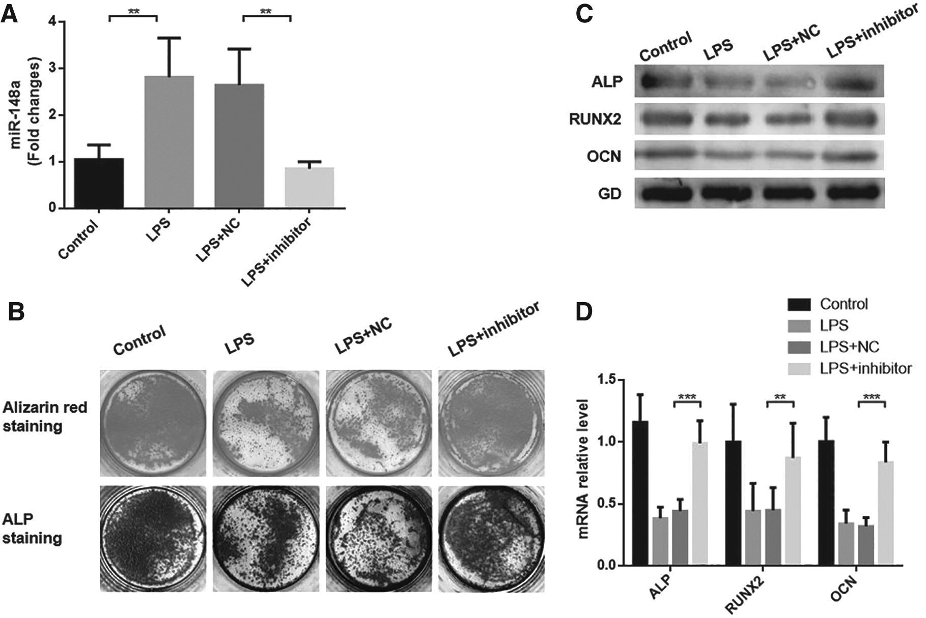
Influence of transfection with miR-148a inhibitor on osteoblastic differentiation of LPS-stimulated PDLSCs.
After 14 days, mineralization induction, alizarin red staining, and ALP staining showed that mineralized bone matrix formation and ALP activity were increased in miR-148a-inhibited LPS-stimulated PDLSCs compared with negative control, and were similar to normal PDLSCs (Fig. 2B). Subsequently, we used RT-qPCR and Western blot analysis to determine the mRNA and protein levels of osteogenic marker genes, including ALP, RUNX2, and OCN (Fig. 2C). We clearly discovered that the downregulation of miR-148a significantly increases the mRNA and protein expression of bone formation-related gene in LPS-stimulated PDLSCs (Fig. 2D). The above results imply that under chronic exposure to LPS, miR-148a participates in the reduction of osteogenic differentiation of PDLSCs.
NRP1 participates in osteoblastic differentiation and is regulated by miR-148a
We gain further insight into the mechanism by which miR-148a influences the osteogenic differentiation of PDLSCs exposed to LPS. According to the previous literature and online databases of target prediction, we predicted NRP1 as a candidate target of miR-148a to regulate osteogenesis.
First, we found out the mRNA and protein expression of NRP1 were increased in PDLSCs and LPS-stimulated PDLSCs after mineralization induction (Fig. 3A, B), indicating that NRP1 may exert an effect in the osteogenic differentiation of PDLSCs. In addition, in both undifferentiated and differentiated conditions, NRP1 was lower in LPS-stimulated PDLSCs than in normal PDLSCs (Fig. 3A, B).

NRP1 positively regulates osteogenic differentiation and is regulated by miR-148a.
These data suggest that NRP1 may take part in LPS-inhibited osteogenesis of PDLSCs. To make it clearer whether the decrease of LPS-stimulated PDLSC mineralization capacity is due to the downregulation of NRP1 level, we overexpressed NRP1 in LPS-stimulated PDLSCs. As expected, NRP1 protein and mRNA expression increased visibly following NRP1 full-length plasmid transfection (Fig. 3D, E). Furthermore, alizarin red and ALP staining indicated overexpression of NRP1-enhanced mineralized matrix deposition and ALP activity of LPS-stimulated PDLSCs (Fig. 3C).
In addition, Western blot and RT-qPCR analysis showed that osteogenic markers ALP, RUNX2, and OCN protein and mRNA levels were also upregulated after NRP1 overexpression (Fig. 3D, E). These results imply that the overexpression of NRP1 improves the osteogenic differentiation in LPS-stimulated PDLSCs. Furthermore, we downregulated miR-148a in LPS-stimulated PDLSCs and found that the expression of NRP1 protein dramatically increased (Fig. 3F). Also, we found the expression of NRP1 protein decreased significantly after upregulated miR-148a in normal PDLSCs (Fig. 3G). MiR148a in PDLSCs can negatively regulate the expression of NRP1. Hence, we hypothesized that the inhibitory effect of miR-148a on osteogenesis is achieved by inhibiting NRP1.
MiR-148a inhibits osteogenic differentiation of PDLSCs in inflammatory environment by targeting NRP1
To clarify whether the upregulation of miR-148a in the LPS-treated PDLSCs can result in the decrease of osteoblastic potential by inhibiting NRP1, we co-transfected miR-148a inhibitor and si-NRP1 into LPS-stimulated PDLSCs. After 14 days of osteogenic differentiation, the increasing extent of the protein and mRNA levels of osteogenic markers was far less than that affected by miR-148a inhibitor transfection alone (Fig. 1A, C). Also, alizarin red and ALP staining revealed that co-transfected miR-148a inhibitor and si-NRP1 cannot significantly reverse the mineralization capacity of LPS-stimulated PDLSCs (Fig. 1B).
These results clarify that under LPS stimulation, the upregulation of miR-148a in PDLSCs leads to the downregulation of NRP1, which results in the decline of osteogenesis. Interestingly, we also discovered that the expression of NRP1 in periodontitis tissues and their source of PDLSCs (P-PDLSCs) was lower than that in a healthy one (Fig. 1D, E). Combined with all the above experiments, we speculate that the periodontal inflammatory environment brings about the increase of miR-148a in PDLSCs, and which further results in the decrease of NRP1 and ultimately inhibits osteogenesis.
Discussion
It is generally known that periodontitis can cause the loss of alveolar bone and consequently the loss of teeth (Winning and Linden, 2017). The imbalance between osteoblasts and osteoclasts leads to the decrease in bone formation and the increase in bone resorption, which is one of the factors contributing to the reduction of bone mass in periodontitis (Kaiser, 2018; Modinger et al., 2018). Recent studies have found that PDLSCs also play an important role in maintaining periodontal bone mass owing to its osteogenic capacity (Du et al., 2016). However, the inflammatory microenvironment will inhibit the osteogenic capacity of PDLSCs, which in turn becomes vital pathogenesis of periodontitis (Du et al., 2016; Li et al., 2018).
Generous studies have shown that LPS produced by dominant bacteria in gingival crevicles plays an indispensable role in the occurrence and development of periodontitis (Hienz et al., 2015; Shirasugi et al., 2018). Our previous research found that 1 μg/mL LPS-stimulated PDLSCs can release high concentrations of proinflammatory factors, such as interleukin 6 (IL-6), interleukin 8 (IL-8), and tumor necrosis factor-α. Therefore, 1 μg/mL LPS exposure was adopted to stimulate the microenvironment of chronic periodontitis in PDLSCs (Zheng et al., 2019).
In our research, we tested the osteogenic capacity of PDLSCs under stimulation with five different LPS concentrations by alizarin red staining and Western blot, including 0, 0.01, 0.1, 1, and 10 μg/mL. We found that LPS at 1 and 10 μg/mL had a strong inhibitory effect on osteogenic differentiation. Thus, we chose 1 μg/mL LPS as the final experimental concentration. Besides, these results indicate that the LPS-mediated inflammatory environment has a distinct inhibitory effect on the osteogenic capacity of PDLSCs, and the mechanism is the focus of our future research.
In the process of changes in cell characteristics and functions, miRNAs are essential regulators of signal network and gene reprogramming (Carthew and Sontheimer, 2009; Ha and Kim, 2014). Recently, many reports have revealed that various miRNAs act as modulators of cell differentiation. MiRNAs such as miR-21, miR-146a, and miR-1294 have been reported to participate in osteogenic differentiation of stem cells (Fan et al., 2018; Kuang et al., 2017; Yang et al., 2017). Other studies have displayed that targeting specific miRNA may be a promising treatment for certain inflammatory diseases (Kearney et al., 2018; Xiang et al., 2017). In addition, researchers have recently suggested that periodontitis might possess a miRNA expression profile that is different from the normal physiological condition (Luan et al., 2018).
As shown in our results, the level of miR-148a in PDLSCs was positively correlated with the stimulation concentration of LPS. In addition, when the LPS concentration reached 1 μg/mL, the expression level of miR-148a no longer increased. Previous studies have indicated that miR-148a can inhibit the mineralization potential of bone marrow MSCs (Liu et al., 2018). We also found the level of miR-148a was negatively correlated with the mineralization ability of PDLSCs, which indicated that miR-148a could take part in PDLSC osteogenic differentiation. Furthermore, the inhibition of miR-148a in PDLSCs could reverse the osteogenic differentiation ability approximately to normal level. Thus, miR-148a also has a negative regulatory effect on osteogenesis of PDLSCs.

NRP1 is involved in the miR-148a-regulated osteogenic differentiation and reduced under inflammatory environment.
The occurrence and development of various diseases, including inflammatory diseases, can be generally regulated by miRNAs through the control of target genes (Friedman et al., 2009). NRP1 is one of the members among neuropilin family, which involved in regulating cell proliferation, apoptosis, and differentiation (Shi et al., 2018; Sun et al., 2018; Zhang et al., 2015). Previous studies have reported that the absorption of bone tissue around the inflammation of apical periodontitis is associated with the decreased expression of NRP1 (Lin et al., 2017). Our previous study also found that NRP1 can promote odontoblast differentiation of dental pulp stem cells through the classical Wnt/β-catenin pathway (Song et al., 2017). In general, NRP1 has been proved to be a vital regulator related to mineralization formation.
Our data show that the level of NRP1 in LPS-stimulated PDLSCs is reduced abnormally. Besides, we surprisingly found that the osteogenic capacity was significantly reversed when NRP1 was overexpressed in LPS-stimulated PDLSCs. Therefore, it can be seen from our experiments that the reduction of NRP1 is closely associated with a decrease in PDLSC osteogenic capacity under the inflammatory condition. In addition, we discovered that the inhibition of miR-148a in LPS-stimulated PDLSCs can cause a rise in the level of NRP1. Also, the overexpression of miR-148a in normal PDLSCs leads to decreased expression of NRP1.
Hence, we suspected that miR-148a inhibits the osteogenic differentiation of PDLSCs by downregulating the target gene NRP1. Based on this hypothesis, we co-transfected miR-148a inhibitor and si-NRP1 into LPS-stimulated PDLSCs and then detected si-NRP1 can eliminate the ability of miRNA-148a inhibitor to reverse osteogenic differentiation. These experiments validate our conjecture that under LPS stimulation conditions, miR-148a is elevated in PDLSCs, which can lead to the inhibition of NRP1, ultimately resulting in decreased osteogenic potential.
We also verified that miR-148a was upregulated, while NRP1 was downregulated in periodontitis tissues and their source of PDLSCs, and these performances were consistent with LPS-stimulated PDLSCs. This result further validates that miR-148a/NRP1 functional axis may play a key role in PDLSC differentiation under an inflammatory microenvironment. We mentioned earlier that NRP1 regulates the odontogenic differentiation through canonical Wnt/β-catenin pathway (Song et al., 2017). Moreover, many literatures have reported that this pathway is closely related to osteogenic differentiation. However, whether NRP1 regulates the osteogenic differentiation of PDLSCs through this pathway requires further investigation.
This study clarified the molecular mechanisms of governing PDLSC osteogenic differentiation potential in a local chronic inflammation microenvironment, providing us with better knowledge of periodontitis and how to improve PDLSC-mediated inflammatory bone defects. Our data suggest that in the case of PDLSC dysfunction, inhibition of miR-148a may have a significant impact on improving bone repair in periodontitis. However, further studies should be designed to confirm the results.
Footnotes
Author Disclosure Statement
The authors declare they have no conflicting financial interests.
Funding Information
The study was partly supported by Postgraduate Research & Practice Innovation Program of Jiangsu Province (KYCX18_2408; KYCX18_2431), Nantong City Science and Technology Projects Funds (MS1201712-2).