
Abstract
Bone marrow-derived mesenchymal stem cells (BMSCs) from livestock are valuable resources for veterinary therapeutics and animal reproduction. Previous studies have shown that hypoxic conditions were beneficial in maintaining the mesenchymal feature of BMSCs. However, the effects of hypoxia on buffalo BMSCs (bBMSCs) remain unclear. In this study, the effects of hypoxic conditions on cell morphology, migration, polarity, and karyotype of bBMSCs were examined. The results showed that hypoxia (5% oxygen) enhanced colony formation and stress fiber synthesis of bBMSCs. Under the hypoxic culture conditions, the migration capacity and normal karyotype rate of bBMSCs were significantly improved (p < 0.05), which resulted in weakened cell polarity and enhanced karyotype stability in bBMSCs. In addition, it was significantly (p < 0.05) upregulated in the expression levels of HIF-TWIST signaling pathway axis-related genes (Hif-1, Hif-2, Twist, Snail, Slug, Fn1, N-cadherin, Collal). The HIF-TWIST axis of bBMSCs was also activated in hypoxia. Finally, it was more effective and easier to maintain the mesenchymal feature of bBMSCs in hypoxic conditions. These findings not only provide theoretical guidance to elucidate the detailed regulation mechanism of hypoxia on mesenchymal nature maintenance of bBMSCs, but also provide positive support to further establish the stable in vitro culture system of bBMSCs.
Introduction
Bone marrow-derived mesenchymal stem cells (BMSCs) are mesodermally derived stem cells with immunomodulatory, anti-inflammatory, self-renewing, and proliferative abilities (Lee et al., 2004). BMSCs have the potential to differentiate into cells of different origins, including chondrocytes, osteoblasts, adipocytes, and neurocyte (Bruder et al., 1998; Liu et al., 2017; Zhang et al., 2017a). Human BMSCs have been widely used in the fields of cell transplantation, tissue engineering, and organ regeneration for the clinical application of human diseases (Bornes et al., 2017; Wang et al., 2015).
However, the applications of BMSCs are severely limited by the malignant transformation of mesenchymal-to-epithelial transition (MET) in vitro (Baxter et al., 2010; Daniel et al., 2008; Gro Vatne et al., 2009; Miura et al., 2010; Reza et al., 2008; Yi Fu et al., 2006). Therefore, how to solve the problem of MET in vitro culture is of considerable significance for further application of BMSCs.
It is of great value for BMSCs to maintain the mesenchymal characteristics in long-term in vitro culture. Ambient O2 concentrations of 20% were widely used for in vitro cell cultures. However, BMSCs are located in the bone marrow cavity where O2 partial pressure is 1%–7% (Lee and Kemp, 2006). Therefore, it can better reflect the physiological nature of BMSCs in vivo by mimicking the low oxygen tension in vitro culture. Many reports have demonstrated that low oxygen pressure or hypoxia affects various fundamental vital cellular activities of BMSCs, including proliferation, apoptosis, differentiation, metabolism, and migration chemotaxis, subsequently affecting the pluripotency of BMSCs (Bornes et al., 2015; Liu et al., 2017; Roth et al., 2016; Zhang et al., 2017b). Thus, it is necessary to research the effects of hypoxia on mesenchymal feature in the future.
Hypoxic stimulation leads to several signaling transduction pathways activated in mammalian cells, including hypoxia-inducible factor 1 (HIF-1α), which is one of the master transcription factors that promote cellular adaptation to hypoxia through regulating the expression of hundreds of genes (Keith and Simon, 2007; Rocha, 2007; Semenza, 2007). Previous studies showed that HIF-1α was bound to the promoter of Twist, which was adjacent hypoxia reaction element, and upregulated the expression levels of Twist. Besides, HIF-1α and Twist formed an axis of the HIF-TWIST signaling pathways that regulated the expression of upstream and downstream genes (Lee and Kemp, 2006; Yang et al., 2008). These results indicated that the HIF-TWIST signaling pathway axis may play a crucial role in maintaining the mesenchymal nature of BMSCs, but the relationships of hypoxia and HIF-TWIST signaling pathway axis are still unclear.
The BMSCs of domestic animals are valuable cell resources for modeling human cell therapies, animal breeding, and regenerative veterinary medicine. The application of BMSCs in animal breeding has been widely demonstrated, especially in somatic cell nuclear transfer (SCNT). It has been indicated that BMSCs were more suitable for donor cells, and reconstructed embryos derived from BMSC donor cells achieved high embryo cleavage rates compared with fibroblast cells (Flisikowska et al., 2012; Guo et al., 2016; Kumar et al., 2010; Kwong et al., 2014; Li et al., 2013; Simon et al., 2012; Sunglim et al., 2010; Yoko et al., 2004). These reports suggested that the BMSCs were easier to reprogram as donor cells and more suitable for SCNT.
As one of the leading domestic animals in southern china, buffalo (Bubalus bubalis) is widely used for the plough, meat, and milk (Deng et al., 2019; Deshun et al., 2007; Lu et al., 2018). To our knowledge, the effects of hypoxia on buffalo BMSCs (bBMSCs) have not been reported. Also, the underlying mechanism of how hypoxic conditions influence bBMSCs in vitro is unknown. In this study, bBMSC's colony formation, migration ability, cell polarity, and karyotype stability in hypoxia (5% oxygen) were examined. In addition, the effects of hypoxia on mesenchymal marker genes and the HIF-TWIST signaling pathway axis were investigated.
This study provides new insight into how hypoxic conditions affect in vitro culture of bBMSCs and also provides a method for procurement of high-quality bBMSC resources without MET as much as possible for animal reproduction, especially in somatic cell reprogramming-mediated buffalo transgenic breeding.
Materials and Methods
Reagents and culture medium
The culture medium, fetal bovine serum (FBS), osteogenesis differentiation kit, adipogenesis differentiation kit, and other supplements were obtained from Gibco (Carlsbad, CA). The bBMSCs culture medium was purchased from StemCell Technologies (Vancouver, BC, Canada). Molecular detection reagents were obtained from TaKaRa (Osaka, Japan). The primary antibodies and secondary antibodies were purchased from Cell Signaling Technology (Danvers, MA). Cell culture plates were obtained from Eppendorf (Hamburg, Germany). The other reagents were purchased from Sigma-Aldrich (St, Louis, MO) unless otherwise indicated.
The complete medium for bBMSCs contained low-glucose DMEM, and was supplemented with 10% FBS, 10,000 U/mL penicillin, and 10,000 μg/mL streptomycin. The cells in normoxic conditions were cultured under a humidified atmosphere of 5% CO2 in air at 37°C (Biospherix X2 Xvivo system).
Cell isolation and culture
bBMSCs were isolated from limbs' long bone marrow cavity of buffalo fetus (Bubalus bubalis), whose body length was 10–18 cm and cultured in low-glucose DMEM (Gibco) with 10% FBS (Gibco). When bBMSCs were cultured for 24 hours, the small glucose DMEM was altered. When bBMSCs reached 80% to 90% confluence, they were passaged or frozen for later experiments.
Flow cytometry and immunofluorescence staining
For flow cytometry, bBMSCs were digested and washed with PBS. Then the cells were incubated with 1% BSA for 1 hour at room temperature. The flow cytometry antibody included CD29, CD31, CD44, and CD45 were added according to the flow cytometry antibody instructions and the cells were incubated for 1 hour in the dark. Then the cells were collected for flow cytometry detection. For immunofluorescence staining the operational procedure could refer to Sandmaier's method (Sandmaier et al., 2015), the primary antibodies included OCT4, NANOG, CD73, and the appropriate secondary antibody was used for 1 hour in the dark. Nuclei were counterstained with Hochst 33342.
RNA isolation, reverse transcription and PCR, and quantitative RT-PCR
Total RNA was isolated from cells using TRIzol reagent (Life Technologies) according to product instruction, and cDNA synthesis was performed using the Prime Script TMRT Reagent Kit (TaKaRa). Then the cDNA samples were applied for real-time quantitative PCR using SYBR Premix Ex Taq™ II (TaKaRa). The primer sequences used in this study are displayed in Table 1. The expression of genes was normalized to that of the internal control gene GAPDH.
Primer for Quantitative RT-Polymerase Chain Reaction
In vitro differentiation and staining identification
When the cells reached 80% to 90% confluence, the osteoblast induction medium was added to perform osteoblast induction according to the manufacturer's instructions. The induction medium was replaced every 3 days, and the staining identification was performed after 21 days of continuous induction. Adipogenic induction procedure was referred to osteoblast induction's method, and the staining identification was performed after 14 days of continuous induction.
The staining identification of osteoblast induction was detected using the Alizarin Red Detection Kit, and the staining identification of adipogenic induction was identified using the Oil Red Detection Kit according to the manufacturer's instructions. The chondrocytes' induction was also referred to as osteoblast and adipocyte induction procedure, and the staining identification was detected using the Alcian Blue Kit according to the Kit's instruction.
Cell adhesion assay
When reaching 80% to 90% confluence, the cells were gently washed with PBS three times. Then the cells were digested in 0.2% EDTA at room temperature and were gently shaken every 5 minutes. The suspended cells that have been digested were extracted and were washed in PBS three times. The cells were photographed under a microscope. The remaining cells were digested, counted, and the adherent rate subsequently calculated.
Cell stress fiber detecting assay
Cells were gently washed with Triton-BSA-PBS three times and fixed in 4% paraformaldehyde for 40 minutes at room temperature. Then the cells were washed with blockage solution three times and incubated in 1% Triton X-100 for 10 minutes. Then the cells were incubated in 1% BSA for 1 hour and washed with Triton-BSA-PBS three times. Finally, the cells were incubated in 10 μM FITC-labeled phalloidin storage solution for 30 minutes at room temperature and washed with Triton-BSA-PBS three times, then the nuclei were counterstained with Hoechst 33342 for 10 minutes in the dark at room temperature.
Karyotype analysis
The cells that had grown up to 60% to 70% confluence were prevented in metaphase by exposing them to 0.2 μg/mL demecolcine for 3 hours in a humidified atmosphere of 5% CO2 in air at 37°C and then resuspended in 0.8% sodium citrate for 30 minutes at 37°C. After centrifugation at 1200 rpm for 3 minutes, cells were fixed with 4 mL acetic methanol (1:3) solution for 20 minutes at 37°C. Therefore, chromosome spreads were arranged by dropping 20 μL cell suspension onto cold slides and incubated at 75°C for 3 hours. Finally, the slides were stained with 10% Giemsa solution for 15 minutes and analyzed under an Olympus microscope.
Statistical analysis
All data are presented as mean ± standard deviation. Statistical significance was determined using a one-way analysis of variance followed by Student's t-test. Statistical analysis was performed by using SPSS 22.0 software. p values <0.05 were deemed significant, with * representing p < 0.05.
Results
Isolation, culture, and identification of bBMSCs
bBMSCs were isolated from the bone marrow cavity by using the total bone marrow adherent method. During the culture process in normoxic conditions (20% oxygen), some fibroblast-like cells appeared on day 1 (Fig. 1a), subsequently, they grew into typical BMSC-like colonies on day 2 (Fig. 1b), and more came up on day 4 (Fig. 1c). These BMSC-like cells grew into tightly packed monolayer and aggregated into helical colonies at the bottom of the plate on day 7 (Fig. 1d). These primary colonies showed the typical morphology of BMSCs.
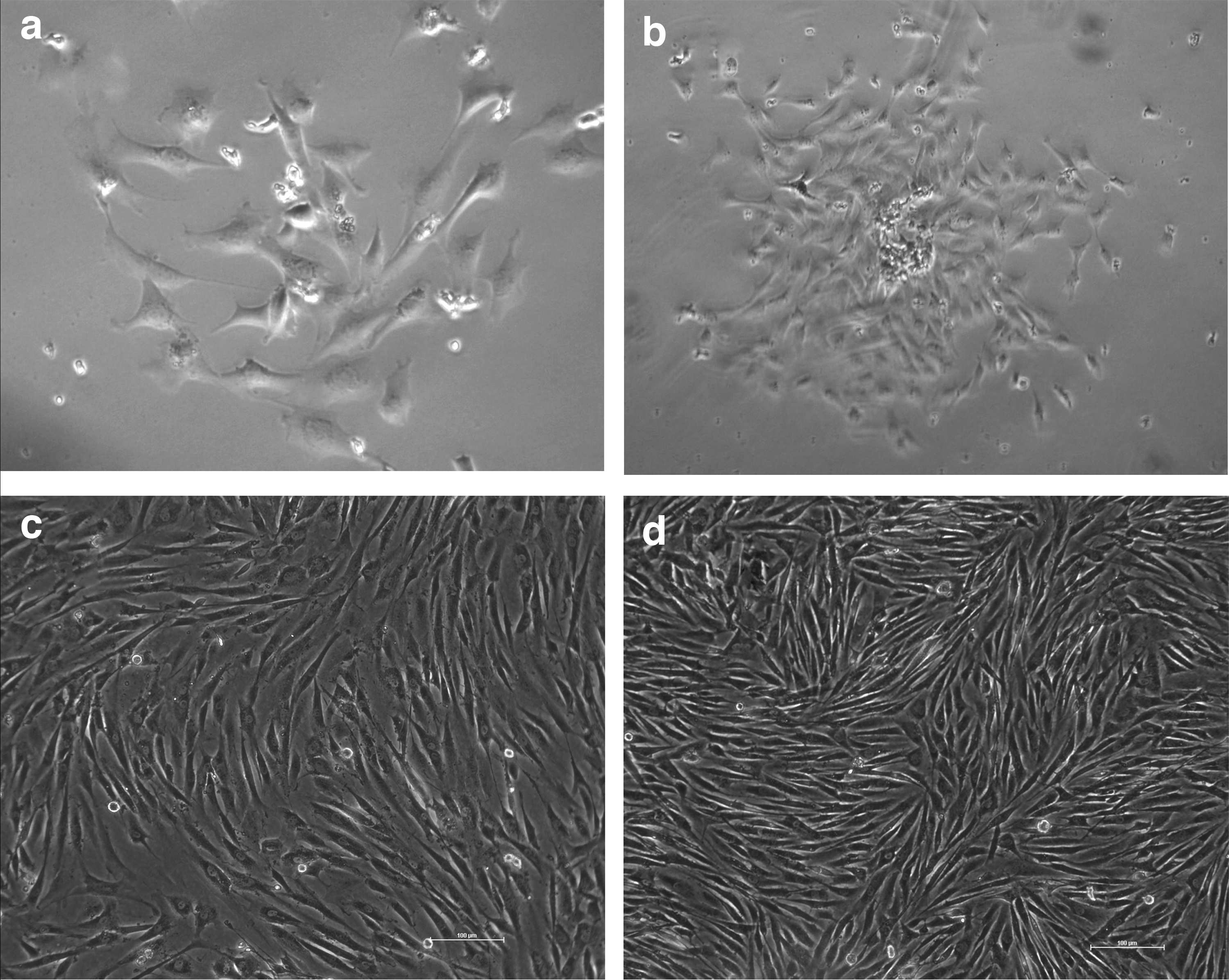
bBMSCs were isolated and cultured in normoxic conditions (20% O2) with morphological change.
By identification, these colonies exhibited mesenchymal stem cell characteristics in vitro. The expression of pluripotency and mesenchymal markers were the main feature of these cells. Flow cytometry results showed that these colonies were marked as CD29 and CD44 positive and CD31 and CD45 negative (Fig. 2a). Immunofluorescence staining results confirmed that these cells were positive for pluripotent stem cell markers: OCT4, NANOG, and CD73 (Fig. 2b).

Expression of mesenchymal markers and pluripotency markers by flow cytometry and immunofluorescence staining, and multipotent differentiation capacity of bBMSCs by induction differentiation assays.
Moreover, the multipotent differentiation capacity of these colonies was analyzed by testing the osteogenic and adipogenic differentiation potential in vitro. In osteogenic induction, it confirmed that calcium nodules were formed and the early osteogenesis marker RUNX2 and OST were expressed in these calcium nodules by Alizarin Red staining and qRT-PCR (Fig. 2c), which were induced from these BMSC-like colonies. The results of adipogenic induction showed that lipid depositions were generated and the adipogenesis-specific gene, PPARγ2, was expressed in these lipid depositions (Fig. 2d). These results suggest that the bBMSCs isolated in the condition exhibits the mesenchymal stemness in vitro.
Hypoxia promotes the colony formation of bBMSCs
The effects of hypoxia on the colony formation of bBMSCs were evaluated through the morphology of bBMSCs. The results showed that there was no significant difference in the colony formation between hypoxia-cultured and normoxia-cultured bBMSCs at passage 1 (Fig. 3a, b). However, the colonies formed from bBMSCs at passage 10 in hypoxic conditions were more compact, three-dimensional, and uniform compared with colonies formed in normoxic conditions (Fig. 3c, d). This was especially obvious after passage 10, which presented some loose, cytoplasmic vacuolization, and epithelial-like cells in normoxia. In addition, bBMSCs cultured in hypoxia maintained typical mesenchymal stem cell morphology up to passage 15 in this study. These results demonstrate that hypoxic conditions enhance bBMSC's colony-forming capacity and maintain the stemness of bBMSCs in vitro.
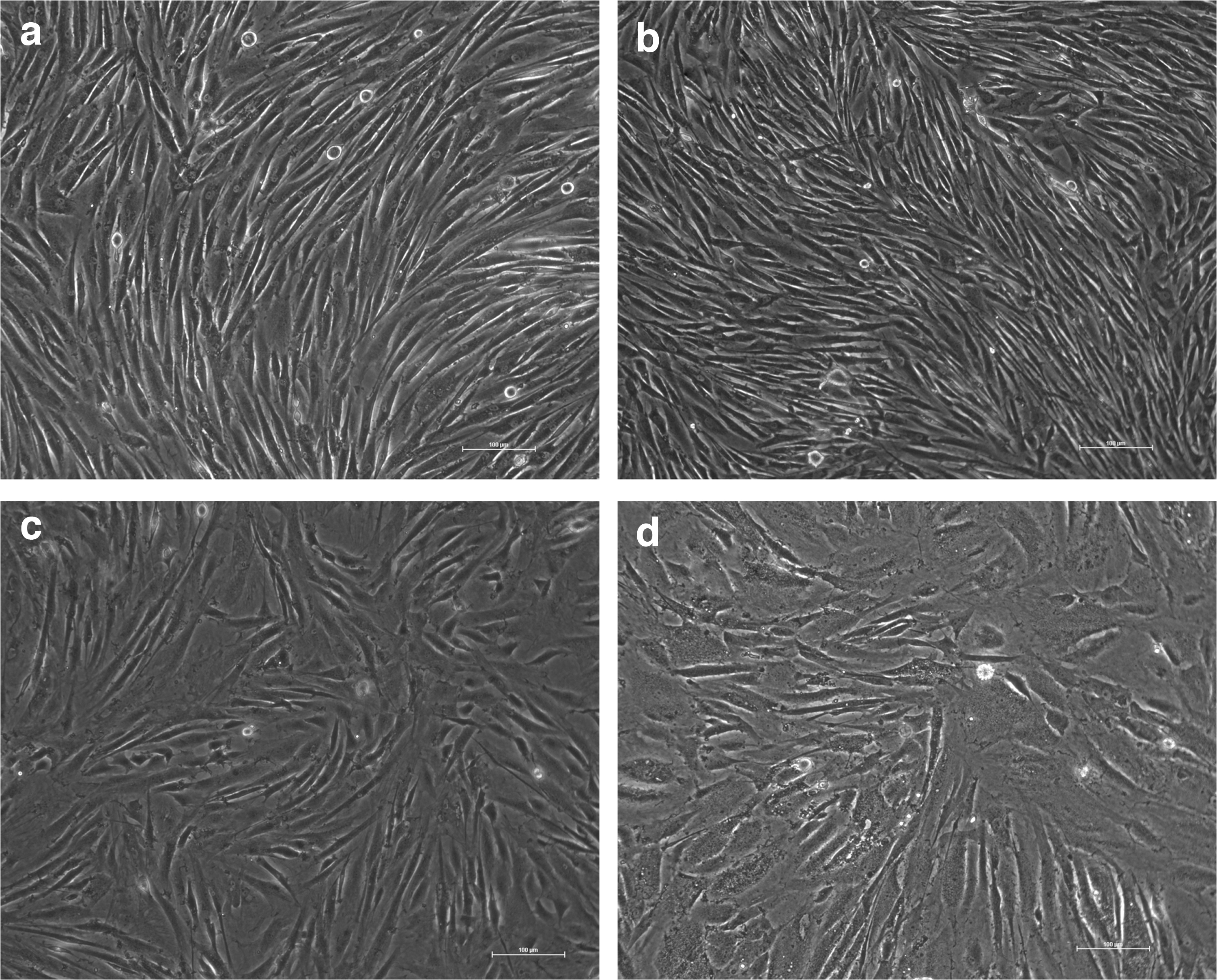
The effects of hypoxia (5% O2) on the colony formation of bBMSCs.
Hypoxia enhances the migration capacity of bBMSCs
The effects of hypoxia on the migration of bBMSCs were analyzed by examining the cell adhesion ability. Around 0.2% EDTA was used to digest bBMSCs to determine cell attachment rate. The results showed that the cell attachment rate of bBMSCs at passage 10 declined with the increase of digestion time, and there was no significant difference in attachment rate at 5 and 10 minutes between hypoxia-cultured bBMSCs and normoxia-cultured bBMSCs (Fig. 4a). However, the attachment rate of bBMSCs in hypoxic conditions was significantly (p < 0.05) decreased compared with bBMSCs in normoxic conditions at 15 and 30 minutes (Fig. 4b). These results suggest that hypoxia promotes the migration capacity of bBMSCs in vitro.
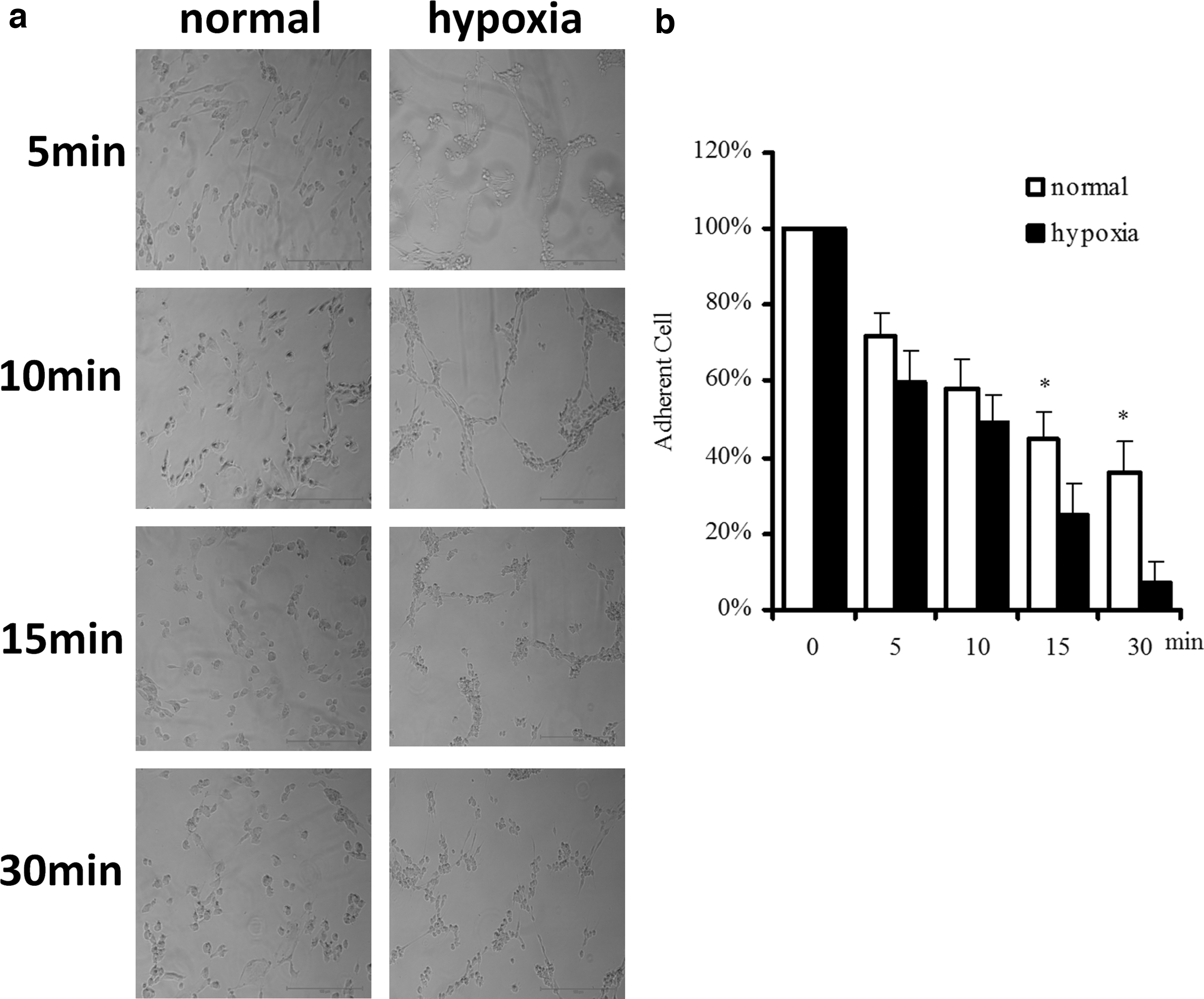
The effects of hypoxia on the migration capacity of bBMSCs.
Hypoxia alters the cell polarity of bBMSCs
The effects of hypoxia on the cell polarity of bBMSCs were investigated by testing the level of cellular stress fiber synthesis. During the MET process, BMSCs transformed from nonpolar to polar cells, and the level of cellular stress fiber synthesis declined. The results showed that stress fiber synthesis levels of bBMSCs at passage 1 had no difference between hypoxic conditions and normoxic conditions (Fig. 5a). However, the levels of stress fiber synthesis of bBMSCs at passage 10 in hypoxia were less compared with bBMSCs in normoxia (Fig. 5b). These results demonstrate that hypoxia changes the cell polarity of bBMSCs in vitro.
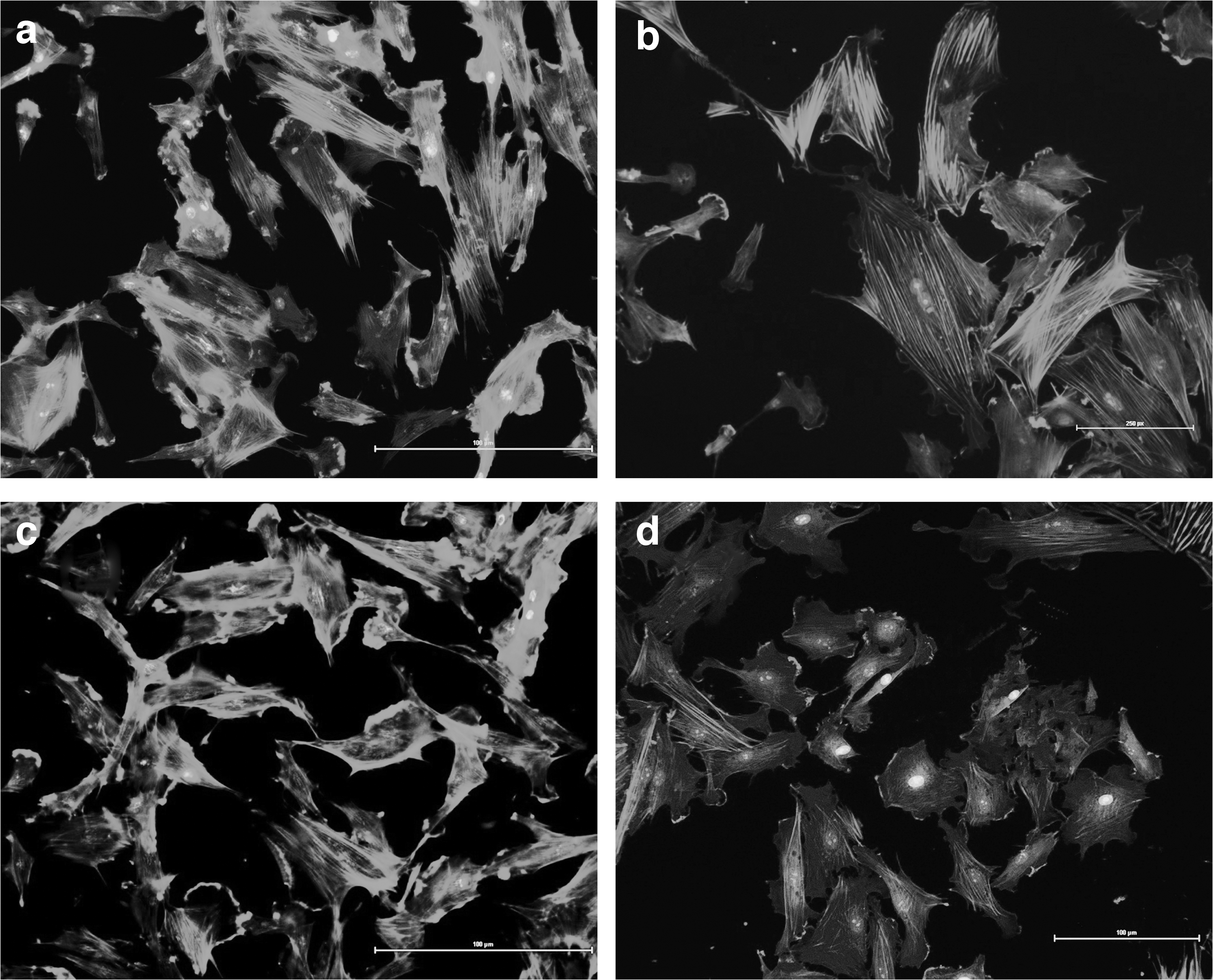
The effects of hypoxia on the cell polarity of bBMSCs.
Hypoxia promotes the stability of karyotype in bBMSCs
The effects of hypoxia on the stability of karyotype in bBMSCs were evaluated by karyotype analysis (Fig. 6). The results showed that the rate of normal karyotype in bBMSCs decreased with the increase of cell passages, and the normal karyotype rate of bBMSCs at passage 1 had no significant difference between hypoxia and normoxia (94.0% vs. 92.0%). However, the normal karyotype rate of bBMSCs at passage 10 and 20 in hypoxic conditions were significantly (p < 0.05) higher compared with bBMSCs in normoxic conditions (72.5% vs.55.5%; 43.3% vs.25.7%) (Table 2). These results suggest that hypoxia promotes the stability of karyotype in bBMSCs in vitro.
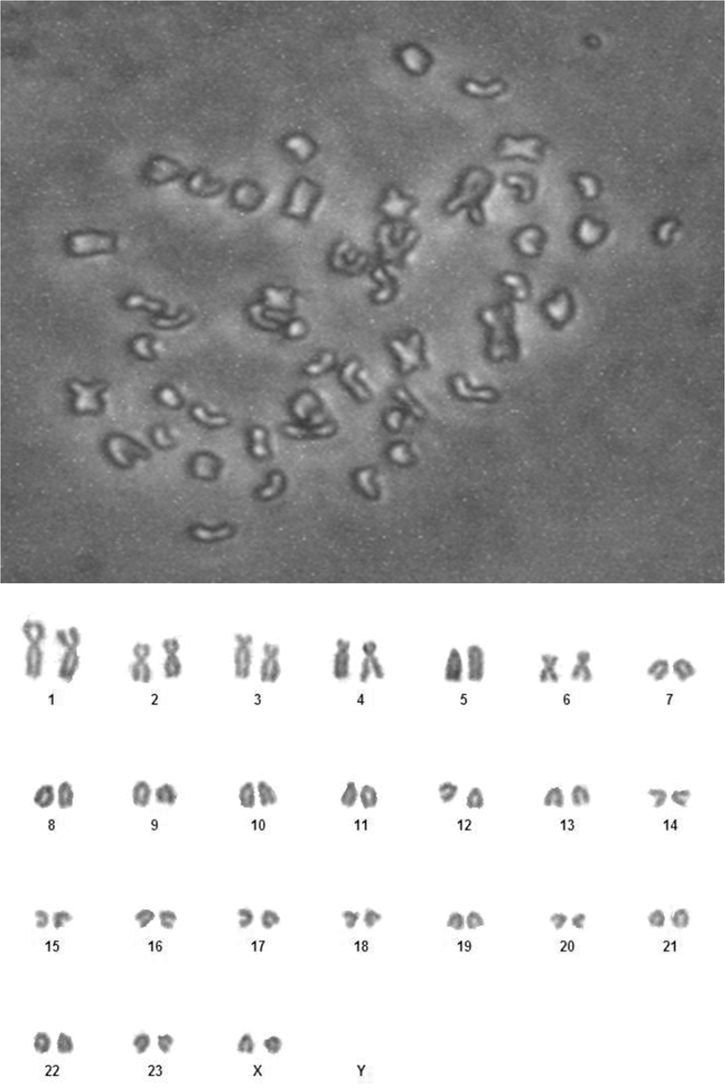
The karyotype analysis of bBMSCs. Scale bars = 100 μm.
Hypoxia (5% O2) Promotes the Stability of Karyotype in Buffalo Bone Marrow-Derived Mesenchymal Stem Cells
Different letters of each row in the chart represent significant differences p < 0.05.
Hypoxia activates the HIF-TWIST signaling pathway axis of bBMSCs
The effects of hypoxia on the HIF-TWIST signaling pathway axis were analyzed by examining the expression levels of the HIF-TWIST signaling pathway axis (Hif-1, Hif-2, Twist) and its downstream-related genes (Snail, Slug, Fn1, N-Cadherin, Collal). The results showed that the expression level of the HIF-TWIST axis and its downstream-related genes in hypoxic conditions were significantly (p < 0.05) upregulated compared with normoxic conditions (Fig. 7a–c). The expression levels of genes, Hif-1, Slug, N-Cadherin, and Collal, exhibited a downward trend. However, the expression levels of genes Hif-2, Twist, Snail, and Fn1 showed an upward, and subsequently, downward trend and the dynamic trends were almost consistent. These results demonstrate that the HIF-TWIST signaling pathway axis of bBMSCs is activated in hypoxic conditions.

The effects of hypoxia on the HIF-TWIST signaling pathway axis of bBMSCs at P1, P10, and P20. The expression levels of HIF-TWIST axis
Discussion
Because of their multipotent nature, capacity of self-renewal, accessible collection, and immune feature in vitro, BMSCs have become an ideal candidate for regenerative medicine, veterinary therapeutics, and animal reproduction (Bornes et al., 2017; Gabrielyan et al., 2017; He et al., 2018; Lin et al., 2019; Wang et al., 2015). In livestock, BMSCs were used as suitable donor cells in SCNT for somatic cell reprogramming-mediated transgenic animal production (Flisikowska et al., 2012; Li et al., 2013; Simon et al., 2012).
At present, there are few reports about the bBMSCs, therefore, it is necessary to conduct a systematic study in the future. It is in consensus among researchers that BMSCs are easy to occur as malignant transition in long-term in vitro culture, and during in vitro culture, they gradually decrease in mesenchymal characteristics, which is accompanied by the changes of cell morphology, proliferation, apoptosis, migration, and karyotype (Izadpanah et al., 2008; Røsland et al., 2009; Zhou et al., 2006). Previous research showed that malignant transition of BMSCs in vitro was strictly related to MET (Daniel et al., 2008). The self-transitioning cells downregulated mesenchymal marker expression, and stress fiber synthesis was inhibited, and the migration capacity was decreased, leading to the loss of mesenchymal features.
Thus, preserving mesenchymal nature in in vitro culture presents a challenge for BMSC researchers. Many factors affect the mesenchymal characteristics of the long-term culture of BMSCs, and manipulating gas composition to represent physiological conditions is a promising opportunity for obtaining high-quality BMSCs in vitro. In in vivo conditions, BMSCs are physiologically adapted to a hypoxic condition where the oxygen is 1%–7%. Atmospheric oxygen concentration (20% oxygen) is typically used in previous BMSC-related studies, however, these conditions do not fit BMSCs' physiological hypoxic conditions and do not maintain their mesenchymal feature. Hypoxia (5% oxygen) was utilized to explore the effects of hypoxic culture on the BMSCs' physiological conditions in this study, using an automatic oxygen control cell culture chamber.
BMSCs were prone to occurring MET in long-term in vitro culture, and lost the mesenchymal characteristics, accompanying the changes of cell morphology. In our previous research, results showed that the morphology of bBMSCs gradually altered with the increase of cell passages in normoxic conditions (5% oxygen), and this was especially obvious after passage 10. In this study, we found that hypoxia contributed to maintaining the morphology of bBMSCs at passage 10, indicating that hypoxic conditions presented herein suitably replicates in vivo physiology. These results are in agreement with the finding of other researchers who reported the effects of hypoxia on MSCs (Andreeva et al., 2015; Deng et al., 2019).
Hypoxic conditions not only affected the morphology of bBMSCs but also promoted the migration ability and altered the cell polarity. BMSCs are characterized by strong migration ability and abundant stress fiber synthesis levels. So, testing cell attachment rate and stress fiber synthesis levels were appropriate methods to evaluate the migration and polarity of BMSCs (Zhao et al., 2019). In this study, the results showed that the attachment rates were significantly lowered and resulted in enhancing the migration capacity of bBMSCs in hypoxic conditions, which is in agreement with the previous research of Rosova et al. (2008). We also found that hypoxia promoted the stress fiber synthesis levels of bBMSCs, leading to altering cell polarity in vitro, which is similar to the work of other researchers (Zhiqian et al., 2003).
Many studies have reported that hypoxia promotes the stability of karyotypes in BMSCs in long-term in vitro culture. In our study, we found that the normal karyotype rates of bBMSCs at passage 10 and 20 were significantly promoted in hypoxic conditions than those in normoxic conditions. These results are in agreement with the findings of other researchers who reported the effects of hypoxia on cell karyotype (Zachar et al., 2010). Our results indicate that hypoxia may decrease the excessive proliferation, and reduce the accumulation of reactive oxygen species and the rate of chromosome aberration, ultimately enhancing the stability of karyotype in bBMSCs. Therefore, our results demonstrate that bBMSCs maintain their biological feature much more robustly when cultured in hypoxic conditions, which mimic their physiological oxygen concentration.
Previous reports have shown that hypoxia enhances the mesenchymal characteristics of BMSCs, but the mechanisms are still ambiguous and are even less known about bBMSCs, although biological conservation would indicate that the mechanism is similar. HIF-1 plays a vital role in mediating transcription response to oxygen deprivation in mammalian cells. HIF-1 is made up of an oxygen-dependent α-subunit and an oxygen-independent beta-subunit. HIF-1 transcriptional activity is mainly regulated by HIF-1α, through binding to a DNA sequence called hypoxia-responsive elements in the promoters of target genes, activating thousands of hypoxia-regulated genes related to various cellular process in MSCs including cell activity, cell proliferation, and cell migration (Palomäki et al., 2013).
Prior research has indicated that tumor cells responded to hypoxia through HIF binding to Twist, forming HIF-TWIST signaling pathway axis to play a crucial role in the process of epithelial-to-mesenchymal transition (Castanon and Baylies, 2002; Lee and Kemp, 2006; Yang et al., 2004, 2008). Thus, we hypothesize that hypoxic conditions affect the mesenchymal nature of bBMSCs through activating the HIF-TWIST signaling pathway axis. The results demonstrate that hypoxia upregulates the expression levels of the HIF-TWIST signaling pathway axis and its downstream-related genes, which is in accordance with the work of Yang et al., (2008).
In this study, the results also showed that the dynamic trend of gene expression levels was almost consistent between gene Hif-2 and Twist, suggesting that Hif-2 and Twist may form the HIF-TWIST signaling pathway axis to maintain the mesenchymal characteristics of bBMSCs in hypoxic conditions. However, it needs to be further studied in the detailed regulation mechanisms.
In conclusion, bBMSCs are initially isolated using total bone marrow adherence methods in this study. Hypoxic conditions enhance colony formation, migration capacity, cell polarity, and karyotype stability of bBMSCs when compared with the normoxic culture conditions. Hypoxia promotes the mesenchymal stemness of bBMSCs mainly through activating the HIF-TWIST signaling pathway axis.
Data Availability Statement
The data that support the findings of this study are available from the corresponding author upon reasonable request.
Footnotes
Author Contributions
Jun Zhang designed the study, performed the experiments, and drafted the article. Deshun Shi and Fenghua Lu designed the study. Chuan Lei performed the experiments and drafted the article. Yanfei Deng helped in performing the experiments, collecting tissue samples, and analyzing the data. Jam zaheer Ahmed edited the article and analyzed the data.
Author Disclosure Statement
The authors declare they have no conflicting financial interests.
Funding Information
This research was supported by the grants from the Chinese National Natural Science Foundation (31560633 and 31760666); Natural Science Foundation of Guangxi (2018JJA130074); Guangxi Innovation-Driven Development Fund Project (AA17204051), and The New Century Guangxi Ten, Hundred, and Thousand Talent Project.