
Abstract
Incomplete DNA methylation reprogramming in cloned embryos leads to poor cloning efficiency. Melatonin has been proven to improve the development of cloned embryos, however, the role of melatonin during somatic cell nuclear transfer remains unclear. This work demonstrated that 10−7 M melatonin significantly enhanced the developmental progress, reduced the arrested rate before zygotic genome activation, and upregulated the blastocyst rate of cloned embryos. Melatonin also promoted the pseudo-pronucleus formation, increased blastocyst cell number, and reduced embryo apoptosis through upregulating the expression of antiapoptosis factors while downregulating the transcription of proapoptosis genes. Further study displayed that DNA methylation reprogramming related genes were greatly improved in cloned embryos when treated with melatonin; then, melatonin effectively promoted genomic DNA demethylation and DNA remethylation, DNA demethylation of pluripotency related gene Oct4, DNA methylation maintenance of imprinted gene H19/Igf2, and DNA remethylation of tissue-specific gene Thy1 in cloned embryos. Thus, zygotic genome activation related gene Eif1a, pluripotency related genes Oct4, Nanog, and Sox2, imprinted genes Igf2 and H19, and blastocyst quality related genes Cdx2 and ATP1b1 were remarkably upregulated, and tissue-specific genes Thy1 and Col5a2 were considerably silenced. In conclusion, melatonin enhanced the development of cloned embryos by ameliorating DNA methylation reprogramming. This work reveals that melatonin can regulate DNA methylation reprogramming and provides a novel insight to improve cloning efficiency.
Introduction
Somatic cell nuclear transfer (SCNT) has been successful in many species, and promotes the development of basic research, animal husbandry, biomedicine, and others. (Matoba and Zhang, 2018). However, cloning efficiency is still low, limiting the wide application of SCNT technology (Czernik et al., 2019).
It is generally believed that low cloning efficiency is mainly due to incomplete DNA methylation reprogramming (Peat and Reik, 2012). DNA methylation predominantly occurs at the fifth carbon of cytosine, and can regulate gene expression by altering the accessibility of transcription factors to gene promoter region (Sepulveda-Rincon et al., 2016). However, DNA methylation reprogramming in cloned embryos is incomplete, leading to ineffective activation of pluripotency related genes, aberrant transcription of imprinted genes, and continuous expression of tissue-specific genes, and finally resulting in the poor cloning efficiency (Huan et al., 2015; Peat and Reik, 2012; Yang et al., 2007).
It has been proven that the culture condition can change DNA methylation pattern of embryos (Meehan et al., 2018). During in vitro culture, embryos are exposed to a hyperoxic environment and become vulnerable to the produced excessive reactive oxygen species (ROS) (Wu et al., 2017). Previous studies have shown that the accumulated ROS disrupts the expression pattern of genes via DNA methylation and causes the developmental delay and arrest of embryo (Bennemann et al., 2018; Takahashi, 2012; Yang et al., 2019b). During SCNT, the excessive ROS also inhibits nuclear reprogramming and causes damage to cloned embryos (Bae et al., 2015). Thus, reducing ROS may improve DNA methylation reprogramming in cloned embryos.
To overcome the oxidative damage, antioxidants including melatonin have been applied to improve embryo development and melatonin has been proved to scavenge ROS, reduce apoptosis, activate gene expression, and promote embryo development (Loren et al., 2017; Tian et al., 2017). In cloned embryos, melatonin is effective to alleviate ROS-mediated apoptosis to improve cloning efficiency and also inhibits repressive epigenetic modification including DNA methylation to promote SCNT-mediated nuclear reprogramming (Liang et al., 2017; Pang et al., 2013 Yang et al., 2019a). Recently, accumulating studies suggest that melatonin could participate in DNA methylation reprogramming in embryo development (An et al., 2019; Yang et al., 2019a, b). However, the effect and potential role of melatonin during SCNT-mediated DNA methylation reprogramming are still unclear.
In this study, the results displayed that melatonin promoted DNA methylation reprogramming, improved gene expression, and enhanced the development of cloned embryos. These results provide a novel insight to improve cloning efficiency.
Materials and Methods
Chemicals were purchased from Sigma Aldrich Corporation (St. Louis, MO), and disposable and sterile plasticware was obtained from Nunclon (Roskilde, Denmark), unless otherwise stated.
All the experiments and treatments were approved and supervised by Animal Care Commission of Qingdao Agricultural University according to animal welfare laws, guidelines, and policies.
Donor cell collection and culture
Donor cell collection and culture have been described previously (Huan et al., 2016). Briefly, fetal tissues were finely minced into pieces, digested, and dispersed in Dulbecco's modified Eagle's medium (GIBCO). Then, the dispersed cells were centrifuged, resuspended, cultured, and used in 3–5 passages.
Oocyte collection and in vitro maturation
Oocyte in vitro maturation system has been established (Huan et al., 2013). Briefly, ovaries were collected, and follicles were aspirated. Cumulus-oocyte complexes were cultured in the maturation medium, and vortexed in hyaluronidase to remove cumulus cells. Only oocytes with the visible polar body, regular morphology, and homogenous cytoplasm were used.
In vitro fertilization and SCNT embryo culture, treatment and collection
The procedures for in vitro fertilization (IVF) and SCNT have been described (Huan et al., 2013). Briefly, for IVF, spermatozoa were diluted with modified Tris-buffered medium, and matured oocytes were transferred into the fertilization medium and co-incubated with spermatozoa, and then, fertilized embryos were cultured in porcine zygote medium-3 (PZM-3). For SCNT, after oocyte enucleation, donor cells were placed into perivitelline space. Fusion and activation of cell-cytoplast complexes were induced by electroporation. Then, reconstructed embryos were cultured in PZM-3 and treated with 0, 10−5, 10−7, 10−9, or 10−11 M melatonin.
For embryo collection, 1-cell, 4-cell, and blastocyst embryos in the IVF, NT-C (control), and NT-M (10−7 M melatonin) groups were collected at 6, 48, and 156 hours postactivation, respectively.
Evaluation of embryo development progress and arrest
Evaluation of embryo development progress and arrest was performed at 12, 24, 48, 72, and 156 hours or at 48 and 156 hours postactivation, respectively, and then, the proportions of 1-cell, 2-cell, 4-cell, 8-cell, morula, blastocyst, and fragmented embryos or embryos blocked before the 4-cell stage were evaluated, respectively.
Examination of pseudo-pronucleus formation and quality of cloned embryos
Embryos at 6 and 156 hours postactivation were treated with acidic Tyrode's solution to remove zona pellucida, fixed in 4% paraformaldehyde, and stained with Hoechst 33342, respectively. Then, nuclear status and blastocyst cell number were examined (Huan et al., 2013).
Assessment of embryo apoptosis
Detection of embryo apoptosis using the TUNEL method with an In Situ Cell Death Detection Kit (Roche) has been described (Huan et al., 2016). Briefly, blastocysts were fixed with 4% paraformaldehyde, permeabilized with 0.5% Triton X-100, and incubated in the terminal deoxynucleotidyl transferase-mediated dUTP nick end labeling reaction medium. Then, blastocysts were stained with Hoechst 33342. The number of apoptotic cells per blastocyst was counted.
Quantitative real-time polymerase chain reaction
Measurement of gene expression with the quantitative real-time polymerase chain reaction (PCR) has been applied (Huan et al., 2015). Briefly, total RNA was extracted from every 50 pooled embryos using a RNeasy Micro Kit (Qiagen). Reverse transcription was performed using a PrimeScript® RT Reagent Kit (TaKaRa). For quantitative real-time PCR, reactions were performed in 96-well optical reaction plate (Applied Biosystems) using a SYBR® Premix ExTaq™ II kit (TaKaRa) and a 7500 Real-Time PCR System (Applied Biosystems). For every sample, the cycle threshold (CT) values were obtained from three replicates. The primers were presented in Supplementary Table S1. Gene relative expression was analyzed using the 2−ΔΔCT method.
Bisulfite sequencing
Bisulfite sequencing has been reported (Huan et al., 2015). Briefly, pooled embryos were digested and treated with sodium bisulfite to convert all unmethylated cytosine to uracil using an EZ DNA Methylation-Direct™ Kit (Zymo Research). For samples of 500, 200, and 50 pooled embryos at the 1-cell, 4-cell, and blastocyst stages, digestion was performed in the M-Digestion Buffer, the CT (cytosine to thymine) conversation condition was 98°C for 10 minutes and 64°C for 2.5 hours, and samples were desalted and purified. Subsequently, nested PCR was carried out to amplify the target regions using the primers described in Supplementary Table S2. The amplified products were verified, cloned into T Vectors, and sequenced.
Statistical analyses
Differences in data (mean ± standard error of the mean) were analyzed with the SPSS statistical software (Huan et al., 2015). Statistical analyses of data concerning embryo development, embryo apoptosis, gene expression, and DNA methylation with one-way analysis of variance or embryo arrest, nuclear remodeling, and blastocyst cell number with t-test were performed. For all analyses, differences were considered to be statistically significant when p < 0.05.
Results
Melatonin improved the development of cloned embryos
After treatment, melatonin with all the concentrations significantly enhanced the development of cloned embryos, and 10−7 M melatonin (NT-M) resulted in the greatest blastocyst rate (Fig. 1A and Supplementary Table S3, p < 0.05). The NT-M group also displayed the significantly lower arrested rate of cloned embryos before the 4-cell stage than the NT-C (0 M melatonin) group (Fig. 1B and Supplementary Table S4, p < 0.05).

Effect of melatonin on the development of cloned embryos.
Then, embryo development progress was examined (Fig. 1C). When compared with IVF embryos, NT-C embryos displayed the significantly greater 1-cell and 2-cell arrested rates, and lower 4-cell, 8-cell, morula, and blastocyst developmental rates, respectively (p < 0.05), while NT-M embryos showed similar development progress and significantly greater 8-cell and blastocyst development rates (p < 0.05). Compared with NT-C embryos, NT-M embryos displayed the significantly downregulated 1-cell and 2-cell arrested rates and upregulated 4-cell, 8-cell, morula, and blastocyst development rates, respectively (p < 0.05). Thus, melatonin overcame the development block, enhanced the development progress, and promoted the blastocyst formation of cloned embryos.
Melatonin promoted nuclear remodeling and blastocyst quality of cloned embryos
Compared with the NT-C group, the NT-M group displayed the significantly upregulated pseudo-pronucleus formation and blastocyst cell number (Fig. 2, p < 0.05). When the apoptosis related indexes were investigated in blastocysts (Fig. 3), compared with the IVF group, the NT-C group showed significantly increased number and rate of apoptotic cells, downregulated expression of antioxidant or antiapoptotic factors (Sod1, Gpx4, and Bcl2l1) but upregulated transcription of proapoptotic genes (Bax, P53, and Caspase-3), while the NT-M group displayed the similar or even better pattern of antiapoptotic related indexes with the significantly upregulated transcription of Gpx4 and Bcl2l1 (p < 0.05).
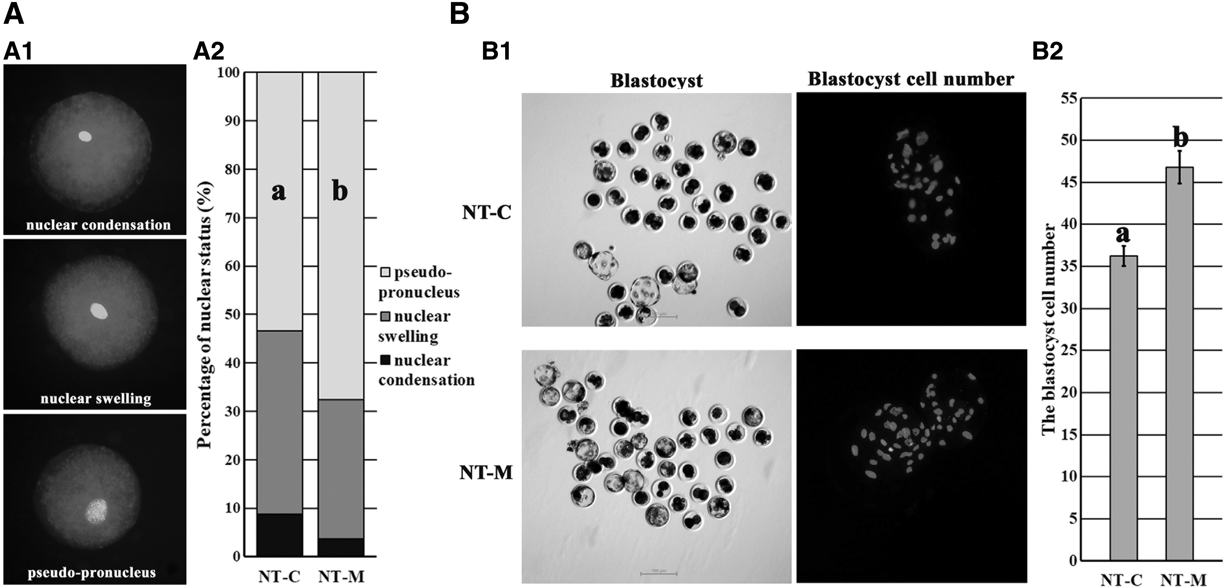
Effect of melatonin on nuclear remodeling and blastocyst cell number of cloned embryos.

Effect of melatonin on the apoptosis of blastocyst cells.
Compared with the NT-C group, the NT-M group displayed significantly reduced number and rate of apoptotic blastocyst cells; upregulated expression of Sod1, Gpx4, and Bcl2l1; and downregulated transcription of Bax, P53, and Caspase-3 (p < 0.05). Thus, melatonin promoted nuclear remodeling, reduced embryo apoptosis, and enhanced blastocyst quality of cloned embryos.
Melatonin enhanced DNA methylation reprogramming in cloned embryos
Here, gene-specific DNA methylation reprogramming was investigated in cloned embryos (Fig. 4). As for genomic (CenRep) DNA methylation changes (Fig. 4A), the trend of genomic DNA remethylation was detected in the IVF and NT-M group, and when compared with IVF group, the NT-C group displayed a significantly lower genomic DNA methylation level at the blastocyst stage (p < 0.05), while no significant differences of genomic DNA methylation were observed in the NT-M group.
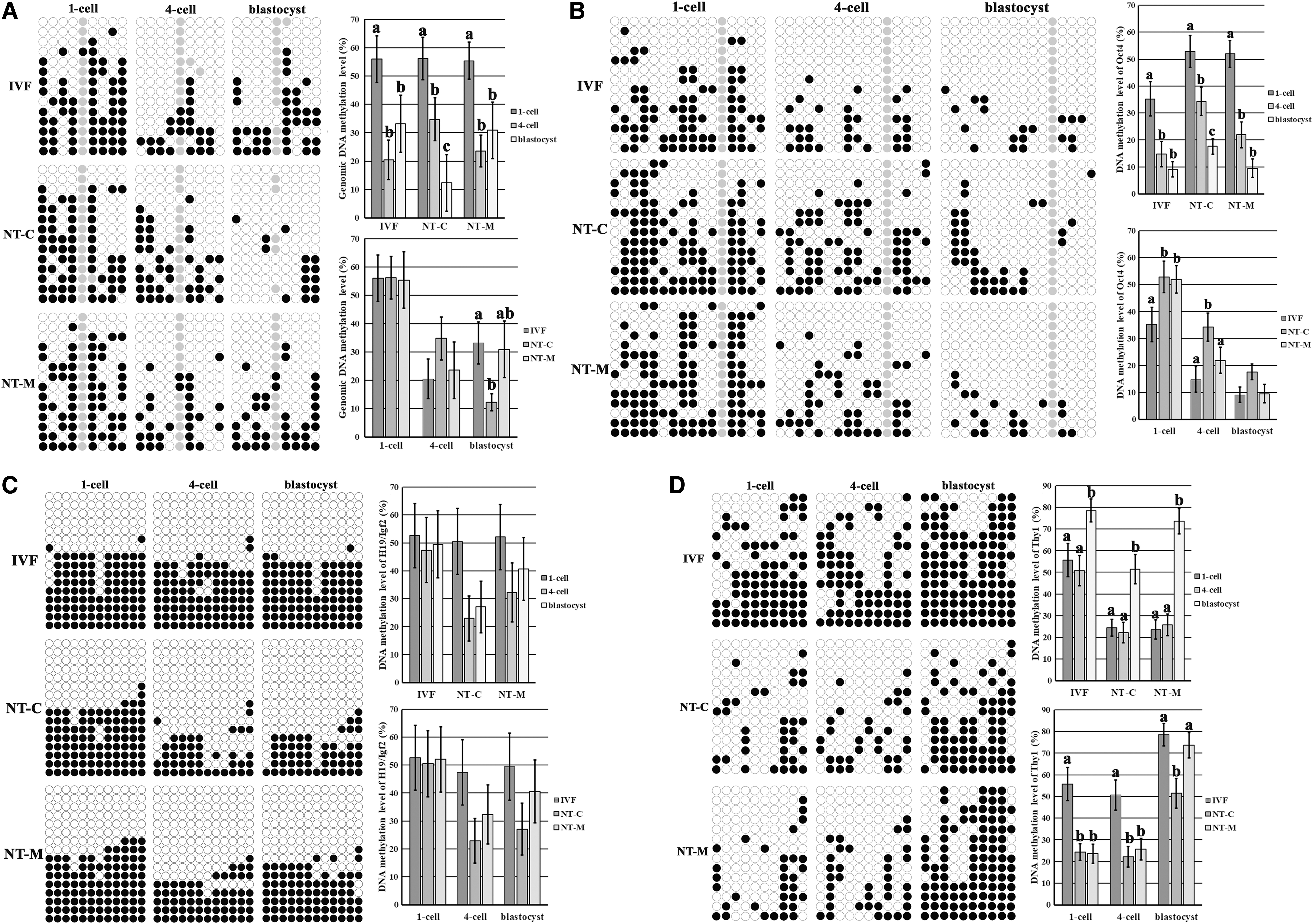
Effect of melatonin on DNA methylation reprogramming in cloned embryos.
When DNA methylation statuses of Oct4 were investigated in cloned embryos (Fig. 4B), low DNA methylation levels of Oct4 were observed at the blastocyst stage of IVF and NT-M groups, and compared with the IVF group, the NT-C group displayed significantly greater DNA methylation levels of Oct4 at the 1-cell and 4-cell stages, while the NT-M group only showed significantly greater DNA methylation level of Oct4 at the 1-cell stage (p < 0.05), and the DNA methylation level of Oct4 at the 4-cell stage in the NT-M group was significantly lower than that in the NT-C group (p < 0.05). As for DNA methylation maintenance of genomic imprinting (H19/Igf2) (Fig. 4C), DNA methylation levels of H19/Igf2 at the 4-cell and blastocyst stages were obviously higher in the IVF and NT-M groups than the NT-C group though no significant differences were observed.
When DNA methylation reprogramming of Thy1 was detected in cloned embryos (Fig. 4D), the significantly upregulated DNA methylation levels of Thy1 at the blastocyst stage were observed in the IVF, NT-C, and NT-M groups (p < 0.05), and compared with the IVF group, the NT-C group displayed significantly lower DNA methylation levels of Thy1 during the whole development progress, while the NT-M group only showed significantly lower DNA methylation levels of Thy1 at the 1-cell and 4-cell stages (p < 0.05), and the DNA methylation level of Thy1 at the blastocyst stage in the NT-M group was significantly greater than that in the NT-C group (p < 0.05). Therefore, melatonin largely ameliorated the disrupted DNA methylation reprogramming in cloned embryos.
Then, the transcription levels of DNA methylation reprogramming related genes were examined in early embryos (Supplementary Fig S1 and Fig. 5). Compared with those in the IVF group, the expression levels of Dnmt1 and Dnmt3a were significantly greater at the 1-cell and 4-cell stages and lower at the blastocyst stage, and the transcription levels of Tet1 and Tet3 were significantly reduced at the 4-cell and blastocyst stages (p < 0.05).
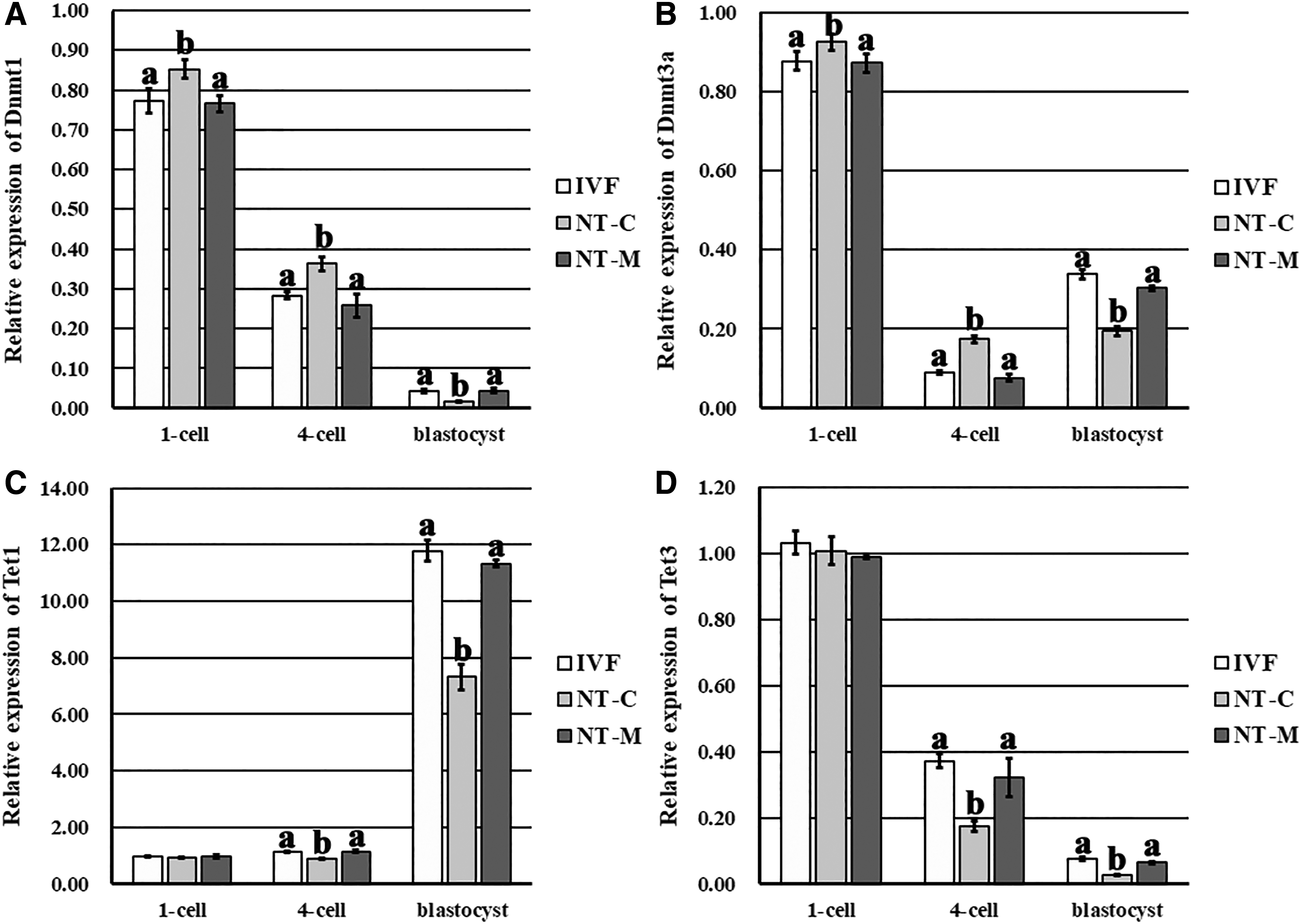
Relative expression levels of DNA methylation reprogramming related genes in early embryos.
In the NT-M group, Dnmt1, Dnmt3a, Tet1, and Tet3 took on the similar expression patterns and levels to those in the IVF groups, and compared with the NT-C group, the significantly reduced transcription at the 1-cell and 4-cell stages and greater expression levels at the blastocyst stage of Dnmt1 and Dnmt3a, and the significantly upregulated expression levels at the 4-cell and blastocyst stages of Tet1 and Tet3 were observed (p < 0.05). Thus, melatonin effectively ameliorated the expression of DNA methyltransferases and ten eleven translocation dioxygenases to regulate DNA methylation reprogramming in cloned embryos.
Melatonin improved the expression patterns of genes related to the development of cloned embryos
Here, zygotic activation related gene Eif1a, pluripotency related genes Oct4, Nanog, and Sox2, imprinted genes Igf2 and H19, blastocyst quality related genes Cdx2 and ATP1b1, and tissue-specific genes Thy1 and Col5a2 were investigated in early embryos (Supplementary Fig S2 and Fig. 6). In the NT-C group, similar gene expression trends to the IVF group were displayed, however, the significantly lower expression levels of Eif1a at the 4-cell stage, Oct4, Nanog, Sox2, and Igf2 at the 4-cell and blastocyst stages, and H19, Cdx2, and ATP1b1 at the blastocyst stage were observed (p < 0.05). In the NT-M group, gene expression trends and levels were similar to those in the IVF group, and ATP1b1 transcription was even significantly upregulated at the blastocyst stage (p < 0.05).
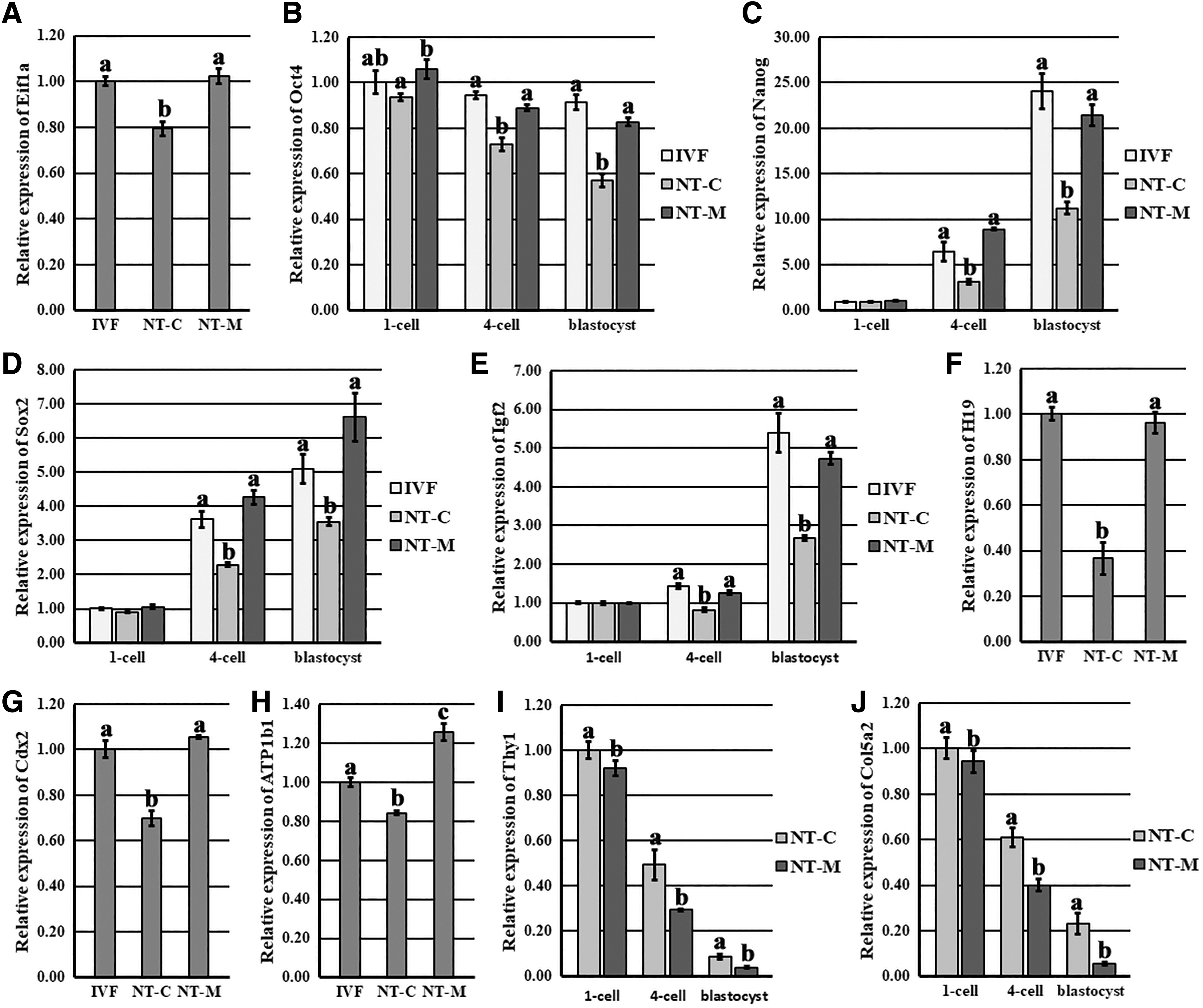
Relative expression levels of genes related to the development of early embryos.
When compared with the NT-C group, the NT-M group showed the significantly greater expression levels of Eif1a at the 4-cell stage, Oct4 at the 1-cell, 4-cell, and blastocyst stages; Nanog, Sox2, and Igf2 at the 4-cell and blastocyst stages; and H19, Cdx2, and ATP1b1 at the blastocyst stage (p < 0.05); moreover, the expression levels of Thy1 and Col5a2 at the 4-cell and blastocyst stages were significantly reduced (p < 0.05). These results displayed that melatonin ameliorated the expression patterns of genes related to cloned embryo development.
Discussion
It is known that epigenetic modification determines the developmental competence of cloned embryos, and environment factors can influence the epigenetic modification reconstruction (Huan et al., 2015; Meehan et al., 2018). In this study, our results demonstrated that melatonin ameliorated DNA methylation reprogramming; rescued the disrupted gene expression patterns, nuclear remodeling, and apoptosis; and resulted in the high developmental competence and quality of cloned embryos (Fig. 7).

Schematic representation of the enhanced development of cloned embryos induced by melatonin through ameliorating DNA methylation reprogramming. Melatonin ameliorated DNA methylation reprogramming, rescued the disrupted gene expression patterns, nuclear remodeling, and apoptosis, and then resulted in the high developmental competence and quality of cloned embryos.
During in vitro culture, embryos are exposed to the high ROS, resulting in the delay and arrest of embryo development (Wu et al., 2017). Melatonin has been shown to protect early embryos against ROS (Wang et al., 2014). Here, 10−7 M melatonin was proved to significantly enhance the development of cloned embryos. The mechanism could be attributed to the antioxidation and ROS reduction capacity of melatonin (Liang et al., 2017). And, the dose effect of melatonin was also shown in this study, in agreement with the previous reports though the appropriate concentration of melatonin is different in each research (Liang et al., 2017; Pang et al., 2013; Yang et al., 2019a). Thus, melatonin with the optimal concentration can enhance the development of cloned embryos.
After SCNT, cloned embryos undergo nuclear remodeling, and the significantly upregulated pseudo-pronucleus formation occurred in the NT-M group, suggesting that melatonin could enhance nuclear reprogramming and the subsequent development of cloned embryos (Huan et al., 2013). Apoptosis is also a criterion for evaluating embryo quality. Here, melatonin was shown to significantly reduce embryo apoptosis by improving the expression of antioxidant and apoptosis related genes, similar to the previous reports (Miao et al., 2018; Tian et al., 2017), further supporting that melatonin can improve the quality of cloned embryos.
Recently, several studies have demonstrated that melatonin participates in epigenetic modification, and most importantly, DNA methylation can be altered in response to melatonin exposure (Fang et al., 2018; Korkmaz and Reiter, 2008; Yang et al., 2019b). As the degree of DNA methylation reprogramming determines the developmental competence of cloned embryos, and the expression levels of genes required for early embryo development are correlated with DNA methylation statuses of their promoters (Calicchio et al., 2014; Huan et al., 2015), here, DNA methylation reprogramming and gene expression patterns were examined during the development of cloned embryos.
The results demonstrated that melatonin promoted genomic and gene-specific DNA methylation reprogramming in cloned embryos, and the improvement of DNA methylation reprogramming could be due to the adjusted expression of DNA methyltransferases and ten eleven translocation dioxygenases by melatonin, as melatonin did not significantly increase the blastocyst rate after Dnmt1 knockdown in cloned embryos (Supplementary Table S5). Then, melatonin significantly upregulated the transcription of zygotic activation gene, pluripotency related genes, imprinted genes, and blastocyst quality related genes, and reduced the expression of tissue-specific genes in cloned embryos, further supporting the theory that DNA methylation regulates gene expression. And, these rescued gene expression patterns resulted in the great development and quality of cloned embryos.
As how melatonin regulated DNA methylation reprogramming related genes, one possible mechanism may be that melatonin binds to the catalytic domain of DNA methylation reprogramming related enzymes and alters the activity of these enzymes, and another possible pathway could be that melatonin regulates the energy metabolism and further improves the expression of DNA methylation reprogramming related genes (Fang et al., 2018; Korkmaz and Reiter, 2008). Of course, the mechanism underlying that melatonin improved genomic and gene-specific DNA methylation reprogramming in cloned embryos and the regulatory network of gene expression are complex, further studies are needed (Canovas et al., 2017). Overall, melatonin enhances DNA methylation reprogramming and gene expression, benefiting the development of cloned embryos.
This study displayed that the mechanism underlying that melatonin enhanced the development and quality of cloned embryos was that melatonin enhanced DNA methylation reprogramming by regulating the expression of DNA methyltransferases and ten eleven translocation dioxygenases. As how melatonin takes the epigenetic regulatory role to regulate the expression of DNA methylation reprogramming related enzymes and further the DNA methylation statuses of apoptosis, blastocyst quality, and DNA methylation reprogramming related genes in cloned embryos requires extensive investigation (Korkmaz et al., 2012).
In conclusion, this study shows that melatonin ameliorates the development and quality of porcine cloned embryos, and the enhanced DNA methylation reprogramming could be the underlying mechanism by which melatonin exerts its beneficial effects on cloned embryos, and then, the rescued gene expression and reduced apoptosis result in the great developmental competence of cloned embryos.
Footnotes
Author Disclosure Statement
The authors declare they have no competing financial interests.
Funding Information
This work was supported by grants from the Key Research and Development Program of Shandong Province (2017NC210007), Entrepreneurship and Innovation Leading Talent Program of Qingdao (19-3-2-2-zhc), Science and Technology Support Program for Youth Innovation in Shandong Colleges and Universities (2019KJF005), Agriculture Variety Project of Shandong Province (2019LZGC011), National Natural Science Foundation of China (31602019), Innovation Program for Graduate Student of Qingdao Agricultural University (QYC201904), Innovation and Entrepreneurship Training Program for College Student of Qingdao Agricultural University (201910435018 and 113), Open Projects of Key Laboratory of Animal Cellular and Genetic Engineering of Heilongjiang Province (KF201704), High-level Talent Research Foundation of Qingdao Agricultural University (6631116029), Key Research and Development Program of Shandong Province (2018GNC110005 and 2017GSF221006), Foundation of Hebei Educational Committee (QN2016008), and National Natural Science Foundation of China (31672506).
References
Supplementary Material
Please find the following supplemental material available below.
For Open Access articles published under a Creative Commons License, all supplemental material carries the same license as the article it is associated with.
For non-Open Access articles published, all supplemental material carries a non-exclusive license, and permission requests for re-use of supplemental material or any part of supplemental material shall be sent directly to the copyright owner as specified in the copyright notice associated with the article.